

What is a sarcomere?
A sarcomere is the basic contractile unit of striated muscle: the stretch of a myofibril from one Z disc to the next. It contains two sets of overlapping protein filaments, thick filaments made of myosin and thin filaments made of actin, which slide past each other to shorten the sarcomere. Thousands of sarcomeres joined end to end make up each myofibril, and all of them shortening together is what makes a skeletal muscle contract.
A resting sarcomere is about 2 µm long in textbook descriptions; in living muscle, lengths of roughly 2 to 3 µm are typical, depending on the muscle, the species and how far the muscle is stretched. That is about one fortieth of the width of a human hair. The banded pattern of sarcomeres lined up across a muscle fibre is what gives skeletal and cardiac muscle their striped, or striated, look under the microscope.
| Feature | Typical value | Note |
|---|---|---|
| Sarcomere length at rest | ≈ 2.0–2.5 µm (frog), 2.4–2.9 µm optimum (human) | Shortens to about 2.0 µm or less during contraction |
| Thick filament (A band) | ≈ 1.6–1.65 µm | Nearly constant across vertebrate muscles |
| Thin filament | ≈ 0.95–1.3 µm (human 1.19–1.37 µm) | Varies between muscles and species |
| Bare zone | ≈ 0.17 µm | Mouse tibialis anterior |
| Z disc thickness | ≈ 0.1 µm | Mouse tibialis anterior |
| Spacing between thick filaments | ≈ 40 nm | From X-ray diffraction |
| Myofibril diameter | ≈ 1.2 µm | Hundreds to thousands per muscle fibre |
Values from OpenStax, Gokhin et al. (2012), Moo et al. (2016) and the MuscleX X-ray documentation; see the references.
Sarcomere diagram labeled
A labeled sarcomere diagram shows, from the outside in: a Z disc at each end, the light I band on either side of each Z disc, the dark A band in the middle, the paler H zone in the centre of the A band, and the M line running down the middle of the H zone. Thin filaments run from each Z disc toward the centre; thick filaments sit in the middle, held by the M line, with myosin heads sticking out everywhere except the bare zone.
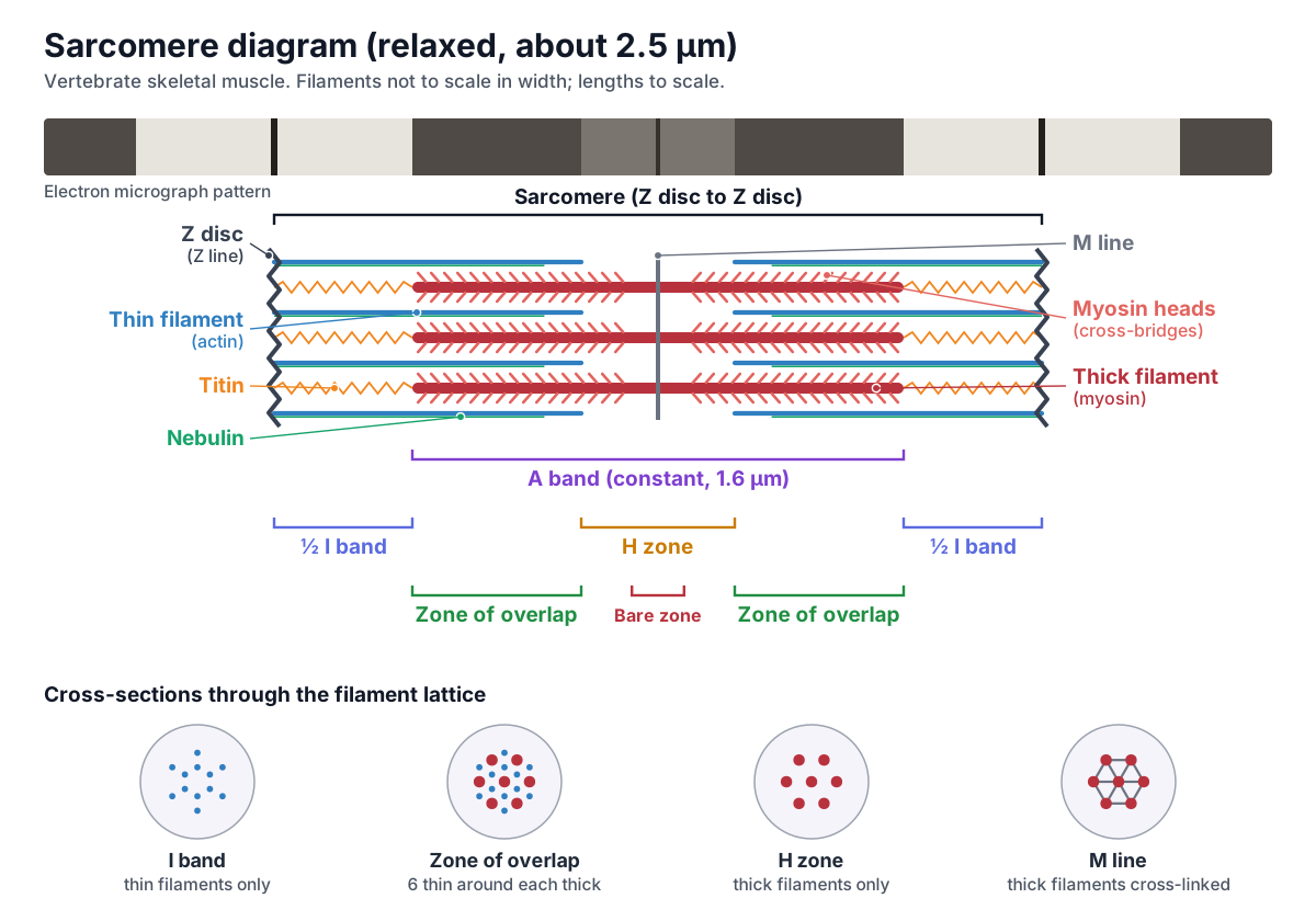
Read the diagram in pairs. The Z disc and M line are the two transverse anchors: the Z disc holds the thin filaments and the M line holds the thick filaments. The A band and I band are the two main stripes: the A band is where the thick filaments are and the I band is where only thin filaments are. Inside the A band, the H zone is where there are only thick filaments, and the two zones of overlap on either side of it are where thick and thin filaments interleave and cross-bridges can form.
Parts of a sarcomere and their functions
A sarcomere is built from six kinds of part: the thick filaments (myosin), the thin filaments (actin with tropomyosin and troponin), the Z discs, the M line, the giant elastic proteins titin and nebulin, and, around it, the membranes that control it, the sarcoplasmic reticulum and the T-tubules. This list is the text version of the 3D model above; click any name in the model's side panel to zoom to it.
| Part | Made of | Main job |
|---|---|---|
| Thick filament | ≈ 300 myosin II molecules, MyBP-C, titin | Motor: heads pull thin filaments toward the M line |
| Thin filament | Actin double helix, tropomyosin, troponin, nebulin | Track the heads pull on; calcium-controlled switch |
| Z disc | α-actinin, CapZ, ends of titin and nebulin | Anchors thin filaments; links sarcomeres end to end |
| M line | Myomesin, M-protein, obscurin, end of titin | Holds thick filaments in a hexagonal lattice |
| Titin | One giant protein, ≈ 3–3.7 MDa | Spring: centres thick filaments, gives passive tension |
| SR and T-tubules | Membrane tubes and sacs | Release and recapture Ca²⁺ to turn contraction on and off |
The figure of about 300 myosins per thick filament follows from the structure: three myosin molecules per 14.3 nm crown along about 1.4 µm of head-bearing filament.
Thick filaments
- Thick filament (myosin) (Myosin filament, myofilament)
- A bundle of several hundred myosin II molecules, about 1.6 µm long, that fills the A band. Its myosin heads grab the thin filaments and pull them toward the M line, which shortens the sarcomere.
- Myosin heads (cross-bridges) (Cross-bridges, myosin motor domains, S1)
- The motor ends of the myosin molecules. Each head has a site that binds actin and a site that binds and splits ATP. Bind to actin to form cross-bridges, then tilt (the power stroke) to slide the thin filament about 10 nm toward the M line. ATP releases them for the next cycle.
- Bare zone
- The head-free middle of the thick filament, about 0.15–0.2 µm long. Here the myosin tails overlap tail to tail and the filament reverses direction. With no heads, no cross-bridges can form here.
- Myosin-binding protein C (MyBP-C, C-protein)
- A thin protein that wraps the thick filament in a set of stripes, the C-zone. Stabilises the thick filament and tunes how fast myosin heads attach to actin.
Thin filaments
- Thin filament (actin) (Actin filament, F-actin)
- A double helix of globular actin (G-actin) subunits, with tropomyosin and troponin along it. About 1.0 µm long in frog muscle. Provides the binding sites that the myosin heads pull on. The thin filaments slide toward the M line during contraction.
- Tropomyosin
- A long, thin coiled-coil protein that lies in the two grooves of the actin helix. At rest, it covers the myosin-binding sites on actin. When Ca²⁺ binds troponin, tropomyosin rolls away from the sites so cross-bridges can form.
- Troponin complex (TnC, TnI, TnT)
- A three-part protein complex bound to tropomyosin about every 38.5 nm. The calcium switch. TnC binds Ca²⁺, TnI inhibits myosin binding, and TnT anchors the complex to tropomyosin. Ca²⁺ binding shifts tropomyosin and exposes the binding sites.
- Tropomodulin
- A capping protein on the free, pointed end of each thin filament. Controls thin filament length by limiting the addition and loss of actin subunits at the pointed end.
Z disc & M line
- Z disc (Z line, Z band)
- The dense protein plate that marks each end of a sarcomere. One sarcomere runs from one Z disc to the next. Anchors the thin filaments and the ends of titin, and passes force from one sarcomere to the next along the myofibril.
- α-actinin (Alpha-actinin)
- Rod-shaped cross-linking proteins that form the zig-zag lattice of the Z disc. Cross-links the actin filaments of neighbouring sarcomeres and binds the end of titin, holding the Z disc together.
- CapZ
- A capping protein that sits on the barbed (plus) end of each thin filament. Anchors the thin filament in the Z disc and stops actin subunits being added or lost at that end.
- M line (M band, Mittelscheibe)
- A band of cross-linking proteins in the exact middle of the sarcomere, inside the H zone. Holds the thick filaments in their hexagonal lattice and in register, and anchors the end of titin. Main proteins: myomesin, M-protein and obscurin.
Titin & nebulin
- Titin (Connectin)
- A giant elastic protein that runs from the Z disc to the M line, about 1 µm long. Acts as a molecular spring: it keeps the thick filament centred in the sarcomere and produces passive tension when a relaxed muscle is stretched.
- Nebulin
- A long, inextensible protein that lies along the thin filament. Stabilises and helps set the length of the thin filament. In human muscle it does not reach the tip, so it is not a strict 'ruler'.
Bands & zones
- Sarcomere
- The contractile unit of striated muscle: the segment of a myofibril from one Z disc to the next. Shortens when its thin filaments slide past its thick filaments. All the sarcomeres shortening together shorten the muscle.
- A band (Anisotropic band)
- The dark band: the full length of the thick filaments, about 1.6 µm. Marks where the thick filaments are. Its width stays the same during contraction.
- I band (Isotropic band)
- The light band, containing thin filaments only. Each I band is cut in half by a Z disc. Gets narrower as the sarcomere contracts and wider as it is stretched.
- H zone (H band, Hensen's zone)
- The lighter middle of the A band, where there are thick filaments but no thin filaments. Gets narrower during contraction and can disappear when the thin filament tips meet at the M line.
- Zone of overlap
- The parts of the A band where thick and thin filaments overlap, one on each side of the H zone. The only place where cross-bridges can form. The amount of overlap sets how much force the sarcomere can make (the length-tension relationship).
SR & T-tubules
- T-tubule (Transverse tubule)
- An inward tube of the muscle cell membrane (sarcolemma) that rings each myofibril. Carries the action potential deep into the fibre. Voltage sensors in its membrane (dihydropyridine receptors) trigger Ca²⁺ release from the SR.
- Terminal cisternae (Lateral sacs, junctional SR)
- The wide end sacs of the sarcoplasmic reticulum on either side of a T-tubule. Store Ca²⁺ and release it through ryanodine receptors (RyR1) when the T-tubule is depolarised.
- Sarcoplasmic reticulum (SR, longitudinal SR)
- A network of membrane tubes, a specialised smooth ER, wrapped around every myofibril. Stores, releases and retrieves Ca²⁺. Its ATP-driven pumps take Ca²⁺ back up after each contraction, which lets the muscle relax.
Sarcomere bands and zones: A band, I band, H zone, Z disc and M line
The bands of a sarcomere are named after how they look under the microscope. The A band is the dark band and the I band the light band. A stands for anisotropic and I for isotropic: the A band is birefringent, so it bends polarised light and shines brightly between crossed polarisers, while the I band is only weakly birefringent. The names go back to the 19th-century physiologist Ernst Brücke, who studied muscle in polarised light.
| Band or line | What it contains | During contraction |
|---|---|---|
| A band (anisotropic) | The full length of the thick filaments, plus the overlapping parts of thin filaments | Stays the same width |
| I band (isotropic) | Thin filaments only, with titin's spring segment; cut in half by a Z disc | Gets narrower |
| H zone | Thick filaments only (centre of the A band) | Gets narrower; can disappear |
| M line | Proteins cross-linking thick filaments, in the middle of the H zone | Stays in the centre |
| Z disc (Z line) | α-actinin lattice holding thin filaments and titin | Discs move closer together |
| Zone of overlap | Thick and thin filaments side by side | Gets wider (until the H zone is gone) |
A quick rule: the A band is a thing, the length of a thick filament, so it cannot change. The I band and H zone are gaps, the spaces where one kind of filament is missing, so they shrink as the filaments slide together. That is why I band width equals sarcomere length minus A band width.
The Z disc is named from the German Zwischenscheibe, "between disc", and the M line from Mittelscheibe, "middle disc". The H zone is usually said to come from hell, German for bright, or from the anatomist Victor Hensen.
Relaxed vs contracted sarcomere diagram
In a relaxed sarcomere the Z discs are far apart and the I band and H zone are wide. In a contracted sarcomere the thin filaments have slid toward the M line: the Z discs are closer, the I band is narrow, and the H zone has shrunk or vanished. The A band has the same width in both. Drag the slider below; it is linked to the 3D model above.
- Sarcomere
- 2.50 µm
- A band
- 1.60 µm
- I band
- 0.90 µm
- H zone
- 0.50 µm
- Active force
- 77% of max
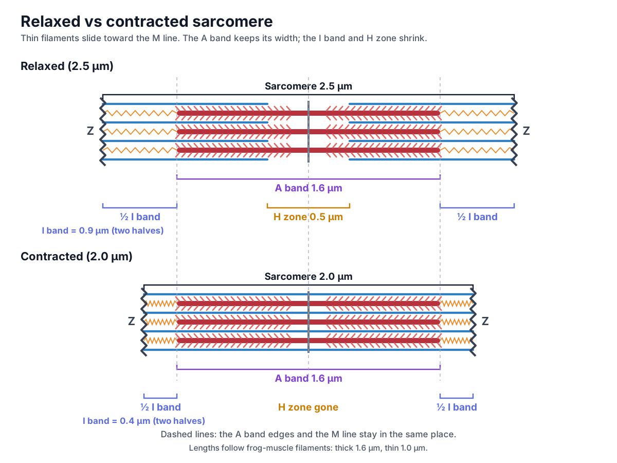
| Measure | Relaxed (2.5 µm) | Contracted (2.0 µm) | Change |
|---|---|---|---|
| Sarcomere (Z to Z) | 2.5 µm | 2.0 µm | Shorter |
| A band | 1.6 µm | 1.6 µm | No change |
| I band | 0.9 µm | 0.4 µm | Shorter |
| H zone | 0.5 µm | 0 µm | Shorter, then gone |
| Zone of overlap (each half) | 0.55 µm | 0.8 µm | Longer |
| Thick and thin filament length | 1.6 and 1.0 µm | 1.6 and 1.0 µm | No change |
Numbers are for the frog-muscle filament lengths used in the model. A sarcomere can shorten further than 2.0 µm: the thin filaments from the two halves then overlap each other in the middle, and below about 1.67 µm the thick filaments start to press against the Z discs. Force falls steeply in that range, as the length-tension section below shows.
Sliding filament theory
The sliding filament theory says that a muscle shortens because its thin filaments slide past its thick filaments, not because any filament gets shorter. Each myosin head binds actin, pulls, lets go and binds again further along, and millions of these cycles in all the sarcomeres in series shorten the whole muscle. The model was proposed in 1954 by two independent pairs of researchers, Andrew Huxley with Rolf Niedergerke and Hugh Huxley with Jean Hanson, who saw that the A band stayed the same width while the I band and H zone narrowed.
Because the two halves of each thick filament point in opposite directions, the heads in the left half pull their thin filaments to the right and the heads in the right half pull theirs to the left. Both sets move toward the M line, so both Z discs are drawn inward. The bare zone in the middle, where the myosin tails overlap tail to tail, is where the polarity reverses.
Sliding can only start once the myosin-binding sites on actin are uncovered, which requires Ca²⁺. Each power stroke moves a thin filament about 10 nm, so a sarcomere shortening by 0.5 µm needs many cycles of every head. For the electrical signal that starts it all, see the action potential diagram; for where the ATP comes from, see cellular respiration.
The cross-bridge cycle step by step
The cross-bridge cycle is the repeating sequence in which a myosin head binds actin, pulls it, releases it and re-cocks. It needs Ca²⁺ to start, because Ca²⁺ moves the troponin-tropomyosin complex off actin, and ATP to keep going, because ATP both releases the head and supplies the energy for the next stroke. Step through it here:
Resting: binding sites covered
Ca²⁺ in the sarcoplasm is low. Tropomyosin lies over the myosin-binding sites on actin, held there by troponin. The myosin head is already 'cocked': it has split an ATP and holds ADP and phosphate (Pi), storing the energy.
- Ca²⁺ binds troponin. An action potential travels down the T-tubules; the SR releases Ca²⁺, which binds troponin C. Troponin shifts tropomyosin away from the myosin-binding sites on actin.
- Cross-bridge formation. The myosin head, already energised and holding ADP and Pi, binds to an exposed site on actin.
- Power stroke. Pi is released, and the head pivots toward the M line, pulling the thin filament about 10 nm. ADP is released after the stroke.
- Detachment. A new ATP binds to the head, which then lets go of actin.
- Re-cocking. Myosin hydrolyses the ATP to ADP and Pi and uses the energy to swing the head back into its high-energy position. The cycle repeats as long as Ca²⁺ and ATP are present.
- Relaxation. When nerve signals stop, ATP-driven pumps return Ca²⁺ to the SR, tropomyosin covers the binding sites again and the cross-bridges stop forming.
| Molecule | Role in contraction |
|---|---|
| Ca²⁺ | On switch: binds troponin C, which moves tropomyosin and exposes the binding sites |
| Troponin | Calcium sensor bound to tropomyosin; TnC binds Ca²⁺, TnI inhibits, TnT anchors |
| Tropomyosin | Covers the myosin-binding sites on actin at rest |
| ATP | Binding releases myosin from actin; hydrolysis re-cocks the head; also powers the SR Ca²⁺ pump |
| ADP + Pi | Held by the cocked head; Pi release triggers the power stroke |
ATP for the cycle comes from three sources. Creatine phosphate regenerates ATP fastest but lasts only about 15 seconds of hard work; anaerobic glycolysis lasts about a minute and produces lactic acid; aerobic respiration in the mitochondria supplies most of the ATP at rest and during moderate activity. Without ATP, heads cannot let go of actin, which is why muscles stiffen in rigor mortis after death.
Length-tension relationship
The length-tension relationship says that a sarcomere produces the most active force at an intermediate length, where every myosin head can reach a thin filament, and less force when it is stretched or squashed. Force is set by filament overlap: only heads that face a thin filament can form cross-bridges.
The classic measurements were made in 1966 on single frog muscle fibres by Gordon, Huxley and Julian, who took care to keep sarcomere length uniform along the part of the fibre they recorded. They found a plateau of maximum force between sarcomere lengths of about 2.05 and 2.2 µm, a straight-line fall in force as the fibre was stretched beyond the plateau, and a sudden steeper drop below about 1.67 µm. The sliding filament theory explains almost the whole curve.
- Plateau (about 2.0–2.2 µm in frog): the thin filament tips lie over the bare zone, so every myosin head overlaps actin. Shortening across the bare zone adds no new heads, so force stays flat.
- Descending limb (longer than the plateau): as the sarcomere is stretched, overlap and the number of cross-bridges fall in proportion. With 1.6 µm thick and 1.0 µm thin filaments, force reaches zero at about 3.6 µm, where the thin filament tips just leave the ends of the thick filaments.
- Ascending limb (shorter than the plateau): the thin filaments cross the M line and overlap the opposite half, getting in each other's way; below about 1.67 µm the thick filaments butt against the Z discs and force drops sharply.
OpenStax describes the same idea in relative terms: the ideal length for maximal tension is about 80 to 120% of resting length. Human muscles have longer thin filaments, so their curves are shifted to longer lengths; see human vs frog sarcomere length below.
Myofibril and sarcomere: from muscle to filament
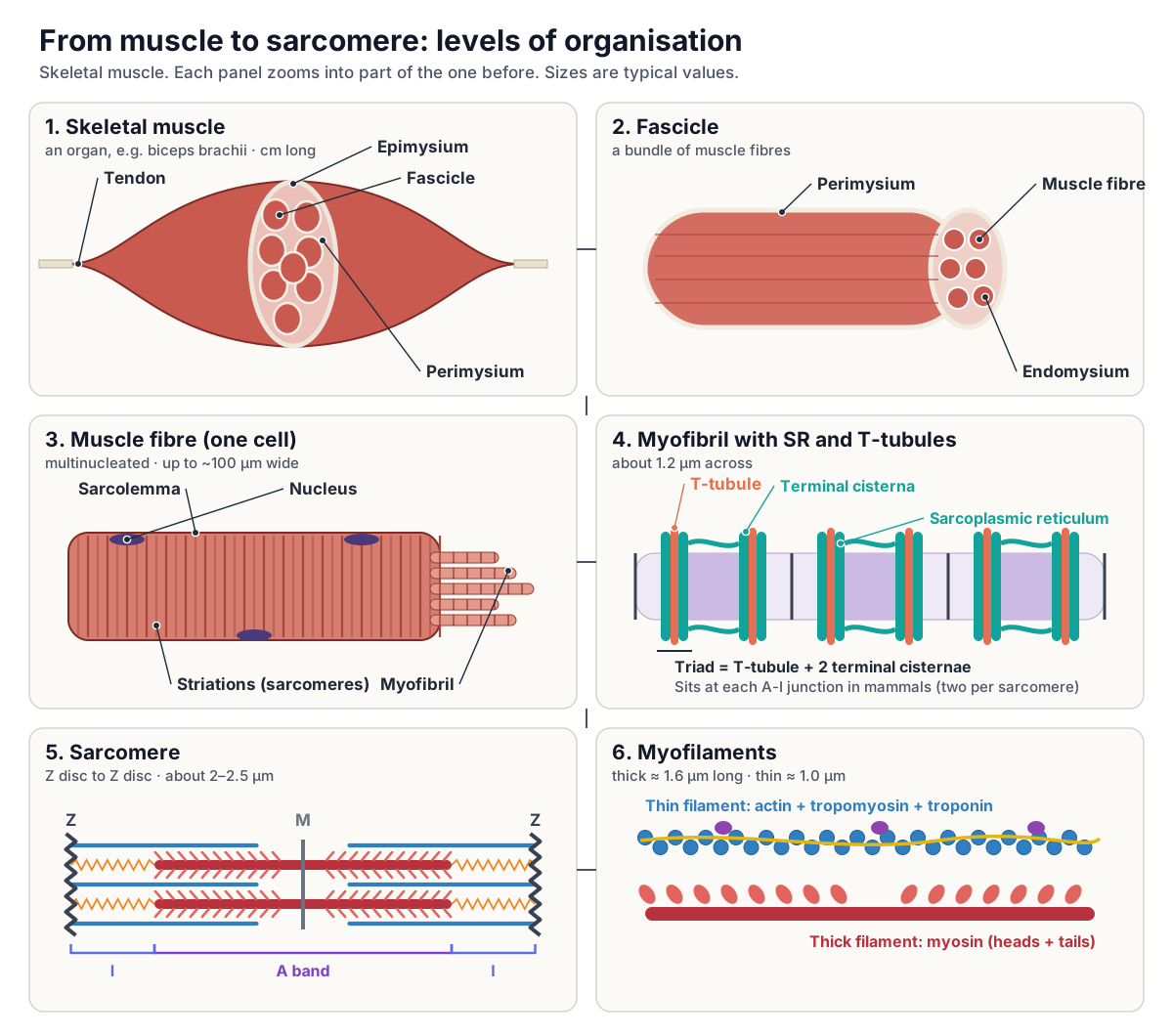
A skeletal muscle is organised like a cable made of smaller cables. The muscle is wrapped in epimysium and divided into bundles called fascicles, each wrapped in perimysium. Each fascicle holds many muscle fibres (muscle cells), each wrapped in endomysium. Each fibre is packed with myofibrils, and each myofibril is a chain of sarcomeres containing the thick and thin myofilaments.
- Skeletal muscle:
- An organ such as the biceps brachii, made of muscle tissue, connective tissue, blood vessels and nerves.
- Epimysium:
- Dense irregular connective tissue that wraps the whole muscle and separates it from neighbouring tissues.
- Fascicle:
- A bundle of muscle fibres inside the muscle.
- Perimysium:
- The connective tissue sheath around each fascicle.
- Muscle fibre (muscle cell, myofibre):
- One long, cylindrical, multinucleated cell, up to about 100 µm wide and up to 30 cm long, formed by the fusion of myoblasts.
- Endomysium:
- A thin layer of collagen and reticular fibres around each muscle fibre.
- Sarcolemma:
- The plasma membrane of the muscle fibre; it dips inward to form the T-tubules.
- Sarcoplasm:
- The cytoplasm of the muscle fibre, packed with myofibrils, mitochondria and glycogen.
- Myofibril:
- A long cylinder of sarcomeres joined end to end, about 1.2 µm across; hundreds to thousands fill each fibre.
- Triad:
- One T-tubule with a terminal cisterna of SR on each side: the site of excitation-contraction coupling.
- Sarcomere:
- The repeating contractile unit of the myofibril, Z disc to Z disc.
- Myofilaments:
- The thick (myosin) and thin (actin) filaments inside each sarcomere, plus titin and nebulin.
The three connective tissue layers are continuous with each other and with the tendon, so the pull of every sarcomere is passed through the fibre, the fascicle and the muscle to the bone. A muscle fibre forms when many embryonic myoblasts fuse, which is why it has many nuclei; a single fibre can be up to about 100 µm wide and, in the sartorius of the thigh, up to 30 cm long.
Around every myofibril lies the sarcoplasmic reticulum (SR), a specialised smooth endoplasmic reticulum that stores Ca²⁺. Its enlarged end sacs, the terminal cisternae, press against the T-tubules, inward extensions of the sarcolemma. One T-tubule with a terminal cisterna on each side forms a triad. In the 3D model, the triads are the orange rings (T-tubules) between teal sacs at the two A-I junctions.
Excitation-contraction coupling: how a nerve impulse switches the sarcomere on
A skeletal muscle fibre contracts only when its motor neuron tells it to. The neuron releases acetylcholine (ACh) at the neuromuscular junction, sodium ions flow in, and an action potential spreads across the sarcolemma and down the T-tubules into the middle of the fibre. That electrical signal makes the sarcoplasmic reticulum release Ca²⁺ into the sarcoplasm around the myofibrils. (OpenStax Anatomy & Physiology 2e, 10.3 Muscle Fiber Contraction and Relaxation.)
The link between the T-tubule and the SR is a pair of proteins. The dihydropyridine receptor (DHPR) is a voltage-gated channel in the T-tubule membrane; the ryanodine receptor (RyR1) is the Ca²⁺ release channel on the facing membrane of the terminal cisterna, seen as 'feet' in electron micrographs. In skeletal muscle the two are physically coupled, so the signal passes in about 2 ms; in cardiac muscle the coupling works through Ca²⁺ entering the cell and takes about 100 ms. (Al-Qusairi & Laporte (2011): T-tubule biogenesis and triad formation in skeletal muscle, Skeletal Muscle.)
Where the triads sit along the sarcomere depends on the animal. In mammalian skeletal muscle the SR junctions form at the A-I junctions, giving a row of triads on each side of every Z line, so two triads per sarcomere. T-tubule networks in other muscles cross the fibre at the level of the Z lines instead. (Lavorato, Iyer & Franzini-Armstrong (2019): non-junctional transverse tubules in skeletal muscle, European Journal of Translational Myology.)
Relaxation is the reverse. When the motor neuron stops firing, the SR channels close and ATP-driven pumps move Ca²⁺ back into the SR. Troponin loses its Ca²⁺, tropomyosin slides back over the binding sites, cross-bridges stop forming and the fibre relaxes. (OpenStax Anatomy & Physiology 2e, 10.3 Muscle Fiber Contraction and Relaxation.)
Titin and nebulin: the sarcomere's spring and its thin-filament guide
Titin is the largest known protein, at about 3 to 3.7 million daltons. A single molecule reaches about 1 µm, from the Z disc (its NH2 end) to the M line (its COOH end), so titin molecules from both halves overlap in every Z disc and every M line. Titin filaments are elastic and attach the thick filaments to the Z discs. (Sanger & Sanger (2001): fishing out proteins that bind to titin, Journal of Cell Biology.)
Along the thick filament, titin lies close to the backbone. Three-dimensional reconstructions of vertebrate cardiac thick filaments suggest about six titin molecules per half filament, running from the centre of the filament to the Z line. (Zoghbi et al. (2008): three-dimensional structure of vertebrate cardiac muscle myosin filaments, PNAS.)
Nebulin is the thin filament's partner in skeletal muscle. The skeletal isoform is about 800 kDa. Its COOH end is anchored in the Z disc and a central region of more than 200 repeats runs along the actin filament; two nebulin molecules lie along each thin filament. (Sanger & Sanger (2001): fishing out proteins that bind to titin, Journal of Cell Biology.)
Nebulin was long called a molecular ruler that sets thin filament length. Measurements of human deltoid and pectoralis muscle found thin filaments 1.19 to 1.37 µm long, longer than nebulin can reach. The nebulin-free end of the filament, regulated by the capping protein tropomodulin, made up as much as a third of its length, so in human muscle nebulin cannot be a strict ruler. (Gokhin et al. (2012): thin-filament length correlates with fiber type in human skeletal muscle, American Journal of Physiology–Cell Physiology.)
Sarcomere length in human muscle compared with frog muscle
Thick filaments are almost the same length, about 1.65 µm, in every vertebrate skeletal muscle studied. Thin filaments are not: published values range from about 0.95 to 1.30 µm, and human thin filaments in the 2012 study ranged from 1.19 to 1.37 µm. Because force peaks when the thin filaments reach the edge of the bare zone, longer thin filaments move the peak to longer sarcomere lengths: the same study predicted optimum human sarcomere lengths of about 2.4 to 2.9 µm. (Gokhin et al. (2012): thin-filament length correlates with fiber type in human skeletal muscle, American Journal of Physiology–Cell Physiology.)
In mouse tibialis anterior, with 1.1 µm thin filaments, 1.6 µm thick filaments, 0.1 µm Z discs and a 0.17 µm bare zone, the calculated optimal range is 2.30 to 2.47 µm. Sarcomere lengths measured in living mouse muscle were not uniform: neighbouring sarcomeres in the same muscle can sit at different lengths and stretch by different amounts. (Moo et al. (2016): in vivo sarcomere lengths and sarcomere elongations are not uniform across an intact muscle, Frontiers in Physiology.)
The 3D model and diagrams on this page use the classic frog values (thick 1.6 µm, thin about 1.0 µm) because the textbook length-tension curve was measured in single frog fibres. Human thin filaments are 0.2 to 0.4 µm longer, so optimal human sarcomere lengths are roughly 0.4 to 0.7 µm longer than the frog values quoted here. (Gordon, Huxley & Julian (1966): the variation in isometric tension with sarcomere length in vertebrate muscle fibres, Journal of Physiology.)
Actin and myosin in the hexagonal filament lattice
In cross-section, the filaments of a vertebrate skeletal sarcomere form a precise hexagonal lattice. Thick filaments sit at the corners of a hexagonal grid, about 40 nm apart, and each thin filament sits at a trigonal point, equidistant between three thick filaments. Each thick filament therefore faces six thin filaments, and there are about twice as many thin filaments as thick ones.
A cross-section looks different depending on where it cuts. Through the I band you see only thin filaments; through the H zone only thick filaments; through the zone of overlap each thick filament is ringed by six thin ones; and at the M line the thick filaments are joined by bridges of M-line protein. The M-band proteins hold the thick filaments in their lattice and also set how each one is rotated relative to its neighbours. In the 3D model, rotate to look straight down the long axis to see the hexagonal pattern.
The myosin heads are not scattered at random. They form rings called crowns, three myosin molecules per crown (each with two heads), with one crown every 14.3 nm and the whole pattern repeating every 43 nm along the filament. On the thin filament, each tropomyosin molecule spans seven actin subunits and carries one troponin complex, and the troponin complexes are spaced 38.5 nm apart.
Sarcomere under the electron microscope
In a transmission electron micrograph of longitudinally cut skeletal muscle, a sarcomere appears as a repeating pattern of stripes: a dark A band in the middle, a slightly paler H zone in the centre of the A band with a thin dark M line through it, and a light I band on each side, cut by a dense, dark Z line. The darkest parts of the A band are the zones of overlap, where both kinds of filament are packed together.
Under a polarising microscope the contrast reverses: the birefringent A band becomes bright and the I band appears dark. The Z line, though it lies in the middle of the isotropic I band, is itself birefringent. In micrographs you can also see mitochondria between the myofibrils and the triads, where dense "feet", the ryanodine receptors, bridge the gap between T-tubule and SR.
To judge contraction from a micrograph, measure from one Z line to the next and compare the widths of the I band and H zone with the A band. If the A band is the same width in two images but the I band and H zone are narrower in one, that one is the more contracted.
Skeletal vs cardiac vs smooth muscle: which have sarcomeres?
Skeletal and cardiac muscle both have sarcomeres, so both are striated. Smooth muscle has actin and myosin but no sarcomeres and no striations: its thin filaments are anchored to dense bodies instead of Z discs, and its contraction is switched on by calmodulin and myosin light-chain kinase instead of troponin. For how cardiac muscle drives the heart, see blood flow through the heart.
| Feature | Skeletal muscle | Cardiac muscle | Smooth muscle |
|---|---|---|---|
| Sarcomeres and striations | Yes | Yes, same banding pattern | No |
| Cell shape | Long cylinders, up to 30 cm | Short, branched | Spindle-shaped, ≈ 30–200 µm |
| Nuclei | Many, at the edge | Usually one, central | One, central |
| Anchors for thin filaments | Z discs | Z discs | Dense bodies |
| Ca²⁺ switch | Troponin | Troponin | Calmodulin and MLCK (no troponin) |
| Main Ca²⁺ source | SR | Much from outside the cell, plus SR | Mostly outside the cell |
| T-tubules | Yes, triads | Yes | No |
| Cell junctions | None between fibres | Intercalated discs (gap junctions, desmosomes) | Gap junctions in single-unit muscle |
| Control | Voluntary | Involuntary, autorhythmic | Involuntary |
Label the sarcomere: unlabeled diagram and quiz
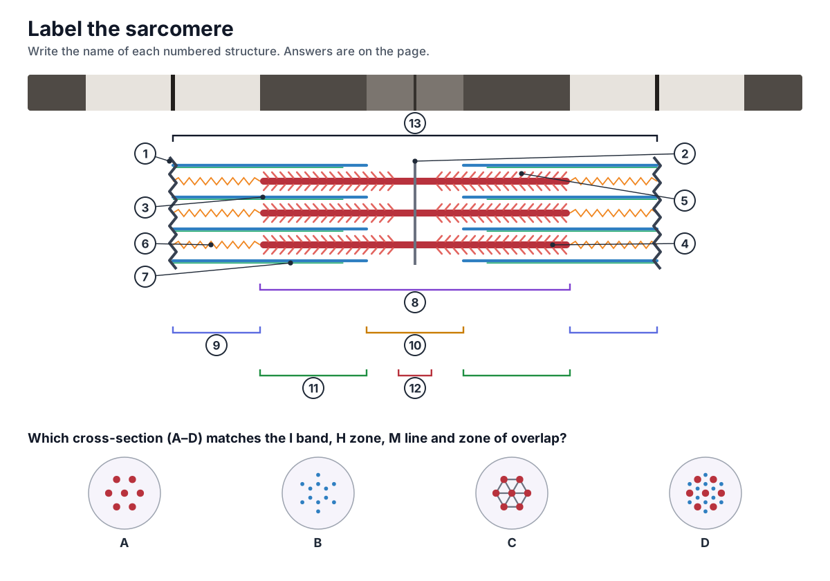
Test yourself with this sarcomere diagram unlabeled. Name each numbered structure, then match each cross-section (A–D) to the part of the sarcomere it cuts through: the I band, the H zone, the M line or the zone of overlap.
Show the answer key
- Z disc (Z line)
- M line
- Thin filament (actin)
- Thick filament (myosin)
- Myosin heads (cross-bridges)
- Titin
- Nebulin
- A band
- I band (half of it; the other half is in the next sarcomere)
- H zone
- Zone of overlap
- Bare zone
- Sarcomere (Z disc to Z disc)
Cross-sections: A = H zone (thick filaments only), B = I band (thin filaments only), C = M line (thick filaments cross-linked), D = zone of overlap (six thin filaments around each thick filament).
Practice questions (MCAT and A-level style)
Below is a diagram of a muscle sarcomere. During contraction, which region keeps the same width: the A band, the I band or the H zone?
Answer
The A band. Its width is the length of the thick filaments, which do not shorten. The I band and the H zone both get narrower.
A sarcomere shortens from 2.5 µm to 2.2 µm. The thick filaments are 1.6 µm long. What happens to the I band?
Answer
I band = sarcomere length − A band. It shrinks from 0.9 µm to 0.6 µm. The A band stays 1.6 µm.
Which structure would you find in the H zone but not in the I band?
Answer
Thick (myosin) filaments, and in the middle the M line. The H zone has thick filaments only; the I band has thin filaments only.
A drug blocks the Ca²⁺ binding site of troponin C. What happens to contraction?
Answer
Contraction is blocked. Without Ca²⁺ on troponin, tropomyosin stays over the myosin-binding sites on actin, so no cross-bridges can form even if ATP is plentiful.
Why do muscles become stiff a few hours after death (rigor mortis)?
Answer
ATP runs out. Myosin heads stay bound to actin after the power stroke because only a new ATP can release them.
A cross-section cuts through six thin filaments around each thick filament. Which part of the sarcomere was cut?
Answer
The zone of overlap, the outer parts of the A band where thick and thin filaments overlap.
Frequently asked questions
What is a sarcomere?
A sarcomere is the basic contractile unit of skeletal and cardiac muscle: the segment of a myofibril between two Z discs. It contains overlapping thick filaments (myosin) and thin filaments (actin, tropomyosin and troponin), held in place by titin, nebulin, the Z discs and the M line. A resting sarcomere is about 2–2.5 µm long.
What happens to the sarcomere during contraction?
The thin filaments slide past the thick filaments toward the M line, pulling the Z discs closer together. The sarcomere, the I band and the H zone get shorter. The A band stays the same width, because the thick filaments themselves do not shorten.
Which bands shorten during muscle contraction?
The I band and the H zone shorten, and the whole sarcomere (Z disc to Z disc) shortens. The A band does not change, because its width is the length of the thick filaments. The zone of overlap gets wider.
What do A band and I band stand for?
A stands for anisotropic and I for isotropic. The A band is birefringent: it changes polarised light and looks bright between crossed polarisers. The I band is only weakly birefringent. In ordinary light and electron micrographs the A band is dark and the I band light.
What is the difference between a Z disc and an M line?
The Z disc marks the two ends of a sarcomere and anchors the thin filaments and titin. The M line is in the middle of the sarcomere and cross-links the thick filaments. The Z discs move toward each other during contraction; the M line stays in the centre.
What is the role of calcium in muscle contraction?
Ca²⁺ is the on switch. An action potential travels down the T-tubules and makes the sarcoplasmic reticulum release Ca²⁺. Ca²⁺ binds troponin C, which moves tropomyosin off the myosin-binding sites on actin, so cross-bridges can form. When Ca²⁺ is pumped back into the SR, tropomyosin covers the sites again and the muscle relaxes.
What is the role of ATP in the cross-bridge cycle?
ATP does two jobs. Binding of ATP makes the myosin head let go of actin. Splitting ATP into ADP and phosphate then re-cocks the head into its high-energy position, ready for the next power stroke. ATP also powers the SR pumps that remove Ca²⁺ during relaxation. Without ATP, heads stay stuck to actin, which causes rigor mortis.
Does smooth muscle have sarcomeres?
No. Smooth muscle has actin and myosin but no sarcomeres, so it has no striations. Its thin filaments are anchored to dense bodies, which do the job of Z discs, and contraction is switched on by calmodulin and myosin light-chain kinase instead of troponin.
Why does a sarcomere produce less force when it is stretched or very short?
Force depends on how many myosin heads can reach actin. When the sarcomere is stretched, thick and thin filaments overlap less, so fewer cross-bridges form. When it is very short, thin filaments overlap each other and thick filaments press on the Z discs, which also lowers force. Force is greatest at about 2.0–2.2 µm in frog muscle.
References
- OpenStax (2022). Anatomy and Physiology 2e, section 10.2: Skeletal Muscle. Rice University.
- OpenStax (2022). Anatomy and Physiology 2e, section 10.3: Muscle Fiber Contraction and Relaxation. Rice University.
- OpenStax (2022). Anatomy and Physiology 2e, section 10.4: Nervous System Control of Muscle Tension. Rice University.
- OpenStax (2022). Anatomy and Physiology 2e, section 10.7: Cardiac Muscle Tissue. Rice University.
- OpenStax (2022). Anatomy and Physiology 2e, section 10.8: Smooth Muscle. Rice University.
- Gordon, A. M., Huxley, A. F. & Julian, F. J. (1966). The variation in isometric tension with sarcomere length in vertebrate muscle fibres. Journal of Physiology 184, 170–192.
- Gokhin, D. S. et al. (2012). Thin-filament length correlates with fiber type in human skeletal muscle. American Journal of Physiology – Cell Physiology 302, C555–C565.
- Moo, E. K., Fortuna, R., Sibole, S. C., Abusara, Z. & Herzog, W. (2016). In vivo sarcomere lengths and sarcomere elongations are not uniform across an intact muscle. Frontiers in Physiology 7, 187.
- Sanger, J. W. & Sanger, J. M. (2001). Fishing out proteins that bind to titin. Journal of Cell Biology 154, 21–24.
- Zoghbi, M. E., Woodhead, J. L., Moss, R. L. & Craig, R. (2008). Three-dimensional structure of vertebrate cardiac muscle myosin filaments. PNAS 105, 2386–2390.
- Paul, D. M., Squire, J. M. & Morris, E. P. (2010). A novel approach to the structural analysis of partially decorated actin based filaments. Journal of Structural Biology 170, 278–285.
- Lange, S. et al. (2020). Review of the sarcomeric M-band. Biochimica et Biophysica Acta – Molecular Cell Research.
- Al-Qusairi, L. & Laporte, J. (2011). T-tubule biogenesis and triad formation in skeletal muscle and implication in human diseases. Skeletal Muscle 1, 26.
- Lavorato, M., Iyer, V. R. & Franzini-Armstrong, C. (2019). A proposed role for non-junctional transverse tubules in skeletal muscle. European Journal of Translational Myology 29, 8264.
- Anatomy Atlases. Atlas of Microscopic Anatomy, Plate 5.65: Striated muscle, polarization microscopy.
- Tune, T. & Sponberg, S. (2024). Nanometer scale difference in myofilament lattice structure of muscle alter muscle function in a spatially explicit model. arXiv:2405.19443.
More interactive anatomy models
- Action potential diagramDepolarization, repolarization and ion channels of a neuron action potential.
- Skin layersEpidermis, dermis and hypodermis with hair follicles, glands and receptors, in 3D.
- Nephron anatomyGlomerulus, Bowman's capsule, tubules, loop of Henle and collecting duct, in 3D.
- Heart blood flowStep-by-step path of blood through the heart's chambers and valves, in 3D.
- Human anatomy1,000+ bones, muscles, organs, vessels and nerves from real anatomical data.
- Octopus anatomyThree hearts, a ring-shaped brain, eight arms and a beak, in 3D.