

Amoeba diagram: the essentials
An amoeba diagram shows a single, shapeless cell with a thin plasma membrane, finger-like pseudopodia, clear outer ectoplasm and granular inner endoplasm, one nucleus, a contractile vacuole and several food vacuoles. Those seven parts are what Class 7 to Class 10 and KS3 diagrams ask you to label. More detailed diagrams of Amoeba proteus add the hyaline cap, uroid, plasmagel and plasmasol, crystals and fat droplets.
This page uses Amoeba proteus as the reference organism. It is a large freshwater amoeba, 220–760 µm long when moving, with a single disc-shaped nucleus 22–62 µm across. It usually has several blunt pseudopodia with one dominant one, and a knobbly rear end called the uroid. (Siemensma, F. Microworld: Amoeba proteus (species description).)
Amoeba is unicellular: one cell carries out every life process. It feeds by engulfing prey (holozoic nutrition), takes in oxygen and loses carbon dioxide and ammonia by diffusion across its surface, pumps out excess water with its contractile vacuole, and reproduces by splitting in two (binary fission).
What is an amoeba? Protozoa, unicellular and eukaryotic
An amoeba is a single-celled (unicellular) eukaryote that has no fixed shape and moves and feeds with temporary bulges of its cell called pseudopodia. Older books call it a protozoan, an animal-like protist. Amoeba is also a genus name: Amoeba proteus is its type species and the amoeba of school diagrams.
Amoeba proteus belongs to the Amoebozoa, the large group of protists that move by amoeboid movement using actin-based pseudopodia. Within it, the lobose amoebae, with blunt, lobe-like or tube-like pseudopodia, include naked forms such as Amoeba and shelled forms (testate amoebae). (OpenStax Biology 2e, 23.3 Groups of protists.)
'Amoeba' is also used loosely for any cell that crawls in this way. Many unrelated protists are amoeboid, and so are human white blood cells. Some organisms called amoebae, such as Naegleria, sit in a completely different branch of the eukaryote tree from Amoeba proteus. (OpenStax Microbiology, 5.1 Unicellular eukaryotic parasites.)
Amoeba proteus lives in fresh water: the bottom of ponds, ditches and slow streams, among decaying leaves and on water plants, and in wet Sphagnum moss. It is common but not abundant. A moving cell is 220–760 µm long, with a mean of about 425 µm, which makes it one of the largest single cells you can study in a school laboratory. (Siemensma, F. Microworld: Amoeba proteus (species description).)
Labeled diagram of an amoeba
A labeled diagram of an amoeba shows, from the outside in: the plasma membrane, the clear ectoplasm, the gel-like plasmagel and the flowing plasmasol. Floating in the plasmasol are the nucleus, the contractile vacuole, food vacuoles at different stages of digestion, crystals and fat droplets. Around the edge are the advancing and lateral pseudopodia, the hyaline cap and the uroid.
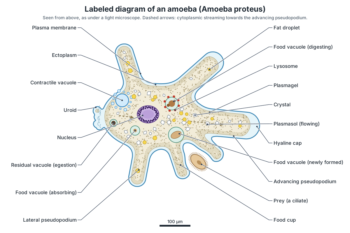
In the 3D model, switch on All labels to name every part at once, or click any part to zoom to it and read what it does. The 2D diagram and the model are drawn from the same outline, so you can compare them directly.
Parts of an amoeba and their functions
Each part of an amoeba does one main job. The table lists the parts that appear on school and first-year university diagrams and what each one does.
| Part | Function |
|---|---|
| Plasma membrane | Encloses the cell; controls entry and exit; gases and ammonia diffuse across it; folds in to engulf food |
| Pseudopodia | Temporary 'false feet' for movement and for catching food |
| Hyaline cap | Clear tip of an advancing pseudopodium; marks where the cell is growing forward |
| Uroid | Rear end; its cortex contracts to push cytoplasm forward; waste is often left here |
| Ectoplasm | Clear, firm outer layer of cytoplasm |
| Plasmagel | Gel-like outer endoplasm; its actin–myosin network contracts to drive streaming |
| Plasmasol | Fluid inner endoplasm that streams forward and carries the organelles |
| Nucleus | Controls the cell's activities, growth and division |
| Contractile vacuole | Collects and expels excess water (osmoregulation) |
| Food vacuole | Holds swallowed food while it is digested and absorbed |
| Lysosomes | Supply digestive enzymes to food vacuoles |
| Mitochondria | Aerobic respiration: release energy as ATP |
| Golgi bodies and ER | Make, modify and package proteins, lipids and membranes |
| Crystals | Probably stored nitrogenous waste (mainly triuret) |
| Fat droplets | Energy store |
Amoeba cell structure: every part in the 3D model
This is the text version of the 3D model: every labeled structure of Amoeba proteus, grouped by region, with its location and function. Use it as a revision list or to check an amoeba labeling worksheet.
Membrane & pseudopodia
- Plasma membrane (Plasmalemma, cell membrane)
- The thin, flexible outer boundary of the amoeba. There is no cell wall, so the cell can change shape. Separates the cell from the pond water and controls what goes in and out. Oxygen, carbon dioxide and ammonia cross it by diffusion; it folds inward to take in food (phagocytosis) and droplets of fluid (pinocytosis).
- Advancing pseudopodium (Dominant pseudopod, lobopodium)
- The largest, leading pseudopodium ('false foot'), a broad, blunt, finger-like lobe pointing the way the cell is moving. Carries the cell forward. Cytoplasm streams into it, so the cell flows in that direction.
- Lateral pseudopodia (Side pseudopods)
- Smaller, blunt pseudopodia on the sides of the cell, each either extending or being pulled back in. Let the amoeba explore and change direction. A new one becomes dominant when the cell turns; old ones are withdrawn into the body.
- Hyaline cap (Hyaline tip, cap of ectoplasm)
- A clear, granule-free zone at the tip of an advancing pseudopodium, where the ectoplasm is thicker than elsewhere. Marks where the pseudopodium is growing. Just behind it the forward-flowing plasmasol turns outward and sets into gel (the fountain zone), building the wall of the pseudopodium.
- Uroid (Tail, posterior end)
- The rear end of a moving amoeba, often bunched into knobs and wrinkles like a mulberry. The rear cortex contracts here and squeezes the cytoplasm forward. Undigested food is often expelled near the uroid.
Ectoplasm & endoplasm
- Ectoplasm (Hyaline layer, hyaloplasm)
- The thin, clear, firm outer layer of cytoplasm just inside the membrane. Gives the cell a firm outer layer and keeps the granular endoplasm off the membrane.
- Plasmagel (Outer endoplasm, gel cortex)
- The outer, stiff, jelly-like part of the granular endoplasm, forming a tube around the flowing plasmasol. Its actin–myosin network contracts, especially at the rear, squeezing the plasmasol forward. At the front new gel forms from sol; at the back gel turns back into sol.
- Plasmasol (Inner endoplasm, fluid endoplasm)
- The inner, fluid, granular cytoplasm that streams forward into the advancing pseudopodia. Carries the nucleus, food vacuoles and organelles along as the cell moves (cytoplasmic streaming), and is where most of the cell's chemistry takes place.
Nucleus
- Nucleus (Discoid nucleus)
- A single, large, disc-shaped nucleus, often slightly hollowed on both faces (biconcave), that holds the cell's DNA. Controls the cell's activities, growth and division. If an amoeba is cut in two, only the half with the nucleus can go on feeding, growing and dividing.
- Nucleolar material (Peripheral nucleoli)
- Many small nucleoli lying just inside the nuclear envelope, around the edge of the nucleus. Makes ribosomal RNA and assembles ribosome subunits, which the cell needs to make proteins.
Feeding & food vacuoles
- Food cup (Phagocytic cup, feeding pseudopodia)
- Two or more pseudopodia that flow around a prey organism, forming a cup that closes over it. Surrounds the prey, often without touching it at first; then the tips meet and fuse, enclosing the prey and a drop of water in a new food vacuole (phagocytosis, ingestion).
- Prey organism (Food: e.g. a ciliate such as Tetrahymena)
- A small ciliate being caught. A. proteus eats other protozoa, algae, rotifers and smaller amoebae. Food. Amoeba is holozoic: it eats whole food organisms and digests them inside the cell.
- Food vacuole (newly formed) (Phagosome)
- A membrane-bound bubble holding a freshly swallowed prey organism in a drop of water. Keeps the live prey and the digestive enzymes that will soon arrive separate from the rest of the cytoplasm.
- Food vacuole (digesting) (Digestive vacuole, phagolysosome)
- A food vacuole that has fused with lysosomes. Its contents turn acidic and the prey is broken down. Digests proteins, fats and carbohydrates of the prey into small molecules such as amino acids, sugars and fatty acids.
- Food vacuole (absorbing) (Late food vacuole)
- A shrinking vacuole whose digested food is passing into the cytoplasm. Digested food diffuses or is carried across the vacuole membrane into the cytoplasm (absorption) and is used for energy, growth and repair (assimilation).
- Residual vacuole (egestion) (Egestion vacuole, residual body)
- A small vacuole holding the indigestible remains of the prey, about to be expelled. Moves to the surface and fuses with the plasma membrane, throwing the waste out of the cell (egestion, by exocytosis).
- Lysosomes (Primary lysosomes, enzyme vesicles)
- Small membrane sacs of digestive enzymes that fuse with food vacuoles. Deliver hydrolytic (digestive) enzymes into food vacuoles; also break down worn-out parts of the cell.
- Pinocytic vesicles (Pinosomes)
- Tiny vesicles of fluid pinched in from the surface when the amoeba 'drinks' dissolved food. Take in dissolved nutrients, such as proteins, with a little of the surrounding water (pinocytosis).
Contractile vacuole
- Contractile vacuole (CV, water expulsion vesicle)
- A clear, round vacuole that slowly fills with excess water and then suddenly empties it outside the cell. Osmoregulation. Pond water is more dilute than the cytoplasm, so water keeps entering by osmosis; the contractile vacuole pumps it out so the cell does not swell and burst.
- Contractile vacuole vesicles (Feeder vesicles)
- Small vesicles around the contractile vacuole that collect water and fuse with it. Gather water from the cytoplasm and feed it into the main vacuole as it fills.
Organelles & inclusions
- Mitochondria (Singular: mitochondrion)
- Small rod-shaped organelles that carry out aerobic respiration. Release energy from digested food by aerobic respiration, making ATP, the cell's energy currency. They use oxygen that diffuses in from the water.
- Golgi bodies (Golgi apparatus, dictyosomes)
- Small stacks of flattened, curved membrane sacs. Modify and sort proteins and lipids made in the ER and package them into vesicles, including lysosomes and membrane for vacuoles.
- Endoplasmic reticulum (ER: rough and smooth)
- A network of membrane tubes and sheets running through the cytoplasm. Rough ER (with ribosomes) makes proteins for membranes and secretion; smooth ER makes lipids.
- Crystals (Bipyramidal crystals, triuret crystals)
- Tiny, glassy crystals shaped like truncated double pyramids or flat plates, scattered through the endoplasm. Thought to be a store of nitrogenous waste. Chemically they are mostly triuret (carbonyl diurea).
- Fat (lipid) droplets (Oil globules, lipid bodies)
- Small, round, shiny droplets of stored fat. Store energy-rich fat made from digested food, used when food is scarce.
Ectoplasm and endoplasm: plasmagel and plasmasol
The cytoplasm of an amoeba has two layers: a thin, clear, firm ectoplasm just under the membrane, and a thick, granular endoplasm inside it. The endoplasm itself has a stiff, jelly-like outer part, the plasmagel, and a fluid inner part, the plasmasol, which flows. Most school diagrams label only ectoplasm and endoplasm.
These are not separate compartments with walls between them. They are the same cytoplasm in two physical states. Measurements of particles inside moving A. proteus show an outer gel layer, the cortex, that behaves like a network of semi-flexible actin filaments, and an inner endoplasm that flows like a liquid down a pipe, fastest in the middle. Particles in the stream move at up to a few micrometres per second. (Rogers, Waigh & Lu (2008): Intracellular microrheology of motile Amoeba proteus, Biophysical Journal.)
Books do not all draw the line in the same place. Some call the whole outer gel layer (hyaline layer plus plasmagel) the ectoplasm and the flowing core the endoplasm; others, as here, keep ectoplasm for the clear hyaline layer and divide the granular endoplasm into plasmagel and plasmasol. Both describe the same cell. If your syllabus uses one version, follow it.
Amoeba pseudopodia and amoeboid movement
An amoeba moves by amoeboid movement: it pushes out a pseudopodium and its cytoplasm flows into it, so the whole cell creeps forward along the surface. The pseudopodia of A. proteus are lobopodia: broad, blunt, finger-like lobes, quite unlike the thin, needle-like pseudopodia of some other amoebae. Step through the diagram to see the cycle.
1. A pseudopodium pushes out
At the front, the plasma membrane bulges forward into a blunt, finger-like pseudopodium. A clear hyaline cap forms at its tip, where granules are kept out. Actin filaments assemble under the new membrane.
What drives it? The engine is the cell’s actin–myosin cytoskeleton, the same system that drives muscle contraction. Two classic explanations were proposed for Amoeba proteus. In the first, the cortex at the rear contracts, using ATP, and pushes the fluid plasmasol forward, rather like squeezing a tube of toothpaste. In the second, the “frontal contraction” model of R. D. Allen (1961), contraction at the front, where sol turns into gel, pulls the cytoplasm forward. Each is supported by experiments, and both may work at once, their relative importance depending on conditions. The view most researchers now loosely accept is that high pressure made at the rear pushes the cytoplasm forward. Calcium is higher in the tail, consistent with contraction there. (Matsumoto, Takagi & Nakagaki (2008): Locomotive mechanism of Physarum plasmodia, Biophysical Journal (introduction reviews Amoeba proteus).)
Physical measurements fit the pressure picture. In moving A. proteus, the drop in pressure along the flowing endoplasm is tiny, so the endoplasm simply passes the pressure forward, as water does in a hose, while the large forces are carried by the actin cortex. Near the tip, the cortex of the pseudopodium flows backward as the endoplasm fountains forward. (Rogers, Waigh & Lu (2008): Intracellular microrheology of motile Amoeba proteus, Biophysical Journal.)
Sol–gel transformation is the textbook name for the change of state that keeps the cycle going: in the fountain zone behind the hyaline cap, plasmasol turns into plasmagel and builds the wall of the growing pseudopodium; in the tail, plasmagel turns back into plasmasol and rejoins the stream. This idea goes back to S. O. Mast and others in the 1920s, and it is still a good description of what you see. What is still being worked out is exactly how the actin network is assembled, cross-linked, contracted and taken apart, and how the cell decides where to put out its next pseudopodium.
- Polypodial vs monopodial: a slow or exploring A. proteus has several pseudopodia; a fast one may put out a single big one, giving a slug-like shape.
- Hyaline cap: found only on pseudopodia that are advancing; it is the quickest way to see which way the cell is going.
- Not just amoebae: human white blood cells (neutrophils, macrophages) and some cancer cells crawl by amoeboid movement too.
Nutrition in amoeba: holozoic feeding and phagocytosis
Amoeba shows holozoic nutrition: it eats whole food and digests it inside its body. The five steps are ingestion, digestion, absorption, assimilation and egestion, and they all happen inside one cell, in food vacuoles, instead of in a digestive system of organs.
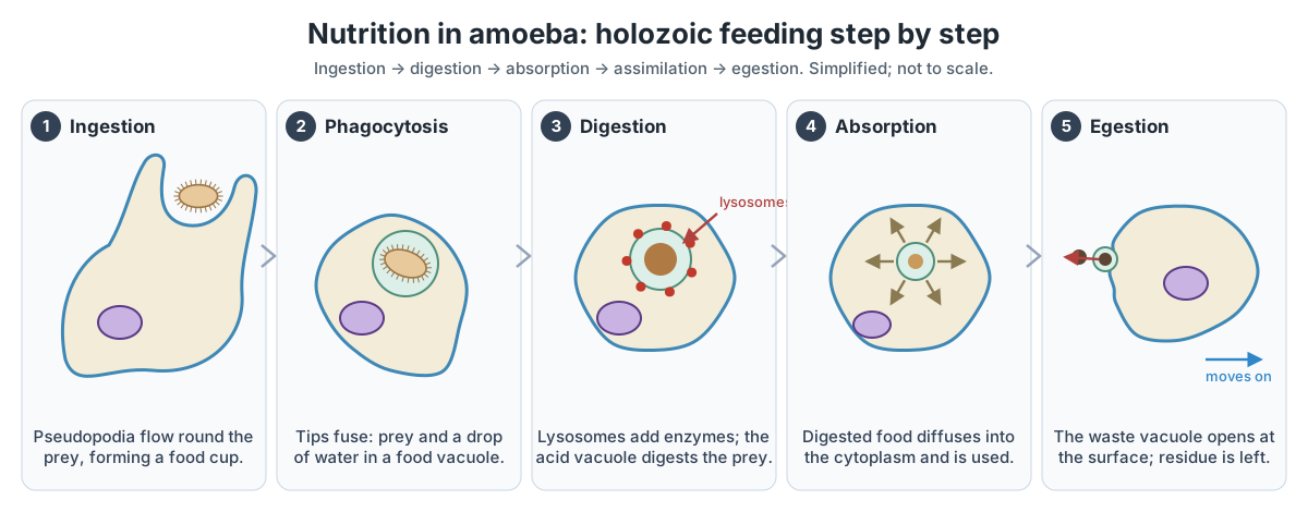
- Ingestion (phagocytosis). When the amoeba senses prey, such as a ciliate, a flagellate or an alga, it pushes out pseudopodia that flow around it, forming a food cup. The tips meet and fuse, and the prey is enclosed with a little water in a food vacuole. A. proteus engulfs prey by an inward fold of its plasma membrane. (Hausmann (2002): Food acquisition, food ingestion and food digestion by protists, Japanese Journal of Protozoology.)
- Digestion. Lysosomes fuse with the food vacuole and add digestive enzymes, which break the prey down into simpler substances. (OpenStax Biology 2e, 5.4 Bulk transport.)
- Absorption. The simple, soluble products (such as sugars, amino acids and fatty acids) diffuse out of the vacuole into the cytoplasm. The vacuole shrinks as it is emptied. (NCERT Science Class 10, Chapter 5: Life processes.)
- Assimilation. The absorbed food is used for energy (in respiration), for growth and repair, and, once the cell is big enough, for division. (NCERT Science Class 7, Chapter 2: Nutrition in animals.)
- Egestion. The vacuole with the undigested remains moves to the surface, fuses with the plasma membrane and throws the waste out. Amoeba has no fixed opening for this; as it crawls on, the waste is usually left behind near the uroid.
Food vacuoles can stay a long time: in A. proteus fed on the ciliate Tetrahymena, remains of the prey stay in the digestive vacuoles for up to about 72 hours. The same amoebae also feed by pinocytosis (“cell drinking”): put in a protein solution, they round up and pinch in fluid through many narrow channels from the surface. (Podlipaeva, Demin & Goodkov (2013): New method for cell cycle synchronization in Amoeba proteus culture, Protistology 8(1).)
Digestive system of amoeba? An amoeba has no digestive organs, mouth or anus. Its “digestive system” is the food vacuole: each one is a temporary stomach that forms around a meal and disappears when the meal is over. Compare Paramecium, which takes in food only at its oral groove and has a fixed anal pore.
Osmoregulation: the contractile vacuole of amoeba
The contractile vacuole keeps an amoeba from bursting. Pond water is much more dilute than the cytoplasm, so water keeps entering the cell by osmosis. The contractile vacuole collects this water, slowly swelling (diastole), then fuses with the plasma membrane and empties it outside in a quick contraction (systole). This control of water content is called osmoregulation. (OpenStax Microbiology, 5.1 Unicellular eukaryotic parasites.)
A. proteus has one contractile vacuole, usually towards the rear. Small vesicles round it collect water from the cytoplasm and merge with it; after it empties, a new vacuole forms from vesicles at the same spot. How often it empties depends on how dilute the water is: the more dilute, the faster it has to work. Watch one under the microscope and you will see it grow, then suddenly vanish.
The contractile vacuole is often called an excretory organelle, but its job is mainly to remove water. Amoeba gets rid of most of its nitrogen waste, as ammonia, by diffusion across the whole cell surface.
Respiration and excretion in amoeba
Respiration in amoeba is aerobic and happens in its mitochondria; gas exchange happens by simple diffusion across the whole plasma membrane. Oxygen dissolved in the pond water diffuses in because there is less oxygen inside the cell, where it is being used up. Carbon dioxide diffuses out the other way. In a single-celled organism the whole surface is in contact with the environment, so no special organs are needed. (NCERT Science Class 10, Chapter 5: Life processes.)
Excretion works the same way. Ammonia, the main nitrogenous waste, is very soluble and diffuses straight out into the water. Many unicellular organisms remove wastes by simple diffusion from the body surface into the surrounding water. (NCERT Science Class 10, Chapter 5: Life processes.)
A. proteus also stores some nitrogen waste inside the cell. Its cytoplasm holds many small crystals, truncated bipyramids and plates. Isolated and analysed, the crystals of A. proteus and related giant amoebae turned out to be mainly triuret (carbonyl diurea), probably an end product of the breakdown of purines, the building blocks of DNA and RNA. (Griffin (1960): The isolation, characterization, and identification of the crystalline inclusions of the large free-living amebae, J. Biophys. Biochem. Cytol..)
Diffusion is fast enough only because the cell is small, so no part of it is far from the surface. That is one reason single cells cannot grow very large; A. proteus, at under a millimetre, is already a giant among cells.
Reproduction in amoeba: binary fission
Amoeba reproduces asexually by binary fission: one parent cell divides into two genetically identical daughter cells. The cell stops feeding, draws in its pseudopodia and rounds up; the nucleus divides by mitosis; then the cytoplasm pinches in two. In Amoeba the cell can split in any plane, unlike organisms such as Leishmania, which divide in a fixed direction. (NCERT Science Class 10, Chapter 7: How do organisms reproduce?.)
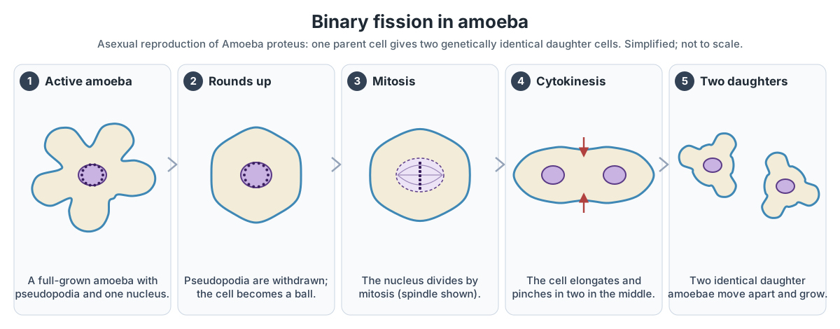
Division takes a while. In a synchronised laboratory culture of A. proteus kept at 20–22 °C, prophase was seen 39 hours after the cells were fed, metaphase at 41 hours and anaphase at 41.5 hours, and mitosis was over by 42 hours. In an ordinary culture only about one cell in a thousand is dividing at any moment. The nucleus has hundreds of tiny chromosomes, and their exact number is still unknown. (Podlipaeva, Demin & Goodkov (2013): New method for cell cycle synchronization in Amoeba proteus culture, Protistology 8(1).)
Does Amoeba proteus form cysts or divide by multiple fission?
Many school textbooks say that in bad conditions an amoeba forms a protective cyst and divides inside it into many small amoebae (multiple fission or sporulation). For Amoeba proteus this is not established. The claim goes back to observers in the 1800s and early 1900s (Carter, Scheel, Calkins, Taylor and others), who reported the nucleus breaking into many small nuclei, cysts, or even sexual stages. Their own contemporaries disagreed: in 1926 Schaeffer wrote that he found “no proof that the life cycle of the common large ameba includes more than … reproduction by fission”. A year later Hulpieu and Hopkins (1927) reported that, after a spell of rapid fission, sluggish large amoebae in some cultures broke up into 150–200 tiny amoeboid forms about 10 µm long, which they believed grew into new large amoebae. They did not describe cysts. (Hulpieu & Hopkins (1927): Observations on the life-history of Amoeba proteus, Biological Bulletin.)
Modern specialists describe A. proteus as obligately asexual and study it in cultures that multiply by binary fission. (Podlipaeva, Demin & Goodkov (2013): New method for cell cycle synchronization in Amoeba proteus culture, Protistology 8(1).) We found no modern study that confirms a cyst stage or multiple fission in this species. Cysts are real and well documented in other amoebae: Entamoeba histolytica spreads as four-nucleate cysts, and Naegleria and Acanthamoeba form resistant cysts. Multiple fission is the textbook example for the malaria parasite Plasmodium, and NCERT Class 10 uses Plasmodium, not Amoeba, as its example. In exams, describe binary fission for Amoeba, and if your syllabus asks about encystment, give the textbook answer but know that it is disputed for A. proteus.
Life cycle of amoeba. The life cycle of A. proteus is therefore simple: a young cell feeds and grows for one to a few days, depending on food and temperature, then divides by binary fission into two young cells, and the cycle repeats. In this sense the parent does not die: it becomes its two daughters.
Amoeba vs Paramecium vs Euglena
Amoeba, Paramecium and Euglena are the three classic single-celled protists of school biology. Amoeba has no fixed shape and crawls with pseudopodia; Paramecium is slipper-shaped and swims with cilia; Euglena is spindle-shaped, swims with a flagellum and can photosynthesise. See the full Euglena diagram for its parts.
| Feature | Amoeba (A. proteus) | Paramecium | Euglena |
|---|---|---|---|
| Shape | No fixed shape | Fixed, slipper-shaped | Fixed, spindle-shaped but can flex |
| Outer covering | Plasma membrane only | Pellicle | Pellicle of protein strips |
| Movement | Pseudopodia (amoeboid movement) | Cilia | Flagellum |
| Nutrition | Holozoic (phagocytosis by food cup) | Holozoic (food swept into oral groove by cilia) | Photosynthesis with chloroplasts; can also absorb food (mixotrophic) |
| Feeding site | Anywhere on the surface | Oral groove and cytostome only | No oral groove; absorbs dissolved food |
| Egestion | Anywhere, usually at the uroid | Fixed anal pore (cytoproct) | No prey remains to egest |
| Nucleus | One large discoid nucleus | Macronucleus and micronucleus | One nucleus |
| Contractile vacuole | One | Usually two | One, near the reservoir |
| Light sensing | None | None | Eyespot (stigma) |
| Reproduction | Binary fission, any plane | Transverse binary fission; conjugation | Longitudinal binary fission |
The ciliate features (cilia, oral groove, macronucleus for everyday functions, micronucleus for sexual reproduction, contractile vacuoles that fill by osmosis) and the euglenoid features (flagella, eyespot, protein pellicle) are those described in OpenStax. (OpenStax Biology 2e, 23.3 Groups of protists.)
Amoeba under a microscope
Under a light microscope, a living Amoeba proteus looks like a slowly flowing, colourless, grainy blob with a clear rim. Look for the blunt pseudopodia, the clear hyaline cap at the tip of the one that is advancing, the granules streaming forward inside it, the round contractile vacuole that swells and then vanishes, food vacuoles holding prey, and, with careful focusing, the grey, disc-shaped nucleus.
Start with the low-power objective (about ×40 to ×100 total magnification). At ×100 the field of view is roughly 1.5–2 mm across, so a large amoeba spans about a quarter of it; switch to ×400 to see the nucleus and vacuoles. Turn the light down and close the condenser iris a little: the cell is almost transparent, and bright light washes it out. Phase-contrast or dark-field illumination shows it much better if your microscope has them. Use a drop of culture under a supported cover slip so the cell is not crushed.
The cytoplasm often holds tiny truncated bipyramidal or plate-shaped crystals, and the posterior uroid often looks bunched and knobbly. Specimens with more than one nucleus are sometimes seen, even though the species is usually described as having one. (Siemensma, F. Microworld: Amoeba proteus (species description).)
On a stained, prepared slide the amoeba no longer moves. Its shape is whatever it had when it was fixed, and the nucleus stains darkly. NCERT's Class 10 activity asks students to compare a permanent slide of Amoeba with one showing binary fission. (NCERT Science Class 10, Chapter 7: How do organisms reproduce?.)
Pathogenic amoebae: Entamoeba histolytica and Naegleria fowleri
Amoeba proteus is harmless. A few other amoebae cause human disease, and exam questions often contrast them with it. Entamoeba histolytica, like Amoeba, is a member of the Amoebozoa. It is the main cause of amoebic dysentery. (OpenStax Microbiology, 5.1 Unicellular eukaryotic parasites.)
People catch E. histolytica by swallowing its cysts in food or water contaminated with faeces. Each mature cyst has four nuclei and is usually 12–15 µm across. In the gut the cysts release trophozoites (feeding cells), which multiply by binary fission. Most infections cause no symptoms, but invasive infection causes amoebic colitis with dysentery, and the commonest complication outside the gut is an amoebic liver abscess. (CDC DPDx: Amebiasis (Entamoeba histolytica).)
Naegleria fowleri, the so-called brain-eating amoeba, is not a true amoeba in the sense of Amoeba proteus. It is classified in a different phylum, Percolozoa, and is only a distant relative of the Amoebozoa. (OpenStax Microbiology, 5.1 Unicellular eukaryotic parasites.)
Unlike Amoeba proteus, Naegleria has three stages: a resistant cyst 7–15 µm across, an amoeboid trophozoite of 10–35 µm that feeds and divides, and a temporary, non-feeding form with flagella that usually turns back into a trophozoite. (CDC DPDx: Free-living amebic infections.)
N. fowleri lives in warm fresh water and soil. Infection happens only when water containing it goes up the nose, usually while swimming; the amoeba then travels to the brain and causes primary amebic meningoencephalitis (PAM). PAM is nearly always fatal: of 167 cases reported in the United States from 1962 to 2024, only four people survived. You cannot be infected by swallowing the water, and it does not spread from person to person. (CDC: About Naegleria fowleri.)
Amoeba diagram for Class 7 to 10 (NCERT) and KS3
For a simple amoeba diagram, draw an irregular blob with three or four blunt pseudopodia, outline it with a firm line for the cell membrane, shade a narrow clear band inside for ectoplasm and dot the rest as granular endoplasm. Add one nucleus, one clear round contractile vacuole and two or three food vacuoles with food inside. Label all seven parts with straight, ruled label lines that do not cross.
- Class 7 (NCERT, Nutrition in animals): Amoeba is a microscopic single-celled organism found in pond water, with a cell membrane, a rounded, dense nucleus and many small bubble-like vacuoles. It pushes out pseudopodia for movement and capture of food, traps food in a food vacuole, and digestive juices break it down. (NCERT Science Class 7, Chapter 2: Nutrition in animals.)
- Class 9 (The fundamental unit of life): amoeba as an example of a unicellular organism whose flexible cell membrane lets it engulf food (endocytosis).
- Class 10 (Life processes): nutrition in amoeba with a labeled figure of the food vacuole forming, digestion, diffusion of food into the cytoplasm and removal of the undigested material. (NCERT Science Class 10, Chapter 5: Life processes.)
- Class 10 (How do organisms reproduce?): binary fission in amoeba, which can take place in any plane; multiple fission in Plasmodium. (NCERT Science Class 10, Chapter 7: How do organisms reproduce?.)
- KS3 (England): amoeba as an example of a unicellular organism, with its parts compared with an animal cell: it has a cell membrane, cytoplasm and nucleus like an animal cell, plus pseudopodia, a contractile vacuole and food vacuoles adapted to life as a whole organism in fresh water.
- Common mistakes: drawing a cell wall; drawing the nucleus with a nucleolus in the middle like a textbook animal cell; calling pseudopodia “legs” or “tentacles”; showing the contractile vacuole full of food; forgetting that amoeba has no mouth or anus.
Amoeba diagram in colour. A living amoeba is colourless. If you colour a diagram, keep the convention used here: pale blue for the membrane and clear ectoplasm, beige for the granular endoplasm, purple for the nucleus, blue for the contractile vacuole and green-grey food vacuoles with brown food. Any colours are fine as long as you use a key.
Label the amoeba: unlabeled diagram and answer key
Test yourself with this amoeba diagram without labels. Write the name of each of the 19 numbered parts, then open the answer key. For a quick quiz, turn off the labels in the 3D model, click parts at random and name them before you read the panel.
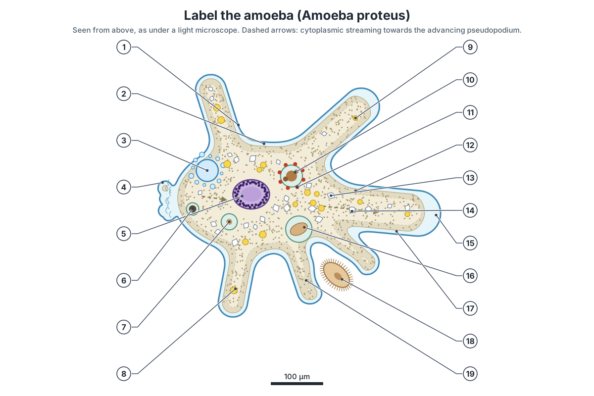
Show the answer key
- Plasma membrane (plasmalemma)
- Ectoplasm (clear hyaline layer)
- Contractile vacuole
- Uroid (posterior end)
- Nucleus
- Residual vacuole (egestion of undigested waste)
- Food vacuole, absorbing digested food
- Lateral pseudopodium
- Fat (lipid) droplet
- Food vacuole, digesting
- Lysosome
- Plasmagel (gel endoplasm)
- Crystal
- Plasmasol (flowing endoplasm)
- Hyaline cap
- Food vacuole, newly formed (phagosome)
- Advancing (dominant) pseudopodium
- Prey (a ciliate)
- Food cup
Short-answer questions:
- Name the structures an amoeba uses to move and to catch food. (Pseudopodia.)
- What is the function of the contractile vacuole? (It removes excess water that enters by osmosis: osmoregulation.)
- List the five steps of holozoic nutrition in amoeba. (Ingestion, digestion, absorption, assimilation, egestion.)
- How does oxygen get into an amoeba? (By diffusion across the whole plasma membrane.)
- What is the clear zone at the tip of an advancing pseudopodium called? (The hyaline cap.)
- Name the type of reproduction in amoeba and say what the daughter cells are like. (Binary fission; two genetically identical cells.)
- Give one difference between amoeba and Paramecium. (For example: amoeba moves with pseudopodia, Paramecium with cilia.)
Frequently asked questions
What are the main parts of an amoeba?
A labeled amoeba diagram shows the plasma membrane, pseudopodia, ectoplasm, endoplasm, nucleus, contractile vacuole and food vacuoles. More detailed diagrams of Amoeba proteus add the hyaline cap, uroid, plasmagel and plasmasol, crystals, fat droplets and mitochondria.
How big is Amoeba proteus?
A moving Amoeba proteus is about 220–760 µm long, with an average of about 425 µm. The largest, at about three-quarters of a millimetre, can just be seen with the naked eye as a tiny white speck. Its disc-shaped nucleus is about 22–62 µm across.
How does an amoeba move?
An amoeba moves by amoeboid movement. It pushes out pseudopodia, and its fluid inner cytoplasm (plasmasol) streams forward into them. The gel-like outer cytoplasm (plasmagel) contracts, mainly at the rear, using actin and myosin, and the pressure drives the flow. At the front, sol turns into gel; at the rear, gel turns back into sol.
How does an amoeba get its food?
An amoeba is holozoic. It surrounds prey with pseudopodia (a food cup) and engulfs it in a food vacuole by phagocytosis. Lysosomes add enzymes that digest the food. The digested food diffuses into the cytoplasm and is assimilated, and undigested waste is expelled through the membrane (egestion).
What is the function of the contractile vacuole in an amoeba?
The contractile vacuole removes excess water. Pond water is more dilute than the cytoplasm, so water enters the amoeba by osmosis. The contractile vacuole fills with this water and empties it outside, stopping the cell from bursting. This is called osmoregulation.
How does an amoeba reproduce?
Amoeba proteus reproduces asexually by binary fission. The cell rounds up, the nucleus divides by mitosis, and the cytoplasm pinches in two, giving two identical daughter amoebae. Sex has never been seen in Amoeba. Claims that A. proteus forms cysts or divides by multiple fission come from nineteenth- and early twentieth-century reports that were disputed at the time; we found no modern study confirming them.
Does an amoeba breathe?
An amoeba has no lungs or gills. Oxygen dissolved in the water diffuses in across the whole plasma membrane and is used in its mitochondria for aerobic respiration. Carbon dioxide and ammonia diffuse out the same way.
Is Amoeba proteus harmful to humans?
No. Amoeba proteus is a harmless pond organism. The amoebae that cause disease are different organisms: Entamoeba histolytica causes amoebic dysentery and liver abscess, and Naegleria fowleri, which is not closely related to Amoeba, causes a rare, almost always fatal brain infection.
Why is it called a 'proteus'?
Proteus was a shape-changing sea god in Greek myth. The name refers to the amoeba's constantly changing shape. Amoeba comes from the Greek amoibē, meaning change.
References
- Siemensma, F. J. Microworld, world of amoeboid organisms: Amoeba proteus (Pallas, 1766) Leidy, 1878.
- Podlipaeva, Y., Demin, S. & Goodkov, A. (2013). New method for cell cycle synchronization in Amoeba proteus culture. Protistology 8(1), 3–7.
- Rogers, S. S., Waigh, T. A. & Lu, J. R. (2008). Intracellular microrheology of motile Amoeba proteus. Biophysical Journal 94(8), 3313–3322.
- Matsumoto, K., Takagi, S. & Nakagaki, T. (2008). Locomotive mechanism of Physarum plasmodia based on spatiotemporal analysis of protoplasmic streaming. Biophysical Journal 94(7), 2492–2504 (introduction reviews the theories of amoeboid movement in Amoeba proteus).
- Griffin, J. L. (1960). The isolation, characterization, and identification of the crystalline inclusions of the large free-living amebae. Journal of Biophysical and Biochemical Cytology 7(2), 227–234.
- Hausmann, K. (2002). Food acquisition, food ingestion and food digestion by protists. Japanese Journal of Protozoology 35(2), 85–95.
- Hulpieu, H. R. & Hopkins, D. L. (1927). Observations on the life-history of Amoeba proteus. Biological Bulletin 52(6), 411–417.
- Clark, M. A., Douglas, M. & Choi, J. (2018). Biology 2e, section 23.3: Groups of protists. OpenStax.
- OpenStax Biology 2e, section 5.4: Bulk transport.
- Parker, N. et al. (2016). Microbiology, section 5.1: Unicellular eukaryotic parasites. OpenStax.
- Centers for Disease Control and Prevention. About Naegleria fowleri.
- Centers for Disease Control and Prevention, DPDx. Free-living amebic infections.
- Centers for Disease Control and Prevention, DPDx. Amebiasis.
- NCERT (2024 reprint). Science, Class 7, Chapter 2: Nutrition in animals (section 2.7, Feeding and digestion in Amoeba).
- NCERT (2026–27 reprint). Science, Class 10, Chapter 5: Life processes.
- NCERT (2026–27 reprint). Science, Class 10, Chapter 7: How do organisms reproduce?
More interactive anatomy models
- Bacterial cell diagramLabeled prokaryotic cell: cell wall, membrane, nucleoid, ribosomes, pili and flagellum.
- Euglena diagramLabeled Euglena: flagellum, eyespot, chloroplasts, pellicle and contractile vacuole.
- Bacteriophage diagramT4 phage structure in 3D: head, collar, tail sheath, baseplate and tail fibres.
- Virus structureCapsid, envelope, spikes and genome of helical, icosahedral and enveloped viruses.
- Human anatomy1,000+ bones, muscles, organs, vessels and nerves from real anatomical data.
- Octopus anatomyThree hearts, a ring-shaped brain, eight arms and a beak, in 3D.