

Skin layers diagram at a glance
The skin has three main layers. From the surface down they are the epidermis, a thin sheet of cells with no blood vessels; the dermis, a thick layer of connective tissue that holds blood vessels, nerves, hair follicles and glands; and the hypodermis (subcutaneous layer), made of fat and loose connective tissue. Strictly, the hypodermis lies under the skin and is not part of it, but nearly every skin layers diagram includes it.
The epidermis is itself made of four or five layers called strata (singular: stratum). From deep to superficial they are the stratum basale, stratum spinosum, stratum granulosum, stratum lucidum (only in thick skin) and stratum corneum. The dermis has two layers: a thin papillary layer under the epidermis and a thick reticular layer below it.
| Layer | Made of | Key contents | Main jobs |
|---|---|---|---|
| Epidermis | Keratinized stratified squamous epithelium | Keratinocytes, melanocytes, Langerhans cells, Merkel cells | Barrier against water loss, microbes and UV light |
| Dermis: papillary layer | Loose areolar connective tissue | Dermal papillae, capillary loops, Meissner corpuscles, lymphatic capillaries | Feeds the epidermis, anchors it, light touch |
| Dermis: reticular layer | Dense irregular connective tissue | Collagen and elastin, hair follicles, sweat and sebaceous glands, vessels, nerves | Strength, stretch and recoil, houses skin appendages |
| Hypodermis | Adipose and loose connective tissue | Fat lobules, large vessels and nerves, Pacinian corpuscles | Insulation, cushioning, energy store, anchors skin to muscle |
Layers of skin diagram, labeled
A labeled diagram of the layers of the skin shows the epidermis at the top, the dermis with its hair follicles, glands, vessels and nerve endings in the middle, and the fatty hypodermis at the bottom. The diagram below labels 26 structures, the same set most biology, anatomy and cosmetology courses ask students to identify.
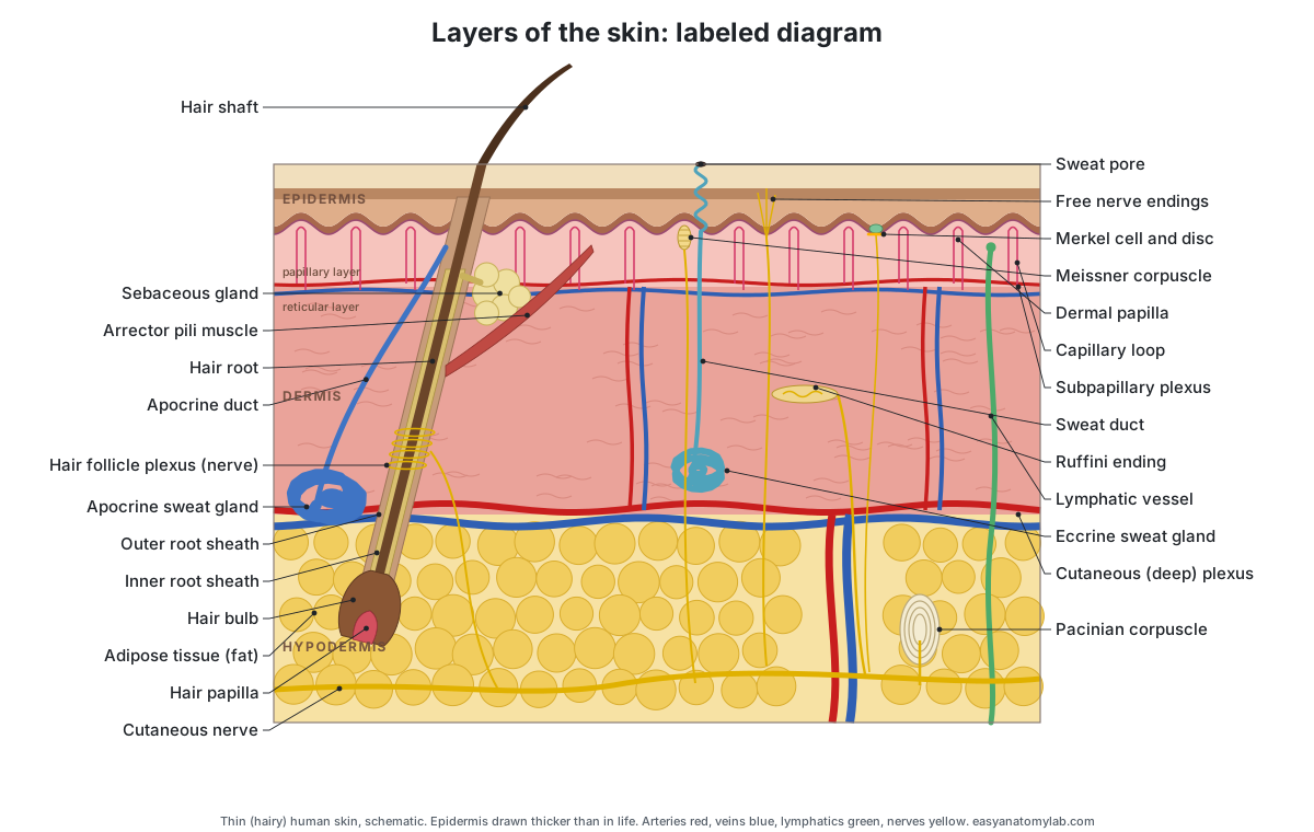
Read a skin diagram from the top down. The hair shaft and sweat pores are on the surface. The wavy line under the epidermis is the dermal–epidermal junction; the bumps pushing up into the epidermis are dermal papillae, each holding a capillary loop or a Meissner corpuscle. In the dermis, the hair follicle runs at a slant, with the sebaceous gland and arrector pili muscle on the side the hair leans toward. The coiled sweat glands sit near the bottom of the dermis, and the onion-like Pacinian corpuscle sits deep, near or in the fat.
How many layers of skin are there? 3, 5 or 7
Anatomists say the skin has two layers, the epidermis and the dermis, resting on a third, the hypodermis. Most school courses simply teach three layers of skin and include the hypodermis. The answer "five" refers to the five strata of the epidermis, not to the whole skin.
You will also see the phrase "seven layers of skin". This is a popular way of counting, not a standard anatomical term. It usually adds the five epidermal strata to the dermis and the hypodermis (5 + 1 + 1). It is a useful memory aid, but it mixes levels: the five strata are sublayers of one tissue, while the dermis and hypodermis are whole layers with their own sublayers. Thin skin, which covers most of the body, has only four epidermal strata, so the count would be six there.
| Count | What is being counted | Layers |
|---|---|---|
| 2 | The skin proper | Epidermis, dermis |
| 3 | Skin plus the layer under it (most textbooks and diagrams) | Epidermis, dermis, hypodermis |
| 4 or 5 | Strata of the epidermis (thin skin / thick skin) | Basale, spinosum, granulosum, (lucidum), corneum |
| 2 | Layers of the dermis | Papillary layer, reticular layer |
| 7 | Popular count: 5 epidermal strata + dermis + hypodermis | Not a formal anatomical division |
Epidermis skin layers: the 5 strata
The five layers of the epidermis, from deepest to most superficial, are the stratum basale, stratum spinosum, stratum granulosum, stratum lucidum and stratum corneum. A common memory aid, reading from the surface down, is "Come, Let's Get Sun Burned": corneum, lucidum, granulosum, spinosum, basale.
| Stratum (deep to superficial) | What it looks like | What happens there |
|---|---|---|
| Stratum basale | One row of cuboidal to columnar cells on the basement membrane | Stem cells divide; melanocytes add pigment; Merkel cells sense touch |
| Stratum spinosum | 8–10 layers of polygonal cells joined by desmosomes | Keratin synthesis begins; Langerhans cells patrol for microbes |
| Stratum granulosum | 3–5 layers of flattened cells with dark granules (1–2 in thin skin) | Keratohyalin and lipids are made; cells die |
| Stratum lucidum | Thin, clear band; thick skin only | Dead cells filled with eleidin add toughness |
| Stratum corneum | 15–30 layers of flat, dead, keratin-filled cells | The barrier; cells are shed and replaced about every 4 weeks |
- Stratum basale: Renews the epidermis. It also holds the melanocytes, which make pigment, and the Merkel cells, which sense touch.
- Stratum spinosum: Gives the epidermis strength and flexibility. Its cells begin making keratin, and it holds the immune Langerhans cells.
- Stratum granulosum: Makes keratin and keratohyalin and releases lipids that seal the spaces between cells, forming the water barrier.
- Stratum lucidum: Adds an extra tough, translucent layer to skin that takes heavy friction and pressure.
- Stratum corneum: Forms the main barrier of the skin. Keratin and lipids between the cells stop water loss and keep out microbes, chemicals and minor abrasion.
Cells of the epidermis
Four cell types live in the epidermis. In the 3D model they are drawn on the front cut face of the thin-skin block, in the window at the front right.
- Keratinocytes: Build the protective, waterproof barrier of the skin from keratin and lipids.
- Melanocytes: Make melanin and pass it in small packets (melanosomes) to nearby keratinocytes, where it shields the cell nuclei from UV light.
- Langerhans cells: Catch microbes and foreign material that get into the epidermis and present them to the immune system.
- Merkel cells: Together with their nerve ending (the Merkel disc) they sense sustained light pressure and the texture and shape of objects.
Dermal–epidermal junction and dermal papillae
The epidermis and dermis are joined at the dermal–epidermal junction, a basement membrane that is folded into ridges and bumps. Finger-like dermal papillae push up into the epidermis, and epidermal (rete) ridges push down between them. The folding increases the contact area, which strengthens the bond and brings capillaries within diffusion distance of the basal cells.
- Dermal–epidermal junction: Anchors the epidermis, resists shearing forces and controls which molecules and cells pass between the two layers.
- Dermal papillae: Increase the contact area between the dermis and epidermis, strengthen the junction and bring capillaries close to the avascular epidermis.
- Capillary loops: Exchange oxygen, nutrients and wastes with the epidermis, which has no blood vessels of its own.
Papillae are taller and more crowded in thick skin, which has to resist friction. In the fingertips the pattern of papillae sets the pattern of the fingerprint before birth. Compare the two blocks in the 3D model: the thick-skin block on the right has much taller papillae.
The dermis: papillary and reticular layers
The dermis is the thick middle layer of the skin, made of connective tissue produced by fibroblasts. It has two layers. The papillary layer is thin, loose areolar tissue just under the epidermis. The reticular layer is much thicker, dense irregular tissue packed with collagen and elastin, and forms most of the skin's thickness. There is no sharp line between them.
| Papillary dermis | Reticular dermis | |
|---|---|---|
| Tissue | Loose areolar connective tissue | Dense irregular connective tissue |
| Thickness | Thin | Much thicker; most of the dermis |
| Fibers | Fine collagen and elastic fibers | Thick bundles of collagen, plus elastin |
| Contents | Dermal papillae, capillary loops, lymphatic capillaries, Meissner corpuscles, free nerve endings | Hair follicles, sebaceous glands, sweat glands, Ruffini endings, larger vessels and nerves |
| Job | Feeds and anchors the epidermis; light touch | Strength, elasticity, hydration; supports appendages |
- Papillary dermis: Feeds the epidermis through its capillaries and houses lymphatic capillaries, nerve endings, Meissner corpuscles and immune cells.
- Reticular dermis: Gives skin its strength (collagen) and its stretch and recoil (elastin), holds water, and supports hair follicles, glands, vessels and nerves.
The hypodermis (subcutaneous layer)
The hypodermis is the layer of fat and loose connective tissue under the dermis. It is also called the subcutaneous layer, subcutis or superficial fascia. It is not strictly part of the skin, but it anchors the skin to the fascia over muscles and bones while letting it slide, and its fat insulates, cushions and stores energy.
- Hypodermis: Anchors the skin to the muscles and bones while letting it slide, and stores fat for insulation, cushioning and energy.
- Adipose tissue (fat lobules): Stores energy as fat, insulates against heat loss and cushions against knocks.
The hypodermis varies more than any other layer. It can be several centimeters thick over the belly and buttocks and is almost absent from the eyelids. The largest blood vessels and nerves that supply the skin run through it, and so do the deep roots of large hair follicles, apocrine glands and Pacinian corpuscles.
Hair follicle structure
A hair follicle is a tube of epidermis that dips deep into the dermis. The hair shaft is the part above the skin; the hair root is the part inside the follicle. At the base, the swollen hair bulb wraps around the hair papilla, a knob of connective tissue with capillaries and nerve endings. Dividing cells in the bulb, the hair matrix, make the hair and the inner root sheath.
- Hair shaft: Insulates, protects the skin from sunlight and abrasion, and senses touch when it is bent.
- Hair root: Anchors the hair; new cells added at the bulb push the root and shaft upward as the hair grows.
- Hair bulb: Contains the hair matrix, a layer of rapidly dividing cells that makes the hair and the inner root sheath. Melanocytes here color the hair.
- Hair papilla: Brings blood capillaries and nerve endings to the hair matrix and sends signals that control hair growth.
- Inner root sheath: Shapes and supports the growing hair, then breaks down so the hair can leave the follicle.
- Outer root sheath: Lines and supports the follicle. Its bulge region holds stem cells that can regrow the hair and repair nearby epidermis after a wound.
- Arrector pili muscle: Contracts when the sympathetic nervous system fires (cold or fear), pulling the hair upright and making goose bumps.
- Hair follicle plexus: Senses when a hair is bent, so you can feel an insect walking on your arm before it touches the skin.
The hair shaft itself has three layers: a central medulla (missing in some hairs), a cortex and a hard outer cuticle. Hair colour comes from melanin made by melanocytes in the bulb. In the 3D model, click the hair bulb or a root sheath to see the follicle on its own; the sheaths are drawn as cut-open sleeves.
Skin glands: sebaceous, eccrine and apocrine
Skin has three main types of gland. Sebaceous glands make oily sebum and empty it into hair follicles. Eccrine sweat glands make watery sweat for cooling and open through their own pores. Apocrine sweat glands, in the armpits and groin, make a thicker secretion and empty into hair follicles.
| Gland | Where | Opens into | Secretion | Main job |
|---|---|---|---|---|
| Sebaceous | Everywhere except palms and soles; densest on scalp and forehead (400–900/cm²) | Hair follicle | Sebum (lipids) | Waterproofs and softens skin and hair; antibacterial |
| Eccrine sweat | Almost all skin; densest on palms and soles (about 500/cm²) | Sweat pore on the surface | Watery, salty sweat with dermcidin | Cooling by evaporation |
| Apocrine sweat | Armpits, groin and genital area | Hair follicle, above the sebaceous gland | Thicker, protein- and lipid-rich | Scent; bacteria break it down into body odour |
- Sebaceous gland: Secretes sebum, which lubricates and waterproofs the skin and hair, keeps the stratum corneum supple and has antibacterial fatty acids.
- Eccrine sweat gland: Makes hypotonic sweat that cools the body as it evaporates. Sweat also carries the antimicrobial peptide dermcidin.
- Sweat duct: Delivers sweat to the surface and reabsorbs some of the salt on the way, so sweat ends up more dilute than blood.
- Sweat pore: Releases sweat onto the surface, where it evaporates.
- Apocrine sweat gland: Makes a thicker, milky secretion rich in proteins and lipids. Skin bacteria break it down, which causes body odour.
- Apocrine duct: Carries apocrine sweat into the follicle, from which it reaches the surface along the hair.
Blood vessels, lymphatics and nerves of the skin
The skin is supplied by two flat networks of vessels in the dermis. The cutaneous (deep) plexus lies at the junction of the dermis and hypodermis; the subpapillary (superficial) plexus lies just under the dermal papillae. Vessels connect the two, and capillary loops rise from the subpapillary plexus into the papillae. Lymphatic capillaries begin as blind-ended tubes in the papillary dermis and drain down to larger lymph vessels.
- Cutaneous arterial plexus: Distributes oxygen-rich blood to the skin and sends branches to hair bulbs, sweat glands and up to the subpapillary plexus.
- Cutaneous venous plexus: Collects oxygen-poor blood from the skin and drains it into the veins of the hypodermis.
- Subpapillary arterial plexus: Supplies the capillary loops of the dermal papillae and so, by diffusion, the whole epidermis.
- Subpapillary venous plexus: Drains blood from the papillae down to the deep venous plexus.
- Lymphatic vessels: Collect excess tissue fluid, proteins and immune cells from the dermis and return them to the blood via the lymph nodes.
- Cutaneous nerve: Carry touch, pressure, vibration, temperature and pain signals to the spinal cord, and autonomic signals to sweat glands, vessels and arrector pili muscles.
Sensory receptors in the skin
Skin has at least six kinds of sensory receptor, each at a typical depth. Free nerve endings and Merkel discs are at or in the epidermis, Meissner corpuscles in the dermal papillae, Ruffini endings in the dermis, and Pacinian corpuscles deep in the dermis and hypodermis. Nerve endings wrapped around hair follicles sense hair movement.
| Receptor | Depth | Senses | Adapts |
|---|---|---|---|
| Free nerve endings | Dermis, reaching into the epidermis | Pain, heat, cold, itch, some touch | Varies |
| Merkel disc (with Merkel cell) | Stratum basale | Sustained pressure, edges, texture (5–15 Hz) | Slowly |
| Meissner corpuscle | Dermal papillae of hairless skin | Light touch, slip, low-frequency vibration (below about 40–50 Hz) | Rapidly |
| Ruffini ending | Dermis | Skin stretch | Slowly |
| Pacinian corpuscle | Deep dermis and hypodermis | Deep pressure, high-frequency vibration (about 200–250 Hz) | Rapidly |
| Hair follicle plexus | Around hair follicles | Hair movement, light touch | Rapidly |
- Free nerve endings: Sense pain, heat and cold, and itch, as well as some touch.
- Merkel discs: Slowly adapting touch receptors: they keep firing during sustained pressure and signal edges, shapes and texture.
- Meissner corpuscle: Rapidly adapting receptor for light touch and low-frequency vibration (below about 40–50 Hz), such as an object slipping in your grip.
- Ruffini ending: Slowly adapting receptor thought to sense skin stretch, which helps you feel finger position and object shape.
- Pacinian corpuscle: Rapidly adapting receptor for deep pressure and high-frequency vibration, best around 200–250 Hz.
Skin layers and their functions
Each layer of the skin has its own main jobs. The epidermis is the barrier; the dermis gives strength, feeling, temperature control and the glands; the hypodermis insulates and cushions. Together they make the skin the body's largest organ.
| Function | Layer and structures responsible | How |
|---|---|---|
| Barrier to water loss and microbes | Stratum corneum, stratum granulosum | Dead keratin-filled cells sealed with lipids |
| Protection from UV light | Melanocytes in the stratum basale | Melanin shields keratinocyte nuclei |
| Immune defence | Langerhans cells; dermal immune cells; sweat (dermcidin) | Catch and present invaders; antimicrobial peptides |
| Vitamin D synthesis | Epidermis | UV light converts a cholesterol derivative into vitamin D3 |
| Temperature control | Eccrine glands; dermal arterioles | Sweat evaporates; vessels widen to lose heat, narrow to save it |
| Touch, pain, temperature | Receptors in epidermis, dermis and hypodermis | Six receptor types at different depths |
| Strength and stretch | Reticular dermis | Collagen resists tearing; elastin recoils |
| Lubrication | Sebaceous glands | Sebum keeps skin and hair supple |
| Insulation, cushioning, energy store | Hypodermis | Fat lobules |
Thick skin vs thin skin
Thick skin covers the palms, the fingertips and the soles of the feet. It has five epidermal strata, including the stratum lucidum, a very thick stratum corneum and friction ridges, but no hair follicles or sebaceous glands. Thin skin covers the rest of the body. It has four strata, no stratum lucidum, and has hair follicles, sebaceous glands and arrector pili muscles.
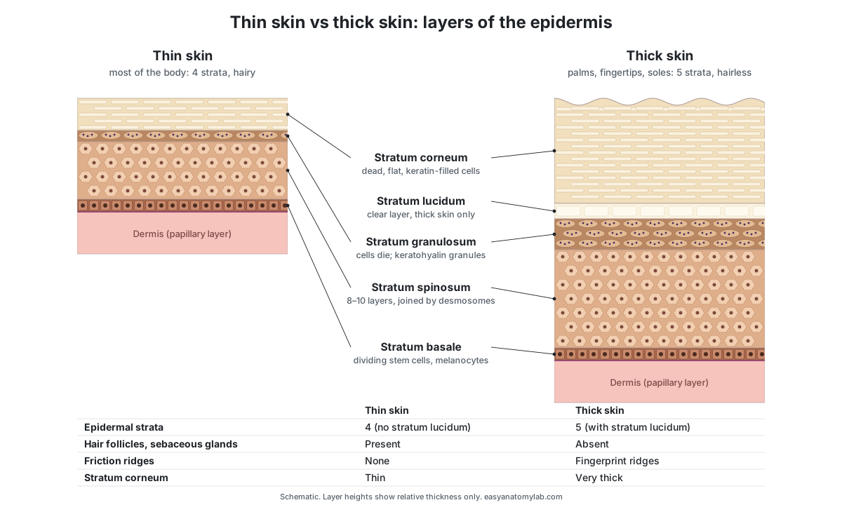
| Feature | Thin skin | Thick skin |
|---|---|---|
| Where | Most of the body | Palms, fingertips, soles |
| Epidermal strata | 4 (no stratum lucidum) | 5 (with stratum lucidum) |
| Epidermis thickness | Mostly well under 0.2 mm | About 0.4–1.4 mm; about 0.6 mm average on the sole |
| Stratum corneum | Thin | Very thick |
| Stratum granulosum | 1–2 cell layers | 3–5 cell layers |
| Hair follicles and sebaceous glands | Present | Absent |
| Eccrine sweat glands | Present | Present and especially dense |
| Dermal papillae and ridges | Low | Tall, in rows under friction ridges |
"Thick" and "thin" describe the epidermis, not the whole skin. The skin of the back has a thin epidermis but a very thick dermis, so it is thin skin even though it is thick to the touch. In the 3D model, the right-hand block is thick skin: look for the clear stratum lucidum, the deep stratum corneum, the friction ridges with pores along their crests, and the Meissner corpuscles in the papillae.
Facial skin layers
Facial skin has the same layers as skin elsewhere, but it is mostly thin skin with a thinner epidermis, many hair follicles and a very high density of sebaceous glands. Sebaceous glands reach about 400–900 per square centimeter on the forehead and scalp, far more than on the limbs, which is why the face is prone to oiliness and acne.
Skin thickness varies a lot across the face. In an ultrasound study of 38 facial sites, the upper eyelid had the thinnest skin (median about 0.57 mm in total) and the tip of the nose the thickest (about 1.9 mm). The eyelid also has almost no subcutaneous fat, which is why it shows swelling and bruising so easily. Facial skin also contains mimetic (expression) muscles that insert into the dermis, which is part of why expression lines form there.
| Facial area | Typical skin features |
|---|---|
| Upper eyelid | Thinnest facial skin (about 0.3–1.1 mm across the lid); epidermis about 0.05 mm; almost no fat |
| Forehead | Very dense sebaceous glands; many eccrine glands |
| Nose tip | Thickest facial skin; large sebaceous glands |
| Lips | Very thin epidermis over a rich capillary bed, so the red colour shows; no hair or sweat glands on the red part |
Skin layers histology: what the layers look like under a microscope
In a standard H&E-stained section, the epidermis is the thin, darker pink-to-purple band of packed cells at the top, the dermis is the paler pink fibrous layer under it, and the hypodermis shows as clear, empty-looking fat cells, because the fat dissolves during processing.
- Stratum basale: one row of small, dark-nucleated cuboidal or columnar cells along the wavy junction. Melanocytes show as pale, clear cells among them.
- Stratum spinosum: several layers of larger pink polygonal cells with round nuclei; the "spines" are desmosomes stretched between cells that shrank during processing.
- Stratum granulosum: flattened cells full of dark purple (basophilic) keratohyalin granules, easy to spot as a dark line.
- Stratum lucidum: in thick skin only, a thin, glassy, pale band with no nuclei.
- Stratum corneum: pink layers of flat dead cells with no nuclei, often split into a basket-weave by processing. It is huge in palm or sole sections and thin in thin skin.
- Dermis: fine, loosely woven fibers in the papillary layer and thick, wavy collagen bundles in the reticular layer, with cut follicles, glands and vessels.
To tell thick from thin skin on a slide, look for a stratum lucidum, a very thick stratum corneum and deep interdigitations between epidermis and dermis (thick skin), or hair follicles and sebaceous glands (thin skin).
Skin layers and burn depth
Burns are classified by how many layers of the skin they destroy. A superficial (first-degree) burn affects only the epidermis. A partial-thickness (second-degree) burn reaches into the dermis: superficial partial-thickness burns stop in the papillary dermis, deep partial-thickness burns reach the reticular dermis. A full-thickness (third-degree) burn destroys the whole epidermis and dermis and reaches the fat. Some systems add a fourth degree for burns into muscle or bone.
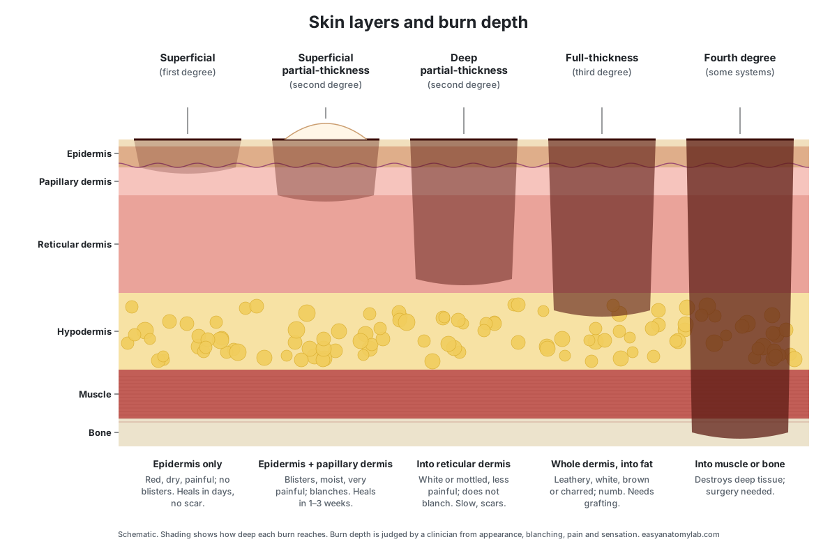
| Burn depth | Layers damaged | Appearance and feeling | Healing |
|---|---|---|---|
| Superficial (first degree) | Epidermis only | Red, dry, tender; no blisters; blanches with pressure | Heals by itself within days, e.g. mild sunburn |
| Superficial partial-thickness (second degree) | Epidermis and papillary dermis | Blisters within 24 hours, pink moist base, painful; blanches | About 1–2 weeks, minimal scarring (some sources give 2–3 weeks) |
| Deep partial-thickness (second degree) | Into the reticular dermis | White, red or mottled; less painful; does not blanch | 2 weeks or more, healing only from hair follicles; scarring common |
| Full-thickness (third degree) | Whole epidermis and dermis, into fat | White, leathery, brown or charred; numb; does not blanch | Heals only from the edges; usually needs excision and grafting |
| Fourth degree | Through the skin into muscle or bone | Deep tissue destroyed | Surgery needed |
The anatomy explains the symptoms. Partial-thickness burns hurt because the nerve endings of the dermis survive and are exposed. Full-thickness burns often hurt less because those nerve endings are destroyed. And a burn can heal on its own only if some epidermal cells survive, in hair follicles and sweat ducts deep in the dermis, to regrow the surface. Once the whole dermis is gone, there is nothing left to regrow from except the wound edges. This page is for learning; anyone with a significant burn needs medical care.
Skin layers labeled: every structure and its function
This is the text version of the 3D model: every labeled structure, grouped by layer and system, with what it does. Use it as a study list or to check a skin diagram worksheet.
Epidermis
- Stratum corneum (horny layer)
- The outermost layer of the epidermis: 15 to 30 layers of flat, dead, keratin-filled cells (corneocytes) that are constantly shed. Forms the main barrier of the skin. Keratin and lipids between the cells stop water loss and keep out microbes, chemicals and minor abrasion.
- Stratum lucidum (clear layer)
- A thin, clear band of flattened dead cells, found only in the thick skin of the palms, fingertips and soles. Adds an extra tough, translucent layer to skin that takes heavy friction and pressure.
- Stratum granulosum (granular layer)
- Three to five layers of flattened keratinocytes filled with dark keratohyalin granules. Here the cells die. Makes keratin and keratohyalin and releases lipids that seal the spaces between cells, forming the water barrier.
- Stratum spinosum (spiny or prickle-cell layer)
- The thickest living layer of the epidermis: eight to ten layers of keratinocytes joined by many desmosomes. Gives the epidermis strength and flexibility. Its cells begin making keratin, and it holds the immune Langerhans cells.
- Stratum basale (stratum germinativum, basal layer)
- The deepest layer of the epidermis: a single row of cuboidal to columnar stem cells that divide to make new keratinocytes. Renews the epidermis. It also holds the melanocytes, which make pigment, and the Merkel cells, which sense touch.
- Dermal–epidermal junction (basement membrane, DEJ)
- A thin sheet of extracellular matrix that glues the epidermis to the dermis and follows its wavy interface. Anchors the epidermis, resists shearing forces and controls which molecules and cells pass between the two layers.
- Friction ridges (epidermal ridges, fingerprint ridges)
- Parallel ridges on the surface of thick skin that make fingerprints, palm prints and footprints. Improve grip and increase sensitivity to touch. Sweat pores open along their crests.
Epidermal cells
- Keratinocytes
- The main cells of the epidermis. They make keratin and change shape as they move up: cuboidal at the base, polygonal in the middle, flat and dead at the top. Build the protective, waterproof barrier of the skin from keratin and lipids.
- Melanocytes
- Pigment cells with long, branching arms (dendrites) scattered among the basal keratinocytes. Make melanin and pass it in small packets (melanosomes) to nearby keratinocytes, where it shields the cell nuclei from UV light.
- Langerhans cells (epidermal dendritic cells)
- Immune cells with long, branching arms that sit mainly in the stratum spinosum. Catch microbes and foreign material that get into the epidermis and present them to the immune system.
- Merkel cells (tactile epithelial cells)
- Touch-sensitive cells in the stratum basale, each in contact with a flattened sensory nerve ending. Together with their nerve ending (the Merkel disc) they sense sustained light pressure and the texture and shape of objects.
Dermis
- Dermal papillae
- Small finger-like bumps of the papillary dermis that push up into the underside of the epidermis. Increase the contact area between the dermis and epidermis, strengthen the junction and bring capillaries close to the avascular epidermis.
- Papillary dermis (papillary layer)
- The thin upper layer of the dermis, made of loose areolar connective tissue with fine collagen and elastic fibers. Feeds the epidermis through its capillaries and houses lymphatic capillaries, nerve endings, Meissner corpuscles and immune cells.
- Reticular dermis (reticular layer)
- The thick lower layer of the dermis: dense irregular connective tissue packed with collagen and elastin. Gives skin its strength (collagen) and its stretch and recoil (elastin), holds water, and supports hair follicles, glands, vessels and nerves.
Hypodermis
- Hypodermis (subcutaneous layer, subcutis, superficial fascia)
- The layer under the skin, made of loose connective tissue and fat. Strictly speaking it is not part of the skin. Anchors the skin to the muscles and bones while letting it slide, and stores fat for insulation, cushioning and energy.
- Adipose tissue (fat lobules)
- Clusters of fat cells (adipocytes) grouped into lobules separated by thin walls of connective tissue. Stores energy as fat, insulates against heat loss and cushions against knocks.
Hair follicle
- Hair shaft
- The part of the hair that sticks out above the skin surface. It is dead keratin. Insulates, protects the skin from sunlight and abrasion, and senses touch when it is bent.
- Hair root
- The part of the hair inside the follicle, below the skin surface. Anchors the hair; new cells added at the bulb push the root and shaft upward as the hair grows.
- Hair bulb
- The onion-shaped swelling at the base of the hair follicle. Contains the hair matrix, a layer of rapidly dividing cells that makes the hair and the inner root sheath. Melanocytes here color the hair.
- Hair papilla (dermal papilla of the hair)
- A small knob of connective tissue that pushes up into the base of the hair bulb. Brings blood capillaries and nerve endings to the hair matrix and sends signals that control hair growth.
- Inner root sheath
- The innermost layer of the follicle wall, tightly wrapped around the growing hair root. Shapes and supports the growing hair, then breaks down so the hair can leave the follicle.
- Outer root sheath (external root sheath)
- The outer epithelial layer of the follicle wall, a downward continuation of the epidermis. Lines and supports the follicle. Its bulge region holds stem cells that can regrow the hair and repair nearby epidermis after a wound.
- Arrector pili muscle
- A small band of smooth muscle that runs from the side of the follicle up to the papillary dermis. Contracts when the sympathetic nervous system fires (cold or fear), pulling the hair upright and making goose bumps.
Glands
- Sebaceous gland (oil gland)
- A grape-like cluster of gland cells that empties oily sebum into the hair follicle. Secretes sebum, which lubricates and waterproofs the skin and hair, keeps the stratum corneum supple and has antibacterial fatty acids.
- Eccrine sweat gland (merocrine sweat gland)
- A tightly coiled tube in the deep dermis that makes watery sweat. Makes hypotonic sweat that cools the body as it evaporates. Sweat also carries the antimicrobial peptide dermcidin.
- Sweat duct (eccrine duct)
- The thin tube that carries sweat from the eccrine coil straight up through the dermis to a pore. Delivers sweat to the surface and reabsorbs some of the salt on the way, so sweat ends up more dilute than blood.
- Sweat pore
- The tiny opening of an eccrine sweat duct on the skin surface. Releases sweat onto the surface, where it evaporates.
- Apocrine sweat gland
- A large coiled sweat gland with a wide lumen, found mainly in the armpits and groin. Makes a thicker, milky secretion rich in proteins and lipids. Skin bacteria break it down, which causes body odour.
- Apocrine duct
- The duct of an apocrine gland. It empties into a hair follicle, not onto the skin surface. Carries apocrine sweat into the follicle, from which it reaches the surface along the hair.
Blood & lymph vessels
- Cutaneous arterial plexus (deep dermal plexus, arteries)
- A network of arteries at the boundary between the dermis and the hypodermis. Distributes oxygen-rich blood to the skin and sends branches to hair bulbs, sweat glands and up to the subpapillary plexus.
- Cutaneous venous plexus (deep dermal plexus, veins)
- The network of veins that runs beside the deep arterial plexus. Collects oxygen-poor blood from the skin and drains it into the veins of the hypodermis.
- Subpapillary arterial plexus (superficial plexus, arterioles)
- A fine network of arterioles just under the dermal papillae, joined to the deep plexus by ascending arterioles. Supplies the capillary loops of the dermal papillae and so, by diffusion, the whole epidermis.
- Subpapillary venous plexus (superficial plexus, venules)
- The fine network of venules that drains the capillary loops, lying with the subpapillary arterioles. Drains blood from the papillae down to the deep venous plexus.
- Capillary loops (papillary capillaries)
- Hairpin-shaped capillaries that rise into the dermal papillae and turn back down. Exchange oxygen, nutrients and wastes with the epidermis, which has no blood vessels of its own.
- Lymphatic vessels
- Blind-ended lymphatic capillaries in the papillary dermis that drain into larger lymph vessels deeper down. Collect excess tissue fluid, proteins and immune cells from the dermis and return them to the blood via the lymph nodes.
Nerves & receptors
- Cutaneous nerve (dermal nerve plexus)
- Bundles of sensory and autonomic nerve fibers that enter the skin through the hypodermis and branch upward. Carry touch, pressure, vibration, temperature and pain signals to the spinal cord, and autonomic signals to sweat glands, vessels and arrector pili muscles.
- Free nerve endings
- Bare nerve-fiber tips with no capsule, which reach up into the epidermis. Sense pain, heat and cold, and itch, as well as some touch.
- Merkel discs (Merkel nerve endings)
- Flattened, disc-shaped nerve endings pressed against Merkel cells in the stratum basale. Slowly adapting touch receptors: they keep firing during sustained pressure and signal edges, shapes and texture.
- Meissner corpuscle (tactile corpuscle)
- An oval, capsule-wrapped nerve ending made of stacked flat cells, sitting inside a dermal papilla. Rapidly adapting receptor for light touch and low-frequency vibration (below about 40–50 Hz), such as an object slipping in your grip.
- Pacinian corpuscle (lamellated corpuscle)
- A large, onion-like receptor: a nerve ending wrapped in many layers (lamellae) of flattened cells. Rapidly adapting receptor for deep pressure and high-frequency vibration, best around 200–250 Hz.
- Ruffini ending (Ruffini corpuscle, bulbous corpuscle)
- A spindle-shaped capsule in the dermis with nerve branches wound among its collagen fibers. Slowly adapting receptor thought to sense skin stretch, which helps you feel finger position and object shape.
- Hair follicle plexus (root hair plexus)
- Nerve endings that wrap around the hair follicle below the sebaceous gland. Senses when a hair is bent, so you can feel an insect walking on your arm before it touches the skin.
From stem cell to surface flake: how the epidermis renews itself
The epidermis is replaced from the bottom up. Stem cells in the stratum basale divide, and each new keratinocyte is pushed upward by the cells made after it. In the stratum spinosum it starts making keratin; in the stratum granulosum it fills with keratohyalin, flattens, and its nucleus and organelles break down. By the time it reaches the stratum corneum it is a dead, flat scale of keratin. The whole stratum corneum is replaced in about four weeks. (OpenStax Anatomy & Physiology 2e, 5.1 Layers of the skin.)
This is why the epidermis can lose its outer cells all day without wearing through. Hair follicles are part of the same system: the outer root sheath of each follicle is a downward extension of the epidermis, and the hair itself is made of keratinized cells produced by the matrix in the hair bulb. (OpenStax Anatomy & Physiology 2e, 5.2 Accessory structures of the skin.)
The epidermis has no blood vessels of its own. Every living epidermal cell depends on oxygen and nutrients that diffuse up from the capillary loops in the dermal papillae. The deepest cells are the best fed; as cells move away from the dermis their supply falls, which is one reason they die as they approach the surface. (OpenStax Anatomy & Physiology 2e, 5.1 Layers of the skin.)
How the skin's touch receptors divide the work
Each type of touch receptor sits at a different depth and answers a different question. Merkel cell–neurite complexes in the basal epidermis are slowly adapting: they keep firing while you press on something, and signal edges and texture. Meissner corpuscles in the dermal papillae of hairless skin adapt quickly and respond to light touch and low-frequency vibration under about 40 Hz, such as an object starting to slip from your fingers. (Zimmerman, Bai & Ginty (2014): the gentle touch receptors of mammalian skin, Science.)
Pacinian corpuscles lie deep in the dermis and in the hypodermis. Their many layers act as a filter, so they respond best to high-frequency vibration around 200 Hz. In the adult human hand they reach 3 to 4 mm long. Ruffini endings in the dermis are thought to sense skin stretch, though the link between stretch-sensing nerve fibers and the Ruffini capsule is still described as a hypothesis. (Zimmerman, Bai & Ginty (2014): the gentle touch receptors of mammalian skin, Science.)
OpenStax lists the same receptors with their classic and modern names: free nerve endings (pain, temperature, some touch), Merkel discs (low-frequency vibration around 5–15 Hz), bulbous corpuscles or Ruffini corpuscles (stretch), tactile or Meissner corpuscles (light touch, vibration below 50 Hz), lamellated or Pacinian corpuscles (deep pressure, vibration around 250 Hz) and the hair follicle plexus (movement of hairs). Different sources give slightly different frequency figures because they come from different experiments. (OpenStax Anatomy & Physiology 2e, 14.1 Sensory perception (Table 14.1, mechanoreceptors).)
Sweat glands by the numbers
A review of regional sweating data estimated that a typical adult with 1.8 square meters of skin has about 2.03 million working eccrine sweat glands. They are not spread evenly: the palm averages about 518 glands per square centimeter and the sole about 497, against about 103 on the back. (Taylor & Machado-Moreira (2013): regional variations in eccrine sweat gland density and sweating, Extreme Physiology & Medicine.)
Each eccrine gland is a single tube. Its secretory coil, deep in the dermis within the first 3 mm of the skin, is about 3.5 mm long and 40 µm wide. The duct runs fairly straight up through the dermis, then becomes a spiral as it crosses the epidermis to open at a pore. (Taylor & Machado-Moreira (2013): regional variations in eccrine sweat gland density and sweating, Extreme Physiology & Medicine.)
Even when you are not visibly sweating, about 500 mL of water a day leaves the body as insensible perspiration. During hard exercise in the heat, an active person can sweat 0.7 to 1.5 liters an hour, and the evaporation of that sweat is the body's main way of shedding heat. (OpenStax Anatomy & Physiology 2e, 5.3 Functions of the integumentary system.)
How thick are the layers of the skin? Real measurements
Skin diagrams, including the 3D model on this page, draw the epidermis far too thick so its layers can be seen. A meta-analysis of 133 studies of healthy people found that epidermal thickness ranges from about 31 µm (0.03 mm) on the penis to about 597 µm (0.6 mm) on the sole of the foot, across 37 body sites. The epidermis gets thinner with age; differences between sexes and ethnic groups were minor. (Lintzeri et al. (2022): epidermal thickness in healthy humans, a systematic review and meta-analysis, JEADV.)
On the face, the upper eyelid has the thinnest skin. A high-frequency ultrasound study of 38 sites on the faces of 45 women found a median total skin thickness of about 574 µm on the upper eyelid, against about 1,907 µm at the tip of the nose. (Korzekwa et al. (2025): human face skin thickness by high-frequency ultrasound, Journal of Clinical Medicine.)
Histology of the upper eyelid of 61 Korean women gave a mean epidermis of 49 µm and dermis of 811 µm at 7 mm above the lashes. Total thickness fell from about 1,127 µm just below the eyebrow to about 320 µm at the lash margin, where the epidermis made up 11% of the total. (Hwang (2013): surgical anatomy of the upper eyelid, Anatomy & Cell Biology.)
The skin's two blood-vessel networks
Blood reaches the skin through vessels that perforate the underlying muscle and fat and form a deep horizontal network at the base of the dermis. Vessels rising from it form a second, superficial network just under the dermal papillae. From this upper network, capillary loops rise into the papillae at right angles to the surface, and side branches from the vessels linking the two networks supply hair bulbs and sweat glands. (Bottino & Bouskela (2023): non-invasive techniques to assess the skin microcirculation, Frontiers in Medicine.)
The dermal blood supply is also a thermostat. When the body is hot, dermal arterioles dilate so more warm blood flows near the surface and loses heat; in the cold they constrict to keep heat in the core. That is why skin flushes in a hot bath and turns pale in the cold. (OpenStax Anatomy & Physiology 2e, 5.3 Functions of the integumentary system.)
Label it yourself: blank skin layers diagram and quiz
Test yourself with this unlabeled diagram of the skin layers. Name the three main layers marked A, B and C, then the 26 numbered structures. Cover the answer key, write your answers, then check. Anatomy, nursing and cosmetology courses (including the textbooks used for esthetics licensing) use this kind of diagram, so it makes a good worksheet.
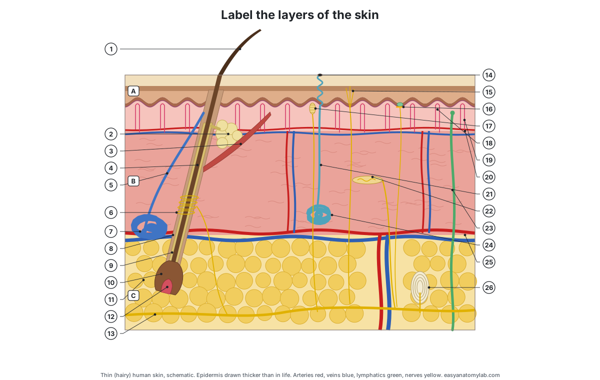
Show the answer key
A Epidermis · B Dermis · C Hypodermis (subcutaneous layer)
- Hair shaft
- Sebaceous gland
- Arrector pili muscle
- Hair root
- Apocrine duct
- Hair follicle plexus (nerve endings around the follicle)
- Apocrine sweat gland
- Outer root sheath
- Inner root sheath
- Hair bulb
- Adipose tissue (fat lobules)
- Hair papilla
- Cutaneous nerve
- Sweat pore
- Free nerve endings
- Merkel cell and Merkel disc
- Meissner (tactile) corpuscle
- Dermal papilla
- Capillary loop
- Subpapillary plexus (arteriole and venule)
- Sweat duct (eccrine duct)
- Ruffini ending
- Lymphatic vessel
- Eccrine sweat gland
- Cutaneous (deep) plexus: artery and vein
- Pacinian (lamellated) corpuscle
Quick quiz
- Which epidermal layer is found only in thick skin? (Stratum lucidum.)
- In which stratum do new keratinocytes form? (Stratum basale.)
- Which layer of the skin has no blood vessels? (The epidermis.)
- Name the two layers of the dermis. (Papillary and reticular.)
- Which gland empties into the hair follicle and makes sebum? (Sebaceous gland.)
- Which receptor senses deep pressure and fast vibration? (Pacinian corpuscle.)
- Which muscle causes goose bumps? (Arrector pili.)
- A burn has blisters and is very painful. How deep is it? (Superficial partial-thickness: into the papillary dermis.)
Related topics: nerve signals from skin receptors are explained on the action potential diagram page, and the rest of the body is in our 3D human anatomy model.
Frequently asked questions
What are the 3 layers of the skin?
The three main layers, from the surface down, are the epidermis (a thin layer of cells with no blood vessels), the dermis (thick connective tissue with blood vessels, nerves, hair follicles and glands) and the hypodermis or subcutaneous layer (fat and loose connective tissue). Strictly, the hypodermis lies under the skin and is not part of it, but most diagrams include it.
What are the 5 layers of the epidermis?
From deep to superficial: stratum basale, stratum spinosum, stratum granulosum, stratum lucidum and stratum corneum. A common memory aid is 'Come, Let's Get Sun Burned' from the top down (corneum, lucidum, granulosum, spinosum, basale). The stratum lucidum is found only in the thick skin of the palms and soles, so thin skin has four layers.
What are the 7 layers of skin?
The '7 layers of skin' is a popular way of counting rather than a standard anatomy term. It usually means the five epidermal strata plus the dermis and the hypodermis. Anatomists describe skin as two layers (epidermis and dermis) with the hypodermis beneath, and the epidermis as having four or five strata.
Which layer of skin has blood vessels?
The dermis. The epidermis has no blood vessels; it gets oxygen and nutrients by diffusion from capillary loops in the dermal papillae. Larger vessels run in two networks, the subpapillary plexus under the papillae and the cutaneous plexus at the base of the dermis, and the biggest vessels pass through the hypodermis.
What is the thickest layer of the skin?
The dermis is the thickest layer of the skin itself; most of it is the reticular dermis. The hypodermis can be thicker still where there is a lot of fat, but it is usually not counted as skin. The epidermis is the thinnest: a review of 142 studies found it ranges from about 0.03 mm on genital skin to about 0.6 mm on the sole of the foot.
Where is the thinnest and the thickest skin on the body?
The upper eyelid has the thinnest skin on the face, under 1 mm in total, and almost no fat beneath it. The thinnest epidermis on the body is on genital skin. The thickest epidermis is on the soles of the feet and the palms, which are covered by thick skin with an extra layer, the stratum lucidum.
Which layers of skin does a burn damage?
A superficial (first-degree) burn damages only the epidermis. A superficial partial-thickness burn reaches the papillary dermis and blisters; a deep partial-thickness burn reaches the reticular dermis. A full-thickness (third-degree) burn destroys the whole epidermis and dermis and reaches the fat. Some systems add a fourth degree for burns into muscle or bone.
Why does the epidermis have no blood vessels?
The epidermis is a stratified epithelium, and epithelia are avascular. Its living cells sit close enough to the capillary loops of the dermal papillae to be fed by diffusion. That is why a shallow scrape that removes only epidermis does not bleed, while a cut that reaches the dermis does.
Is the hypodermis part of the skin?
Not strictly. The skin is the epidermis and dermis. The hypodermis (subcutaneous tissue) lies under the skin and attaches it to the muscles, though the border between dermis and hypodermis can be hard to see. Textbooks usually describe it with the skin because it is part of the integumentary system's supporting tissues and appears in skin diagrams.
References
- OpenStax (2022). Anatomy and Physiology 2e, section 5.1: Layers of the skin. Rice University.
- OpenStax (2022). Anatomy and Physiology 2e, section 5.2: Accessory structures of the skin. Rice University.
- OpenStax (2022). Anatomy and Physiology 2e, section 5.3: Functions of the integumentary system. Rice University.
- OpenStax (2022). Anatomy and Physiology 2e, section 5.4: Diseases, disorders, and injuries of the integumentary system. Rice University.
- OpenStax (2022). Anatomy and Physiology 2e, section 14.1: Sensory perception (Table 14.1, mechanoreceptors of somatosensation). Rice University.
- Merck Manual Professional Edition. Burns: classification by depth.
- Zimmerman, A., Bai, L. & Ginty, D. D. (2014). The gentle touch receptors of mammalian skin. Science 346, 950–954.
- Taylor, N. A. S. & Machado-Moreira, C. A. (2013). Regional variations in transepidermal water loss, eccrine sweat gland density, sweat secretion rates and electrolyte composition in resting and exercising humans. Extreme Physiology & Medicine 2, 4.
- Ludovici, M. et al. (2018). Influence of the sebaceous gland density on the stratum corneum lipidome. Scientific Reports 8, 11500.
- Lintzeri, D. A., Karimian, N., Blume-Peytavi, U. & Kottner, J. (2022). Epidermal thickness in healthy humans: a systematic review and meta-analysis. Journal of the European Academy of Dermatology and Venereology 36, 1191–1200.
- Hwang, K. (2013). Surgical anatomy of the upper eyelid relating to upper blepharoplasty or blepharoptosis surgery. Anatomy & Cell Biology 46, 93–100.
- Korzekwa, S. et al. (2025). Quantitative analysis of the human face skin thickness: a high-frequency ultrasound study. Journal of Clinical Medicine 14, 8401.
- Bottino, D. A. & Bouskela, E. (2023). Non-invasive techniques to access in vivo the skin microcirculation in patients. Frontiers in Medicine 9, 1099107.
- Digital Histology. Skin overview: thick and thin skin, epidermal strata and dermis.
- Grujičić, R. (reviewed by Mytilinaios, D.). Thick skin. Kenhub.
More interactive anatomy models
- Action potential diagramDepolarization, repolarization and ion channels of a neuron action potential.
- Sarcomere diagramZ discs, A and I bands, H zone, actin and myosin of a sarcomere, in 3D.
- Nephron anatomyGlomerulus, Bowman's capsule, tubules, loop of Henle and collecting duct, in 3D.
- Heart blood flowStep-by-step path of blood through the heart's chambers and valves, in 3D.
- Human anatomy1,000+ bones, muscles, organs, vessels and nerves from real anatomical data.
- Octopus anatomyThree hearts, a ring-shaped brain, eight arms and a beak, in 3D.