

Pterodactyl skeleton at a glance
A pterodactyl skeleton is the skeleton of a flying reptile whose wing is a single, enormously long fourth finger. It has a long, low skull full of small pointed teeth, a long neck, a short, compact body with a broad breastbone, a very short tail, strong arms, three small clawed fingers at the wrist, and slender legs with long feet. A skin membrane ran from the tip of the wing finger to the ankle.
"Pterodactyl" is used loosely for any pterosaur, the whole group of Mesozoic flying reptiles. Strictly, it means Pterodactylus, one small genus from the Late Jurassic of Bavaria, and that is the reference animal on this page. Many people searching for a "pterodactyl" mean the big crested Pteranodon or the giant Quetzalcoatlus; both are compared below.
Pterodactylus antiquus lived about 150 million years ago around the lagoons of the Solnhofen archipelago, a scatter of islands in a shallow tropical sea over what is now southern Germany. It was the first pterosaur ever described (by Cosimo Collini in 1784) and is known from more than 30 specimens, most of them juveniles. The largest individuals had a wingspan of just over 1 m.
Pterosaurs are not dinosaurs. They were their close cousins: pterosaurs and dinosaurs are sister branches of the bird-line archosaurs (Avemetatarsalia, also called Ornithodira). Birds are living dinosaurs; pterosaurs left no descendants.
Pterodactyl skeleton labeled: diagram
A labeled pterodactyl skeleton shows the skull with its large nasoantorbital fenestra and orbit, seven neck vertebrae, about fifteen dorsal vertebrae with their ribs, a small fused sacrum and a stub of a tail. Each wing has a humerus with a big deltopectoral crest, a radius and ulna, two syncarpals and a pteroid at the wrist, four metacarpals, three clawed fingers and a wing finger of four phalanges. The legs have a pelvis with prepubic bones, femur, tibia, a short fibula, ankle bones and a five-toed foot with a reduced fifth toe.
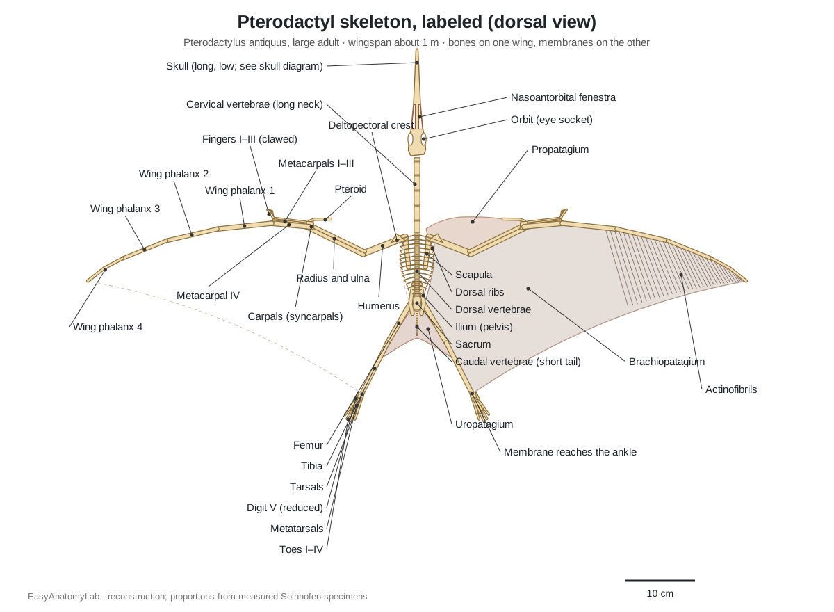
Three proportions make a pterodactyl skeleton look right. The skull is the longest single element, longer than the neck. The wing finger is longer than the whole rest of the arm (about 280 mm against about 210 mm in the model), and its four phalanges get gradually shorter toward the tip. And the shin (tibia) is always longer than the thigh (femur).
Pterodactyl skeleton parts and their functions
This is the text version of the 3D model: every labeled structure of Pterodactylus antiquus, grouped by body region, with what it did.
Skin, crest & wing membranes
- Body (skin and pycnofibres)
- The skin of the neck, trunk and tail, shown faded so the skeleton inside can be seen. Covers the body; in pterosaurs it carried a coat of fine hair-like filaments called pycnofibres.
- Soft-tissue head crest
- A thin crest of soft tissue that rose above the snout opening and eye socket in large individuals. Probably display (showing maturity or species); it was too thin to help with flight.
- Occipital lappet
- A small tab of soft tissue that projected backward from the back of the skull. Unknown; probably display, along with the head crest.
- Propatagium (forewing membrane)
- The membrane in front of the arm, from the shoulder to the wrist, supported at its front edge by the pteroid bone. Forms the front of the wing; moving the pteroid changed its size and angle, like a flap on an aircraft wing.
- Brachiopatagium (main wing membrane, plagiopatagium)
- The main flight membrane, stretched from the wing finger to the side of the body and the hind leg. Produces most of the lift; its outer part was stiffened by actinofibrils.
- Uropatagium (cruropatagium) (tail membrane, cruropatagium)
- A membrane between the hind legs. May have helped with pitch control and braking in flight.
- Actinofibrils (aktinofibrils)
- Stiffening fibres in the outer part of the wing membrane. Strengthened the membrane and may have let the animal fine-tune its tension in flight.
Skull & jaws
- Premaxilla (premaxillae)
- The bone forming the tip of the long, low snout and the bar over the large skull opening. Holds the front, largest teeth and roofs the snout.
- Maxilla
- The upper-jaw bone behind the premaxilla that forms the lower edge of the nasoantorbital fenestra. Carries the rear upper teeth and forms part of the palate.
- Nasoantorbital fenestra (NAOF)
- One large opening on each side of the skull, made by the nostril and the antorbital opening merging. Lightens the long skull; it also housed air spaces and jaw-muscle space.
- Lacrimal
- The slender bar of bone between the nasoantorbital fenestra and the eye socket. Braces the skull roof against the jaw bar and separates the two openings.
- Braincase and skull roof (frontal, parietal, postorbital, occipital)
- The rounded back of the skull that holds the brain, with the frontal roof over the eye sockets. Protects the brain, eyes and inner ears and anchors jaw and neck muscles.
- Bony crest (sagittal crest)
- A low ridge of bone along the top of the skull in large individuals. Supported the taller soft-tissue crest.
- Sclerotic ring
- A ring of small overlapping bony plates inside the eye. Supported the eyeball and helped it keep its shape.
- Jugal
- The cheek bone, a bar below the eye socket. Completes the upper jaw bar and braces the skull.
- Quadrate
- The bone at the back of the skull that the lower jaw hinges on. Forms the upper half of the jaw joint.
- Mandible (lower jaw)
- The long, straight lower jaw, its two halves fused at the front into a long symphysis. Carries the lower teeth; the mouth is shown slightly open.
- Hyoid bones
- A pair of thin, slightly bowed rods between the halves of the lower jaw. Supported the tongue and throat.
Teeth
- Upper teeth
- Low, conical, pointed teeth in the premaxilla and maxilla, largest at the front. Seizing small prey such as invertebrates and small fish.
- Lower teeth
- Conical teeth along the front of the lower jaw, decreasing in size towards the back. Interlock with the upper teeth to grip prey.
Vertebrae, ribs & sternum
- Atlas and axis
- The first two neck vertebrae, which carry the skull. Let the head nod and turn.
- Cervical vertebrae (C3–C7) (neck vertebrae)
- Five long, low neck vertebrae that make Pterodactylus's neck longer than in most of its relatives. Hold the long head out in front and let the animal strike at prey.
- Dorsal vertebrae (trunk vertebrae)
- About fifteen short vertebrae of the back, which carry the ribs. Support the trunk between the wings and the legs.
- Sacrum
- A few fused vertebrae joined to the ilia of the pelvis. Transfers weight from the spine to the hind legs.
- Caudal vertebrae (short tail)
- A very short tail of tiny vertebrae. Little or no role in steering; a short tail is a hallmark of pterodactyloids.
- Dorsal ribs
- Thin, slightly curved ribs on the dorsal vertebrae; the front ones meet the sternum. Enclose the lungs and heart and help with breathing.
- Gastralia (belly ribs)
- Thin, overlapping rods in the belly wall, not attached to the spine. Supported the belly and probably helped with breathing.
- Sternum (breastbone)
- A broad, shallow, shield-shaped breastbone with a low keel. Anchors the large flight muscles that pull the wings down.
- Cristospine (cristospina)
- A spine-like process that projects forward from the front of the sternum. Extends the attachment area for flight and neck muscles.
Shoulder girdle
- Scapula (shoulder blade)
- A long, strap-like bone that runs from the shoulder joint back over the ribs. Forms the upper half of the shoulder socket and anchors muscles that raise the wing.
- Coracoid
- A strut from the shoulder joint down to the front of the sternum. Braces the shoulder against the breastbone during the downstroke.
Wing skeleton (forelimb)
- Humerus
- The short, stout upper-arm bone. Takes the power of the flight muscles; it was also used to push off in a quadrupedal launch.
- Deltopectoral crest
- A large flange on the front of the upper humerus. Attachment for the pectoralis and deltoid muscles that power the wing.
- Radius
- The front bone of the forearm, as long as the ulna and lying parallel to it. Supports the inner wing and the propatagium in front of it.
- Ulna
- The thicker rear bone of the forearm. Carries most of the load of the forearm.
- Proximal syncarpal
- The fused first row of wrist bones. Forms the joint between forearm and hand.
- Distal syncarpal
- The fused second row of wrist bones that carries the metacarpals. Joins the wrist to the hand and the wing metacarpal.
- Preaxial carpal (medial carpal)
- A small extra wrist bone on the front of the wrist. Acts as a strut that carries the pteroid.
- Pteroid bone
- A thin, rod-like bone found only in pterosaurs, jutting from the wrist. Supported and controlled the propatagium.
- Metacarpals I–III
- Three thin hand bones lying along the front of the wing metacarpal. Carry the three clawed fingers.
- Metacarpal IV (wing metacarpal)
- The stout hand bone of the wing finger, ending in a pulley-like joint. Supports the wing finger, which folds back at its pulley joint.
- Fingers I–III (with claws) (manual digits, manual unguals)
- Three small fingers with 2, 3 and 4 phalanges, each ending in a curved claw. Used for walking on all fours and probably for climbing and handling food.
- Wing phalanx 1
- The first and longest bone of the wing finger (digit IV). Supports the inner part of the wing finger; its base has the extensor tendon process.
- Wing phalanx 2
- The second bone of the wing finger. Carries the leading edge of the outer wing.
- Wing phalanx 3
- The third bone of the wing finger. Supports the outer wing.
- Wing phalanx 4
- The last, tapering bone of the wing finger, with no claw. Forms the tip of the wing.
Pelvis & hindlimb
- Ilium
- The long upper bone of the pelvis, fused to the sacrum. Joins the legs to the spine and anchors thigh muscles.
- Pubis and ischium (ischiopubic plate)
- The lower bones of the pelvis, fused into a plate below the hip socket. Closes the pelvis and carries the prepubis.
- Prepubis (prepubic bones)
- A pair of separate bones that project forward from the pelvis under the belly. Supported the belly; their mobility may have helped breathing.
- Femur
- The slender thigh bone. Supports the body when walking; in flight the leg held the back of the wing membrane.
- Tibia (tibiotarsus)
- The long shin bone; in adults the upper ankle bones fuse to it. Main bone of the lower leg; the wing membrane attached near its lower end.
- Fibula
- A very thin bone along the outside of the tibia, only half its length. Attachment for lower-leg muscles.
- Tarsals (ankle bones)
- Small, tightly fitted ankle bones: two proximal and three distal. Form a simple hinge ankle.
- Metatarsals I–IV
- Four long, slender foot bones; metatarsal II is the longest. Form the long, narrow foot.
- Metatarsal V and digit V
- A tiny splint-like fifth metatarsal with a single short phalanx and no claw. Reduced in pterodactyloids; in early pterosaurs a long fifth toe supported the uropatagium.
- Toes I–IV (with claws)
- Four clawed toes with 2, 3, 4 and 4 phalanges in adults. Walking and gripping.
Pterodactyl head and skull
The skull of Pterodactylus is long, low and straight, with a slender snout and up to about 25 small, conical teeth on each side of each jaw in large individuals, roughly 90 in all. The teeth are largest at the front and shrink toward the back, and they fill about three-quarters of the jaw. Behind the snout, one huge opening on each side, the nasoantorbital fenestra, takes up a fifth to a third of the skull length. Then comes a round eye socket with a bony sclerotic ring, and a rounded braincase.
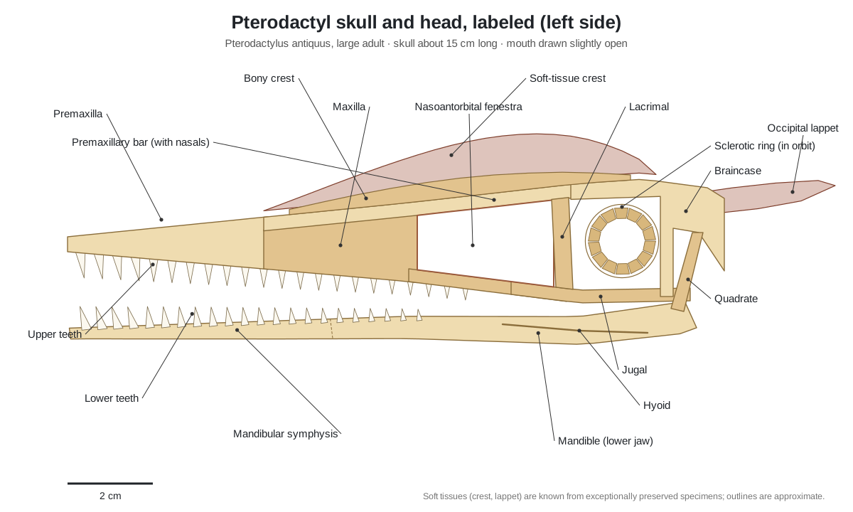
The nasoantorbital fenestra is a defining feature of pterodactyloids: the nostril and the antorbital opening, separate in early long-tailed pterosaurs, merged into one. The bars of bone left around it are the premaxilla on top, the maxilla below and the lacrimal behind. The jugal forms the cheek under the eye, and the lower jaw hinges on the quadrate at the back.
Large adults also had a head crest. A low bony ridge ran along the midline above the fenestra and eye; on top of it stood a taller crest of soft tissue, and a small soft flap, the occipital lappet, stuck out from the back of the head. These soft parts are known only from exceptionally preserved specimens. They were most likely for display, since young animals lacked them.
- Premaxilla: Holds the front, largest teeth and roofs the snout.
- Nasoantorbital fenestra: Lightens the long skull; it also housed air spaces and jaw-muscle space.
- Sclerotic ring: Supported the eyeball and helped it keep its shape.
- Mandible (lower jaw): Carries the lower teeth; the mouth is shown slightly open.
- Upper teeth: Seizing small prey such as invertebrates and small fish.
- Bony crest (sagittal crest): Supported the taller soft-tissue crest.
Its diet fits its teeth. A study of microscopic tooth wear across 17 pterosaur genera found Pterodactylus to be a generalist that ate mostly invertebrates, while its Solnhofen neighbour Rhamphorhynchus was a fish-eater (Bestwick et al. 2020).
Neck, backbone, ribs and tail
The pterodactyl backbone has a long neck of seven cervical vertebrae, about fifteen dorsal vertebrae that carry the ribs, a few fused sacral vertebrae joined to the hips, and a very short tail. The middle neck vertebrae (C3–C7) are long and low, and in the largest individuals the neck is as long as or longer than the whole trunk column. Pterodactyloids have no neck ribs.
Two features of big pterosaurs are missing here. Pterodactylus has no notarium, the fused block of front dorsal vertebrae that braces the shoulder blades in large species such as Pteranodon (where six vertebrae fuse), and its tail is a stub of tiny bones instead of the long, stiff tail of Rhamphorhynchus.
The chest is short and deep. Thin ribs curve down to a broad, shallow sternum with a low keel; a spine-like cristospine projects forward from its front edge. The belly was supported by thin gastralia, and a pair of separate prepubic bones projected forward from the pelvis under the belly, a distinctive pterosaur feature.
- Cervical vertebrae (C3–C7): Hold the long head out in front and let the animal strike at prey.
- Dorsal vertebrae: Support the trunk between the wings and the legs.
- Sternum (breastbone): Anchors the large flight muscles that pull the wings down.
- Cristospine: Extends the attachment area for flight and neck muscles.
- Prepubis: Supported the belly; their mobility may have helped breathing.
- Caudal vertebrae (short tail): Little or no role in steering; a short tail is a hallmark of pterodactyloids.
Pterodactyl wing skeleton
The pterodactyl wing skeleton is the whole arm and hand. From the shoulder outward it has a short, stout humerus with a large deltopectoral crest for the flight muscles; a radius and ulna of equal length lying side by side; a wrist of two fused rows of carpals (the proximal and distal syncarpals) plus a preaxial carpal; the pteroid, a bone found only in pterosaurs; four metacarpals; three small clawed fingers; and the wing finger, digit IV, with four long phalanges and no claw.
In Pterodactylus the humerus is a quarter to a third shorter than the radius, the wing metacarpal is about as long as the humerus, and the first wing phalanx is clearly longer than the metacarpal. The finger formula of the hand is 2-3-4-4-0: two, three and four phalanges in fingers I–III, four in the wing finger, and no fifth finger. The wing finger folded backward along the body when the animal walked, and the three small fingers pointed forward for walking and climbing.
The pteroid is controversial. It is a thin rod that jutted from the wrist and held up the front edge of the propatagium. Bennett and most reconstructions point it toward the body, giving a narrow forewing (the 3D model shows this). Wilkinson, Unwin and Ellington argued it could swing forward in flight, giving a much broader forewing that acted like the leading-edge flap of an airliner; their wind-tunnel models produced very high lift that way. Neither view has been settled.
- Humerus: Takes the power of the flight muscles; it was also used to push off in a quadrupedal launch.
- Deltopectoral crest: Attachment for the pectoralis and deltoid muscles that power the wing.
- Radius: Supports the inner wing and the propatagium in front of it.
- Ulna: Carries most of the load of the forearm.
- Proximal syncarpal: Forms the joint between forearm and hand.
- Pteroid bone: Supported and controlled the propatagium.
- Metacarpal IV (wing metacarpal): Supports the wing finger, which folds back at its pulley joint.
- Wing phalanx 1: Supports the inner part of the wing finger; its base has the extensor tendon process.
- Wing phalanx 4: Forms the tip of the wing.
The wing membrane is divided into named parts:
| Membrane | Where it is | Supported by |
|---|---|---|
| Propatagium | In front of the arm, from the shoulder to the wrist | Pteroid, humerus, radius |
| Brachiopatagium | From the wing finger to the side of the body and the ankle | Wing finger, forearm, body, hindlimb |
| Uropatagium (cruropatagium) | Between the hind legs | Thighs and shins (extent uncertain in Pterodactylus) |
| Actinofibrils | Fibres inside the outer brachiopatagium | Stiffen the membrane; at least three crossing layers |
Where the membrane is completely preserved in fossils, its trailing edge reaches the lower leg or ankle, so the wing was broad near the body rather than a narrow sail tied to the hip.
Wing skeleton of bats, birds and pterodactyls compared
Pterosaurs, bats and birds all fly with a modified forelimb, so their wing bones are homologous: the same humerus, radius, ulna, wrist and hand bones as in a human arm. The wings themselves are analogous, evolved separately. A pterosaur hangs its wing on one huge finger, a bat stretches skin between four long fingers, and a bird carries feathers on a short, fused hand.
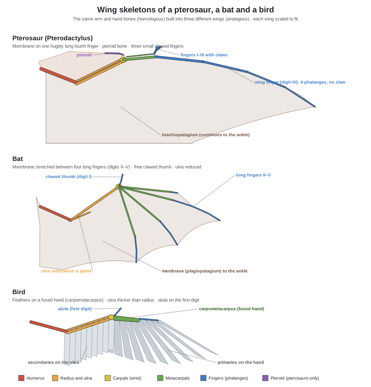
| Feature | Pterosaur (Pterodactylus) | Bat | Bird |
|---|---|---|---|
| Flight surface | Skin membrane | Skin membrane | Feathers |
| Main wing support | Digit IV only (4 phalanges) | Digits II–V | Ulna and fused hand |
| Free clawed fingers | Three (I–III) | Thumb (and digit II in fruit bats) | Usually none; alula on the first digit |
| Special wing bone | Pteroid | None (the calcar is on the heel) | Carpometacarpus (fusion) |
| Forearm | Radius and ulna equal | Radius large, ulna reduced | Ulna thicker than radius |
| Collarbones | None | Two separate clavicles | Fused into a wishbone |
| Breastbone | Broad, low keel, cristospine | Small keel on the manubrium | Large keel |
| Hollow, air-filled bones | In many pterosaurs, limited in small ones | No (marrow-filled) | Yes |
| Hind legs in the wing | Membrane reaches the ankle | Membrane reaches the foot | Legs free |
See the full skeletons on the bat skeleton and bird skeleton pages. The repeated evolution of similar solutions, such as a keeled breastbone, is an example of convergent evolution.
Hollow, pneumatic bones
Pterodactyl bones were hollow: the long bones of the wings and legs were thin-walled tubes. In many pterosaurs they were also pneumatic, meaning air sacs from the lungs grew into them through small openings, as in birds. Pneumaticity tracks size. Claessens, O'Connor and Unwin found it mostly confined to the vertebrae in early pterosaurs, spreading into the limbs in big pterodactyloids, generally absent in small species, almost always present above a 2.5 m wingspan and universal above 5 m.
So a 1 m Pterodactylus had light, hollow bones but probably little of the extensive air-sac invasion seen in Pteranodon or Quetzalcoatlus. Pneumatic openings imply a bird-like, flow-through breathing system with air sacs, which the same study dates some 70 million years before the comparable bird system. Air-filled bones lowered body density and may have been one key to pterosaur gigantism.
Hollow does not mean weak. In giant pterosaurs the humerus was up to about three times more resistant to breaking than the humerus of a bird of the same size, while the femur was relatively weaker (Witton & Habib 2010).
Pterodactyl foot and the quadrupedal launch
The pterodactyl foot is long and narrow, with four slender metatarsals (the second is the longest), four clawed toes and a tiny fifth toe. In adults the toe formula is 2-3-4-4-1; juveniles have 2-3-3-3-1, so the toes gain bones as the animal grows. The fifth metatarsal is a short splint with one small phalanx and no claw. In early long-tailed pterosaurs this toe was long and helped spread the membrane between the legs; its reduction is a pterodactyloid feature.
- Femur: Supports the body when walking; in flight the leg held the back of the wing membrane.
- Tibia (tibiotarsus): Main bone of the lower leg; the wing membrane attached near its lower end.
- Fibula: Attachment for lower-leg muscles.
- Tarsals (ankle bones): Form a simple hinge ankle.
- Metatarsals I–IV: Form the long, narrow foot.
- Metatarsal V and digit V: Reduced in pterodactyloids; in early pterosaurs a long fifth toe supported the uropatagium.
Trackways show that pterosaurs walked on all fours. The most common pterosaur trackway, Pteraichnus, was named by Stokes in 1957 from the Upper Jurassic of Arizona. Its tracks pair the hindfoot with a distinctive handprint made by the small fingers. More than 50 tracksites are known, and many Late Jurassic tracks are small (foot length about 2–10 cm), a fair match for animals of Pterodactylus size, though trackmakers can rarely be named.
The quadrupedal launch hypothesis says pterosaurs took off like pole-vaulters: crouching on all fours, then pushing off mainly with the powerful arms before the first wingbeat. Birds take off with their legs. Habib found that pterosaur humeri are far stronger relative to their femora than in any bird he sampled, which is what a forelimb-powered launch predicts. Many pterosaur researchers now favour this model, and it helps explain how giants with 10 m wingspans could get airborne.
Pterodactyl size: how big was a pterodactyl?
Pterodactylus antiquus was small. The largest known skull is about 15 cm long, and the largest individuals had wingspans of just over 1 m (Bennett 2013). Most fossils are much smaller juveniles: skull lengths in the sample run from 23 mm to 149 mm, and the second pterosaur specimen ever found, a baby, had a wingspan of only about 25 cm. The specimens fall into size groups that Bennett interpreted as year classes.
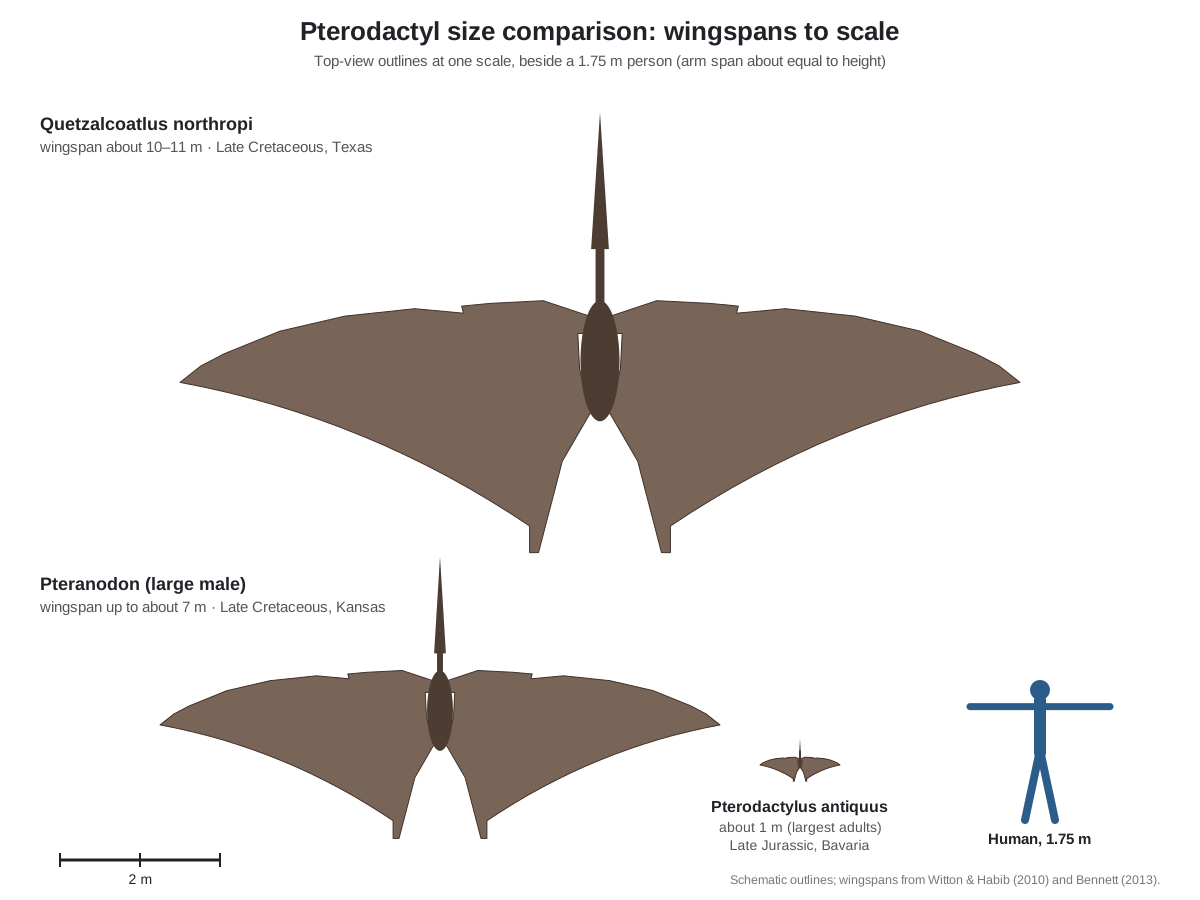
The "giant pterodactyl skeletons" in museums are other pterosaurs. Pteranodon reached a wingspan of about 7 m in large males, and the azhdarchid Quetzalcoatlus northropi about 10–11 m, making it one of the largest flying animals ever. A tall human's arm span is about 1.75 m.
Pterodactylus vs Pteranodon vs Quetzalcoatlus
Pterodactylus, Pteranodon and Quetzalcoatlus are all pterodactyloid pterosaurs, but they lived tens of millions of years apart and look very different. Pterodactylus is a small, toothed Jurassic animal; the other two are large, toothless Cretaceous giants. If you picture a "Cretaceous pterodactyl skeleton" with a long backward crest, you are picturing Pteranodon.
| Pterodactylus antiquus | Pteranodon | Quetzalcoatlus northropi | |
|---|---|---|---|
| Age | Late Jurassic, about 150 million years ago | Late Cretaceous (late Coniacian–Campanian) | Late Cretaceous (Maastrichtian) |
| Where found | Plattenkalk (Solnhofen limestone), Bavaria, Germany | Niobrara Formation and Pierre Shale, Kansas, USA | Javelina Formation, Big Bend, Texas, USA |
| Wingspan | Up to just over 1 m | Up to about 7 m in large males; smaller females | About 10–11 m |
| Teeth | Yes, up to about 25 per jaw side | None (the name means 'wing without tooth') | None |
| Crest | Low bony crest with a soft-tissue crest | Large bony crest, pointing backward in large males | — |
| Notarium | No | Yes (six fused dorsal vertebrae) | — |
| Limb-bone pneumaticity | Limited (small species) | Extensive | Extensive |
| Fossil record | 30+ specimens, many complete, mostly juveniles | Well over 1,000 specimens | Partial skeletons |
Pteranodon was named by Othniel Marsh in 1876 from the Niobrara Chalk of Kansas, where the first toothless pterosaurs and the first truly giant flying animals were found. Quetzalcoatlus was found in Big Bend National Park, Texas, in 1971 and named by Douglas Lawson in 1975; a reconstructed skeleton hangs in the Texas Memorial Museum in Austin.
Long-tailed "rhamphorhynchoids" vs pterodactyloids
Pterosaurs are traditionally split into two grades: the early, long-tailed "rhamphorhynchoids" and the short-tailed pterodactyloids, named after Pterodactylus. The split goes back to Hermann von Meyer and was formalised by Plieninger in 1901. "Rhamphorhynchoids" are not a natural group, because pterodactyloids evolved from among them, so the name is used in quotation marks today.
| Feature | 'Rhamphorhynchoids' (e.g. Rhamphorhynchus, Dimorphodon) | Pterodactyloids (e.g. Pterodactylus, Pteranodon) |
|---|---|---|
| Time range | Late Triassic to Jurassic | Late Jurassic to the end of the Cretaceous |
| Tail | Long, often stiffened, some with a vane | Very short |
| Skull openings | Separate nostril and antorbital fenestra | One nasoantorbital fenestra |
| Neck | Shorter | Longer, without neck ribs |
| Wing metacarpal | Relatively short | Long (about humerus length in Pterodactylus) |
| Fifth toe | Long, helps spread the uropatagium | Reduced to a stub |
So a "pterodactyloid skeleton" is recognisable by its short tail, single big skull opening, long neck and long hand. Pterodactylus is an early pterodactyloid.
Is a pterodactyl a dinosaur? Classification
No. A pterodactyl is a pterosaur, and pterosaurs are a separate branch of reptiles. Both pterosaurs and dinosaurs belong to the bird-line archosaurs (Avemetatarsalia, or Ornithodira), so they are close relatives, and crocodiles sit on the other main archosaur branch. In 2020, Ezcurra and colleagues showed that the closest known relatives of pterosaurs are the lagerpetids, small, ground-running, non-flying dinosaur precursors. That narrowed the gap between pterosaurs and their ancestors and confirmed that pterosaurs belong on the bird line.
| Rank | Group |
|---|---|
| Clade | Archosauria (crocodiles, pterosaurs, dinosaurs and birds) |
| Clade | Avemetatarsalia / Ornithodira (the bird line) |
| Order | Pterosauria |
| Suborder | Pterodactyloidea |
| Genus | Pterodactylus Cuvier, 1809 |
| Species | Pterodactylus antiquus (Soemmerring, 1812) |
Pterosaurs were the first vertebrates to evolve powered flight. They appeared in the Late Triassic and died out at the end of the Cretaceous, 66 million years ago, together with the non-bird dinosaurs. For a dinosaur skeleton to compare with, see the Velociraptor skeleton.
Real pterodactyl fossils and where to see them
Real Pterodactylus fossils come almost entirely from the Late Jurassic Plattenkalk of Bavaria, the fine limestone quarried around Solnhofen and Eichstätt for building stone and lithographic plates. Animals that fell into the stagnant, salty bottom waters of the lagoons were buried quickly, so many specimens are complete, articulated skeletons, and some preserve soft tissues such as the head crest and wing membranes. More than 500 pterosaur specimens are known from these deposits.
- Munich. The holotype of Pterodactylus antiquus (BSP AS I 739), the very first pterosaur described, has been in the Bavarian State Collection of Palaeontology and Geology (SNSB-BSPG) since 1802, when it moved there from Mannheim. The collection's Paleontological Museum at Richard-Wagner-Str. 10 is free to enter and displays flying reptiles and the Munich Archaeopteryx; check the museum's website for which pterosaur specimens are on view.
- Solnhofen and Eichstätt. The Bürgermeister-Müller-Museum in Solnhofen holds the largest known skull (BMMS 7), and the Jura-Museum in Eichstätt also holds Solnhofen pterosaurs.
- Elsewhere. Pterodactylus specimens are also held by the Teylers Museum (Haarlem), the Senckenberg Museum (Frankfurt), the State Museum of Natural History (Stuttgart) and, for the Painten skeleton, the Dinosaurier Museum Altmühltal (Denkendorf).
Casts and replicas of Solnhofen pterosaurs and of Pteranodon are widely made for museums and teaching. A good cast is moulded from a named specimen; when you see one, the label should say which original it copies.
History: from Collini to Cuvier
The pterodactyl was the first pterosaur known to science. Cosimo Alessandro Collini, keeper of the Elector Karl Theodor's natural history collection in Mannheim, published an accurate engraving of the Eichstätt specimen in 1784. He saw that its arms could fold and even suspected a membrane, but thought it was a sea creature. In 1801 Georges Cuvier recognised it as a flying reptile with a membrane on one long finger, working only from Collini's engraving, and in 1809 he named it "Ptero-Dactyle", wing finger, later Latinised to Pterodactylus.
Others disagreed. Blumenbach thought it was a waterbird, and Samuel Thomas von Soemmerring, who studied the actual fossil in Munich, decided it was a bat, naming it Ornithocephalus antiquus in 1812; his species name antiquus is still used. Soemmerring mistook the humeri for collarbones, which pterosaurs do not have. Hermann von Meyer later described one specimen as Pterodactylus crassipes; more than a century later it was recognised as an Archaeopteryx, the Haarlem specimen.
How Pterodactylus grew: year classes in the Solnhofen limestone
Most Pterodactylus fossils are young animals. Skull lengths run from 23 mm in the smallest specimen to 149 mm in the largest complete skull (BMMS 7, in the Bürgermeister-Müller-Museum, Solnhofen), and the specimens are not spread evenly across that range. They cluster into a small size class with skulls of about 15–45 mm and a large class of about 55–95 mm, separated by gaps. Bennett interpreted these as year classes: animals that died in their first and second years. (Augustin et al. (2022), The geologically oldest specimen of Pterodactylus, Fossil Record 25: 331–343.)
The skeleton changes as the animal grows. Tooth count rises with skull length (about 12 upper teeth per side in the smallest skull, 18 in the 108 mm holotype), the eye socket becomes relatively smaller, the fused front of the lower jaw becomes relatively longer, and the toes gain phalanges, from a formula of 2-3-3-3-1 in juveniles to 2-3-4-4-1 in adults. A low bony crest appears only in large individuals. A nearly complete skeleton from Painten, with a 48 mm skull, is a rare animal from the gap between the two size classes and is also the geologically oldest Pterodactylus, from the latest Kimmeridgian. (Augustin et al. (2022), The geologically oldest specimen of Pterodactylus, Fossil Record 25: 331–343.)
These growth changes caused a century of confusion. Small juveniles were named as separate species, and the second pterosaur specimen ever found, a baby with a wingspan of only about 25 cm, was even given its own genus (Ptenodracon). Hermann von Meyer described more than 40 Solnhofen pterosaur specimens under 20 species names. (Wellnhofer (2008), A short history of pterosaur research, Zitteliana B28: 7–19.)
One species or several? The Pterodactylus debate
Vidovic and Martill argued that some specimens once called Pterodactylus belong in other genera. They placed P. scolopaciceps in a new genus, Aerodactylus, which has a more elongate snout, a straight thin pteroid and no notarium, and they found that Bennett's broad P. antiquus sample contained at least two different forms. (Vidovic & Martill (2014), Pterodactylus scolopaciceps and the problem of cryptic pterosaur taxa in early ontogeny, PLoS ONE.)
In a later study they kept Diopecephalus kochi as a valid genus separate from Pterodactylus, using geometric morphometrics of the prepubic bones and a cladistic analysis. Other workers, including Bennett, treat P. kochi as a young P. antiquus, and the 2022 Painten study followed that view because the two were hard to tell apart. There is no consensus yet, so this page describes P. antiquus in the broad sense. (Vidovic & Martill (2018), The taxonomy and phylogeny of Diopecephalus kochi and 'Germanodactylus rhamphastinus', Geol. Soc. Spec. Publ. 455.)
What the wing membrane was made of
The best evidence for the inside of a pterosaur wing comes from Jeholopterus, a small Chinese pterosaur preserved with soft tissue. Its outer membrane (the actinopatagium) contains at least three layers of stiff actinofibrils, each layer running in a different direction, while the inner membrane (the tenopatagium) lacks them. The fibres are about 0.1 mm thick and packed 4–7 per millimetre. The body was covered in hair-like pycnofibres. (Kellner et al. (2009), The soft tissue of Jeholopterus and the structure of the pterosaur wing membrane, Proc. R. Soc. B (PMC).)
How far back the membrane reached was debated for decades. Elgin, Hone and Frey reviewed every specimen with preserved membrane and found that, wherever the brachiopatagium is complete, its trailing edge reaches the lower leg or ankle. Reconstructions with a narrow wing ending at the hip or knee rest on incomplete or shrunken membranes. (Elgin, Hone & Frey (2011), The extent of the pterosaur flight membrane, Acta Palaeontologica Polonica 56: 99–111.)
Mechanical models add a further constraint. Palmer and Dyke argue that the trailing edge must have been concave, because a convex edge would flutter and lose tension, and that actinofibrils mainly resisted spanwise tension in a membrane less than 1 mm thick. (Palmer & Dyke (2012), Constraints on the wing morphology of pterosaurs, Proc. R. Soc. B (PMC).)
The pteroid debate
The pteroid is a rod-like bone found only in pterosaurs. Most researchers reconstruct it pointing towards the body, with a narrow forewing. Wilkinson, Unwin and Ellington tested the alternative, a pteroid pointing forwards in flight, with scale models in a wind tunnel. The forward position gave better lift-to-drag ratios and exceptionally high maximum lift, which they suggested could have helped even the largest pterosaurs take off and land. (Wilkinson, Unwin & Ellington (2006), High lift function of the pteroid bone and forewing of pterosaurs, Proc. R. Soc. B.)
Bennett's reconstruction of the pterodactyloid forelimb describes most carpals fused into two large syncarpals, a strut-like preaxial carpal and a medially directed pteroid, with digits I–III (2, 3 and 4 phalanges) flexing forward for walking and climbing, and digit IV folding backward. The 3D model on this page follows the medial orientation. (Bennett (2008), Morphological evolution of the wing of pterosaurs: myology and function, Zitteliana B28: 127–141.)
Quadrupedal launch: taking off with the arms
Habib tested one prediction of the quadrupedal launch hypothesis: if pterosaurs vaulted into the air using mainly the forelimbs, their humeri should be far stronger than their femora. In three pterosaurs (Anhanguera, Zhejiangopterus and Dorygnathus) the ratio of humeral to femoral bending strength was well above that of every bird sampled, including the tropicbird Phaethon, which can hardly use its legs to take off. (Habib (2008), Comparative evidence for quadrupedal launch in pterosaurs, Zitteliana B28: 159–166.)
Witton and Habib extended this to giant pterosaurs. They found pterosaur humeri up to about three times more resistant to failure than those of similar-sized birds, and argued that a 200–250 kg azhdarchid with a 10 m wingspan could launch from a standing start by vaulting over its forelimbs. (Witton & Habib (2010), On the size and flight diversity of giant pterosaurs, PLoS ONE.)
Pteraichnus and other pterosaur trackways
At least 50 pterosaur tracksites are known, from the Late Jurassic to the Late Cretaceous. Pteraichnus, named by Stokes in 1957 from the Upper Jurassic of Arizona, is by far the most common ichnogenus. Its trackways combine hindfoot prints with distinctive handprints, showing that pterosaurs walked on all fours. A 1984 paper argued that Pteraichnus was made by crocodilians, but later finds confirmed the pterosaur makers. (Lockley, Harris & Mitchell (2008), A global overview of pterosaur ichnology, Zitteliana B28: 185–198.)
Many Late Jurassic Pteraichnus footprints are small (foot length about 2–10 cm), a good size match for animals like Pterodactylus, although trackmakers are rarely identified to genus. Sites often contain dense tracks of many sizes alongside invertebrate traces, suggesting flocks feeding on shorelines, and some show swim tracks made by floating pterosaurs touching the bottom with their feet. (Lockley, Harris & Mitchell (2008), A global overview of pterosaur ichnology, Zitteliana B28: 185–198.)
Air-filled bones and pterosaur breathing
Claessens, O'Connor and Unwin mapped pneumatic openings across pterosaurs. In early pterosaurs pneumaticity is mostly limited to the dorsal vertebrae. It spreads into the limbs in large pterodactyloids such as Pteranodon and Anhanguera and, independently, in azhdarchoids. It is generally absent in small species, almost always present above 2.5 m wingspan and universal at 5 m or more. (Claessens, O'Connor & Unwin (2009), Respiratory evolution facilitated the origin of pterosaur flight and aerial gigantism, PLoS ONE.)
Pneumatic openings imply air sacs connected to the lungs, and the authors infer a bird-like flow-through breathing system some 70 million years before the comparable avian system. Lower body density from air-filled bones may have been one key to pterosaur gigantism. (Claessens, O'Connor & Unwin (2009), Respiratory evolution facilitated the origin of pterosaur flight and aerial gigantism, PLoS ONE.)
Pterodactyl skeleton worksheet: label it yourself
Use this unlabeled pterodactyl skeleton diagram as a worksheet or template. Write the name of each numbered part, then check your answers against the key. All the answers appear in the labeled diagram and the 3D model above.
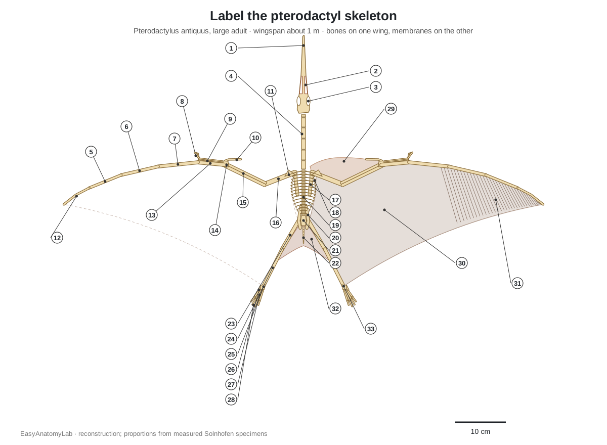
Answer key
- Skull
- Nasoantorbital fenestra
- Orbit (eye socket)
- Cervical vertebrae
- Wing phalanx 3
- Wing phalanx 2
- Wing phalanx 1
- Fingers I–III (clawed)
- Metacarpals I–III
- Pteroid
- Deltopectoral crest
- Wing phalanx 4
- Metacarpal IV (wing metacarpal)
- Carpals (syncarpals)
- Radius and ulna
- Humerus
- Scapula
- Dorsal ribs
- Dorsal vertebrae
- Ilium (pelvis)
- Sacrum
- Caudal vertebrae (short tail)
- Femur
- Tibia
- Tarsals
- Digit V (reduced)
- Metatarsals
- Toes I–IV
- Propatagium
- Brachiopatagium
- Actinofibrils
- Uropatagium
- Ankle (where the brachiopatagium attaches)
- Which finger forms the pterosaur wing? (Digit IV, the fourth finger.)
- How many phalanges are in the wing finger? (Four.)
- What bone is found only in pterosaurs? (The pteroid.)
- Which skull opening marks a pterodactyloid? (The single nasoantorbital fenestra.)
- Where did the main wing membrane attach at the back? (The lower leg or ankle.)
- Name one difference between Pterodactylus and Pteranodon. (For example: teeth vs toothless; about 1 m vs 7 m wingspan; no notarium vs a notarium.)
- Are pterosaurs dinosaurs? (No; they are close relatives within Avemetatarsalia.)
How to draw a pterodactyl skeleton
To draw a pterodactyl skeleton, start with the long, low skull and a neck a little shorter than the skull. Add a small, egg-shaped rib cage and a tiny tail. For each wing, draw a short thick humerus, a longer forearm, a metacarpal about as long as the humerus, and then a wing finger of four bones that together are longer than the rest of the arm. Put three small clawed fingers at the wrist and a thin pteroid pointing in from the wrist. Finish with slender legs (shin longer than thigh) and long feet, and stretch the membrane from the wingtip to the ankles.
Frequently asked questions
Is a pterodactyl a dinosaur?
No. Pterodactyls (pterosaurs) were flying reptiles that lived alongside dinosaurs, but they are a separate group. Both belong to the bird-line archosaurs (Avemetatarsalia or Ornithodira), and the closest known relatives of pterosaurs are the lagerpetids, small, ground-running dinosaur precursors. Birds are dinosaurs; pterosaurs are not.
What is the difference between a pterodactyl and a pterosaur?
Pterosaur is the name of the whole group of flying reptiles. Pterodactylus is one genus, the first pterosaur ever described, from the Late Jurassic of Bavaria. In everyday English 'pterodactyl' is used loosely for any pterosaur, including Pteranodon and Quetzalcoatlus, which are not Pterodactylus.
How big was a pterodactyl?
Pterodactylus antiquus was small. Most fossils are juveniles with wingspans of about 25–50 cm, and the largest individuals reached a wingspan of just over 1 m, with a skull about 15 cm long. The giant 'pterodactyls' people imagine are other pterosaurs: Pteranodon had a wingspan of up to about 7 m and Quetzalcoatlus about 10–11 m.
What bones make up a pterodactyl wing?
The wing is the whole arm and hand: the humerus with its large deltopectoral crest, the radius and ulna, the wrist bones (proximal and distal syncarpals and a preaxial carpal carrying the pteroid), four metacarpals, three small clawed fingers, and the huge fourth finger made of four long phalanges. The membrane stretched from that fourth finger to the ankle.
Did pterodactyls have hollow bones?
Yes. Pterosaur limb bones were hollow tubes with very thin walls. In many pterosaurs air sacs from the lungs invaded the bones (pneumaticity), as in birds, but in small forms like Pterodactylus postcranial pneumaticity was limited; it became almost universal in pterosaurs with wingspans above about 2.5 m.
Did pterodactyls have teeth?
Pterodactylus did: up to about 25 small conical teeth on each side of each jaw in large individuals, largest at the front. Many later pterosaurs, including Pteranodon ('wing without tooth') and Quetzalcoatlus, were toothless.
How did pterodactyls take off?
Most researchers now favour a quadrupedal launch: the animal crouched on all fours and vaulted into the air mainly with its powerful arms, then started flapping. Pterosaur humeri are far stronger relative to their femora than in any bird, which fits this idea, and trackways show that pterosaurs walked on all four limbs.
Where can I see a real pterodactyl skeleton?
The holotype of Pterodactylus antiquus, the first pterosaur ever described, belongs to the Bavarian State Collection of Palaeontology and Geology in Munich. Other Solnhofen specimens are held in museums in Solnhofen, Haarlem, Frankfurt, Stuttgart and Denkendorf. Many natural-history museums show casts; check the museum's own website for what is on display.
What did Pterodactylus eat?
It probably ate small animals caught along the shores of the Solnhofen lagoons. A tooth-wear study suggests a generalist diet dominated by invertebrates, and its many small pointed teeth suit grabbing small, slippery prey.
References
- Augustin, F. J., Kampouridis, P., Hartung, J., Albersdörfer, R. & Matzke, A. T. (2022). The geologically oldest specimen of Pterodactylus: a new exquisitely preserved skeleton from the Upper Jurassic (Kimmeridgian) Plattenkalk deposits of Painten (Bavaria, Germany). Fossil Record 25, 331–343. (Measurements, diagnosis, phalangeal formulae, size classes, collections.)
- Bennett, S. C. (2013). New information on body size and cranial display structures of Pterodactylus antiquus, with a revision of the genus. Paläontologische Zeitschrift 87, 269–289. (Wingspan over 1 m, bony and soft-tissue crest; abstract.)
- Wellnhofer, P. (2008). A short history of pterosaur research. Zitteliana B28, 7–19. (Collini, Cuvier, Soemmerring, the Munich holotype, Pteranodon and Quetzalcoatlus discoveries.)
- Bennett, S. C. (2008). Morphological evolution of the wing of pterosaurs: myology and function. Zitteliana B28, 127–141.
- Habib, M. B. (2008). Comparative evidence for quadrupedal launch in pterosaurs. Zitteliana B28, 159–166.
- Lockley, M., Harris, J. D. & Mitchell, L. (2008). A global overview of pterosaur ichnology: tracksite distribution in space and time. Zitteliana B28, 185–198.
- Claessens, L. P. A. M., O'Connor, P. M. & Unwin, D. M. (2009). Respiratory evolution facilitated the origin of pterosaur flight and aerial gigantism. PLoS ONE 4, e4497.
- Kellner, A. W. A., Wang, X., Tischlinger, H., Campos, D. A., Hone, D. W. E. & Meng, X. (2009). The soft tissue of Jeholopterus (Pterosauria, Anurognathidae, Batrachognathinae) and the structure of the pterosaur wing membrane. Proceedings of the Royal Society B 277, 321–329.
- Elgin, R. A., Hone, D. W. E. & Frey, E. (2011). The extent of the pterosaur flight membrane. Acta Palaeontologica Polonica 56, 99–111.
- Palmer, C. & Dyke, G. (2012). Constraints on the wing morphology of pterosaurs. Proceedings of the Royal Society B 279, 1218–1224.
- Wilkinson, M. T., Unwin, D. M. & Ellington, C. P. (2006). High lift function of the pteroid bone and forewing of pterosaurs. Proceedings of the Royal Society B 273, 119–126.
- Ezcurra, M. D. et al. (2020). Enigmatic dinosaur precursors bridge the gap to the origin of Pterosauria. Nature 588, 445–449.
- Witton, M. P. & Habib, M. B. (2010). On the size and flight diversity of giant pterosaurs, the use of birds as pterosaur analogues and comments on pterosaur flightlessness. PLoS ONE 5, e13982.
- Vidovic, S. U. & Martill, D. M. (2014). Pterodactylus scolopaciceps Meyer, 1860 (Pterosauria, Pterodactyloidea) from the Upper Jurassic of Bavaria, Germany: the problem of cryptic pterosaur taxa in early ontogeny. PLoS ONE 9, e110646.
- Vidovic, S. U. & Martill, D. M. (2018). The taxonomy and phylogeny of Diopecephalus kochi (Wagner, 1837) and 'Germanodactylus rhamphastinus' (Wagner, 1851). Geological Society, London, Special Publications 455.
- Bestwick, J. et al. (2020). Dietary diversity and evolution of the earliest flying vertebrates revealed by dental microwear texture analysis. Nature Communications 11, 5293.
- Kellner, A. W. A. (2017). Rebuttal of Martin-Silverstone et al. 2017. Vertebrate Anatomy Morphology Palaeontology 3, 81–89. (Pteranodon sample, stratigraphy and notarium.)
- University of California Museum of Paleontology: Introduction to the Pterosauria; Pterosaur flight.
- Jackson School of Geosciences, University of Texas at Austin: Pterosaurs of Big Bend (Quetzalcoatlus).
- City of Munich (muenchen.de): Paleontological Museum Munich.
More interactive anatomy models
- Velociraptor skeletonEvery bone of Velociraptor mongoliensis in 3D: sickle claw, wishbone, stiff tail and feathered arms.
- Human anatomy1,000+ bones, muscles, organs, vessels and nerves from real anatomical data.
- Octopus anatomyThree hearts, a ring-shaped brain, eight arms and a beak, in 3D.
- Squid anatomyPen, eight arms, two tentacles, giant axons and three hearts of the longfin squid.
- Squid dissectionStep-by-step guided squid dissection with every structure labeled.
- Fish anatomyExternal and internal anatomy of a bony fish: fins, gills, swim bladder, heart and brain.