

Leaf cross section at a glance
A leaf cross section shows, from top to bottom: a waxy cuticle, the upper epidermis, a row of tall palisade mesophyll cells, loosely packed spongy mesophyll with air spaces, and the lower epidermis with stomata and their guard cells, covered by a thinner lower cuticle. Veins (vascular bundles) run between the palisade and spongy layers; each has xylem on the upper side, phloem on the lower side and a bundle sheath around them.
A cross section (also called a transverse section, T.S. or C.S.) is a thin slice cut at right angles to the surface of the blade and across the main vein. It shows all the tissue layers at once, which is why it is the standard view of a leaf in textbooks and exams. A slice cut parallel to the surface, through just one layer, is called a paradermal section.
The reference leaf on this page is common lilac, a dicot shrub. Lilac is the textbook example of a mesomorphic leaf, one adapted to a place with enough water in the soil: broad, flat and thin, with a single-layered epidermis, palisade above spongy mesophyll, and veins with xylem above phloem. Most broad-leaved trees, shrubs and garden plants have leaves built the same way. Further down, the page compares it with a monocot (maize), a C4 grass, a CAM succulent (aloe) and a conifer needle (pine).
Labeled cross section of a leaf
In a labeled cross section of a dicot leaf, you should be able to find 15 to 20 structures. They belong to three tissue systems: the dermal tissue (cuticle, epidermis, stomata, guard cells, hairs), the ground tissue or mesophyll (palisade and spongy layers, air spaces), and the vascular tissue (veins with xylem and phloem).
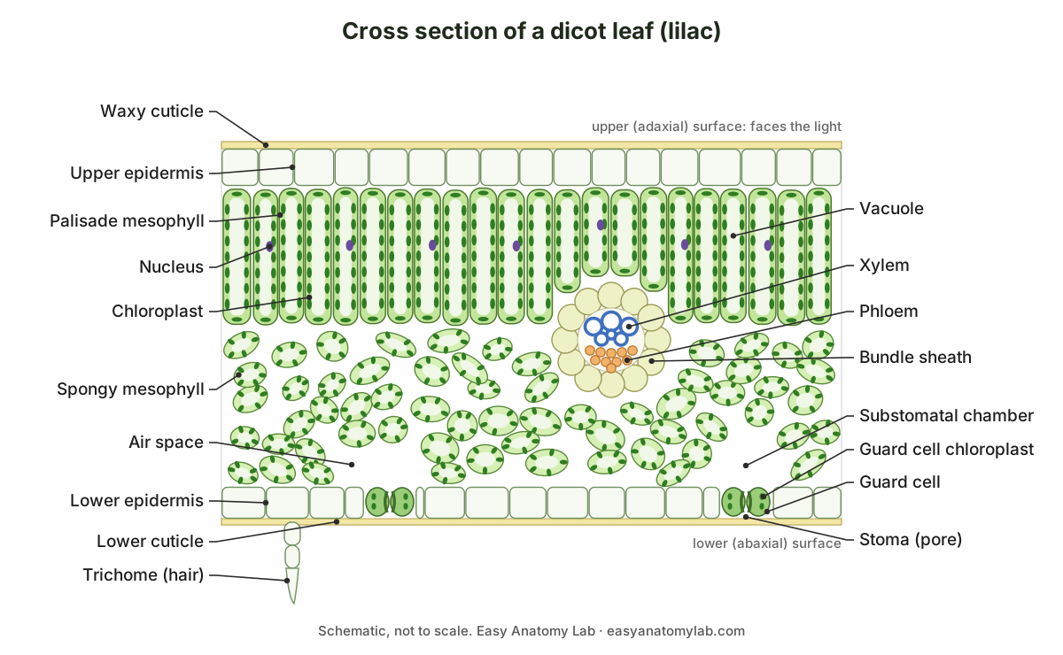
| Layer, top to bottom | What it looks like | Main function |
|---|---|---|
| Waxy cuticle | Thin, clear, non-cellular film | Cuts water loss; lets light through |
| Upper epidermis | One layer of flat, clear cells; few or no stomata | Protection; transparent so light reaches the mesophyll |
| Palisade mesophyll | Tall, column-shaped cells packed side by side, full of chloroplasts | Main site of photosynthesis |
| Vein (vascular bundle) | Xylem on top, phloem below, ring of bundle sheath cells | Brings water in, takes sugar out, supports the blade |
| Spongy mesophyll | Rounded, irregular cells with large air spaces | Gas exchange and some photosynthesis |
| Lower epidermis | One layer of clear cells with many stomata | Protection; gas exchange through the stomata |
| Stomata and guard cells | Pores between pairs of kidney-shaped, green guard cells | Let CO₂ in and O₂ and water vapour out; open and close |
| Lower cuticle | Thin waxy film | Cuts water loss from the underside |
Two quick ways to tell top from bottom in any slide: the palisade layer is always on the side facing the light (the upper, or adaxial, side), and in each vein the xylem faces the upper side while the phloem faces the lower (abaxial) side.
Leaf cross section labeled: every structure and its function
This is the text version of the 3D model: every labeled structure in the lilac leaf block, grouped by tissue system, with what it does. Use it as a study list or to check a labeling worksheet.
Dermal tissue (epidermis)
- Waxy cuticle (upper) (Adaxial cuticle)
- A thin, clear, waxy film secreted by the upper epidermis and lying on its outer surface. Waterproofs the leaf and cuts water loss by evaporation, while letting light straight through to the photosynthetic cells below.
- Upper epidermis (Adaxial epidermis)
- A single layer of flat, tightly fitting, transparent cells covering the top of the leaf. Protects the leaf, secretes the cuticle and lets light pass to the palisade layer. Its cells have no chloroplasts, so they do not shade the cells below.
- Lower epidermis (Abaxial epidermis)
- A single layer of epidermal cells on the underside of the leaf, broken at intervals by stomata. Protects the underside and carries most of the stomata, the pores that let carbon dioxide in and water vapour and oxygen out.
- Waxy cuticle (lower) (Abaxial cuticle)
- The waxy film on the lower surface, usually thinner than the upper cuticle. Reduces water loss from the underside. Because it is waterproof, gases must pass through the stomata instead.
- Stoma (stomatal pore) (Stomate; plural stomata)
- A small, adjustable pore in the lower epidermis, framed by two guard cells. Lets carbon dioxide diffuse into the leaf for photosynthesis and lets oxygen and water vapour out. It closes when the plant is short of water.
- Guard cells
- Two kidney-shaped epidermal cells that surround each stoma and change shape to open or close it. When they take up water and swell, the pore opens; when they lose water and go limp, the pore closes. This balances carbon dioxide uptake against water loss.
- Guard cell chloroplasts
- Small chloroplasts inside the guard cells. Make sugars and supply energy that helps the guard cells change their water content to open and close the pore.
- Trichome (Leaf hair, epidermal hair)
- A small outgrowth of an epidermal cell. Drawn here as a short multicellular hair on the underside. Trichomes deter plant-eating insects and slow the air moving over the leaf surface, which reduces transpiration. Glandular trichomes secrete oils or sticky substances.
Mesophyll (photosynthetic tissue)
- Palisade mesophyll (Palisade parenchyma, palisade layer)
- One or two rows of tall, column-shaped cells packed side by side like a fence (palisade) under the upper epidermis. The main site of photosynthesis. The cells stand upright and close together so that a large number of chloroplasts can catch the light entering from above.
- Chloroplasts (palisade)
- Green, lens-shaped organelles that line the walls of each palisade cell. Absorb light with chlorophyll and use its energy to turn carbon dioxide and water into sugar, releasing oxygen.
- Central vacuole (Large permanent vacuole)
- A large, sap-filled sac that takes up most of the volume of each mesophyll cell. Keeps the cell firm (turgid) by holding water under pressure, and pushes the chloroplasts out to the edge of the cell, close to the air spaces and the light.
- Nucleus
- The cell's control centre, holding its DNA, pressed against the wall in a mesophyll cell. Controls the cell's activities, including making the proteins needed for photosynthesis.
- Spongy mesophyll (Spongy parenchyma, spongy layer)
- Loosely arranged, irregular, rounded or lobed cells with large air spaces between them. Does some photosynthesis and, with its air spaces, lets carbon dioxide, oxygen and water vapour diffuse between the stomata and every mesophyll cell.
- Chloroplasts (spongy)
- Chloroplasts in the spongy mesophyll cells: fewer per cell than in the palisade layer. Carry out photosynthesis using the light that has passed through the palisade layer.
- Intercellular air spaces
- The connected network of air between the mesophyll cells, largest in the spongy layer. Give a huge internal surface for gas exchange and a short diffusion path: carbon dioxide spreads through the air spaces and dissolves in the wet cell walls.
- Substomatal chamber (Substomatal cavity, air chamber)
- A large air space immediately above each stoma. Connects the stomatal pore to the air spaces of the mesophyll, so gases entering or leaving the stoma can spread through the leaf.
Vascular tissue (veins)
- Xylem (minor vein)
- Thick-walled, hollow water-conducting cells on the upper side of the small vein. Brings water and dissolved minerals from the roots into the leaf. The water is used in photosynthesis and replaces water lost by transpiration.
- Phloem (minor vein)
- Small, thin-walled sieve tubes and companion cells on the lower side of the vein. Carries sugars made in photosynthesis (as sucrose) out of the leaf to the rest of the plant, a process called translocation.
- Bundle sheath
- A ring of tightly packed parenchyma cells wrapped around each small vein. Controls what passes between the vein and the mesophyll and keeps the vein's conducting cells out of the air spaces. In C4 plants such as maize it is enlarged and does the Calvin cycle.
- Xylem (midrib)
- The large, crescent-shaped band of xylem in the main vein, with rows of vessels. The main water supply line of the blade, which branches into the smaller veins.
- Phloem (midrib)
- A curved band of phloem below the midrib xylem. Collects sugar from the smaller veins and carries it out of the leaf through the petiole.
Midrib support tissue
- Collenchyma
- Living cells with unevenly thickened, flexible walls, in a band just inside the epidermis of the midrib. Strengthens the midrib while letting it bend, so the leaf can flex in the wind without snapping.
- Midrib parenchyma (Ground parenchyma)
- Large, round, thin-walled colourless cells that fill the midrib around the vascular bundle. Fills and supports the midrib, stores water and starch, and passes substances between the vein and the blade.
How the leaf is adapted for photosynthesis and gas exchange (GCSE)
A leaf is an organ built to absorb light and exchange gases while losing as little water as possible. For GCSE and IGCSE biology, the key adaptations are: a large, thin, flat shape; a transparent epidermis and cuticle; palisade cells packed with chloroplasts near the top; air spaces in the spongy mesophyll; stomata controlled by guard cells; and a network of veins that supplies water and removes sugar.
| Feature | How it helps photosynthesis or gas exchange |
|---|---|
| Broad, flat blade | Large surface area to absorb light and take in carbon dioxide |
| Thin blade | Short diffusion distance: no cell is far from the air or from the light |
| Clear cuticle and epidermis | Waterproof, but transparent, so light passes straight to the mesophyll |
| Palisade cells near the top, tightly packed | As many chloroplasts as possible where the light is strongest |
| Many chloroplasts per palisade cell | More chlorophyll to absorb light |
| Large vacuole pushes chloroplasts to the cell edge | Chloroplasts sit close to the air spaces and to the light |
| Air spaces in the spongy mesophyll | Carbon dioxide diffuses quickly to every cell; oxygen and water vapour diffuse out |
| Stomata, mostly on the underside | Openings for gas exchange, kept out of direct sun to reduce water loss |
| Guard cells | Close the stomata when water is short and open them in light |
| Xylem in the veins | Brings water, a raw material of photosynthesis, and keeps cells turgid |
| Phloem in the veins | Carries away the sugar made, so it does not build up in the leaf |
Exams often ask why a leaf is a compromise. The same stomata and air spaces that let carbon dioxide in also let water vapour out (transpiration). The plant controls this trade-off with the cuticle, which blocks water loss everywhere except at the stomata, and with the guard cells, which close the pores when the plant is short of water. The tissue names GCSE specifications use are epidermal tissue, palisade mesophyll, spongy mesophyll, xylem and phloem.
Vascular bundle: xylem and phloem in the midrib and minor veins
Every vein in a leaf is a vascular bundle with xylem on the upper side and phloem on the lower side. The midrib is the largest vein, running down the middle of the blade; the minor veins branch from it into a net (reticulate venation) so that no mesophyll cell is more than a few cells away from a vein.
- Xylem (minor vein): Brings water and dissolved minerals from the roots into the leaf. The water is used in photosynthesis and replaces water lost by transpiration.
- Phloem (minor vein): Carries sugars made in photosynthesis (as sucrose) out of the leaf to the rest of the plant, a process called translocation.
- Bundle sheath: Controls what passes between the vein and the mesophyll and keeps the vein's conducting cells out of the air spaces. In C4 plants such as maize it is enlarged and does the Calvin cycle.
- Xylem (midrib): The main water supply line of the blade, which branches into the smaller veins.
- Phloem (midrib): Collects sugar from the smaller veins and carries it out of the leaf through the petiole.
- Collenchyma: Strengthens the midrib while letting it bend, so the leaf can flex in the wind without snapping.
- Midrib parenchyma: Fills and supports the midrib, stores water and starch, and passes substances between the vein and the blade.
| Midrib (main vein) | Minor vein | |
|---|---|---|
| Size | Largest bundle; makes a ridge on the underside | Small; sits inside the mesophyll |
| Shape of bundle | Crescent or arc, xylem on the inner (upper) side | Small round bundle |
| Xylem | Many vessels in radial rows | A few vessels or tracheids |
| Surrounded by | Large colourless parenchyma, with collenchyma under the epidermis | A single ring of bundle sheath cells |
| Mesophyll | Palisade stops above the midrib | Palisade and spongy layers run right up to it |
| Job | Main supply line and support for the whole blade | Delivers water to, and collects sugar from, the mesophyll |
Why is the xylem on top? A leaf vein is a branch of the vascular system of the stem. In the stem, the xylem lies towards the centre and the phloem towards the outside. Where a bundle bends out into a leaf, the side that faced the centre of the stem becomes the upper side of the leaf, so the xylem ends up on top. Some plants, such as oleander, have phloem on both sides of the xylem (a bicollateral bundle), but lilac has phloem below only.
Stomata and guard cells in a leaf cross section
In a cross section, a stoma looks like a narrow gap in the lower epidermis with a small, rounded guard cell on each side and a large air space, the substomatal chamber, just above it. Guard cells are green because they are the only epidermal cells with chloroplasts.
- Stoma (stomatal pore): Lets carbon dioxide diffuse into the leaf for photosynthesis and lets oxygen and water vapour out. It closes when the plant is short of water.
- Guard cells: When they take up water and swell, the pore opens; when they lose water and go limp, the pore closes. This balances carbon dioxide uptake against water loss.
- Substomatal chamber: Connects the stomatal pore to the air spaces of the mesophyll, so gases entering or leaving the stoma can spread through the leaf.
In the 3D model, the front stoma is cut through the middle, exactly as on a slide, and a second stoma sits further back on the underside, where you can see the two kidney-shaped guard cells around the pore from below. For how guard cells open and close the pore, the different stomatal types and stomatal density, see the stomata diagram. For the inside of a single chloroplast, see chloroplast structure.
Dicot vs monocot leaf cross section
A dicot leaf such as lilac is dorsiventral (bifacial): its two sides differ, with palisade on top, spongy mesophyll below, and most stomata on the underside. A monocot leaf such as maize or a grass is isobilateral: both sides look alike, the mesophyll is not divided into palisade and spongy layers, and stomata are found on both surfaces.
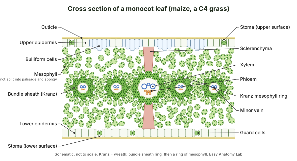
| Feature | Dicot leaf (lilac) | Monocot leaf (maize, grasses) |
|---|---|---|
| Symmetry | Dorsiventral: upper and lower sides differ | Isobilateral: both sides similar |
| Mesophyll | Palisade on top, spongy below | One type of mesophyll, not divided |
| Stomata | Mostly or only on the lower surface | On both surfaces |
| Guard cells | Kidney-shaped | Dumbbell-shaped, with subsidiary cells |
| Venation | Net (reticulate): veins of very different sizes | Parallel: vascular bundles of similar size, except the main veins |
| Bulliform cells | None | Present in grasses, in the upper epidermis |
| Bundle sheath | Thin-walled, pale (C3) | Pale in C3 grasses; large and green in C4 grasses such as maize |
| Sclerenchyma | Little in the blade | Strands or girders above and below the larger veins |
Bulliform cells are large, empty-looking, colourless cells in the upper epidermis of grass leaves, usually in groups between the veins. When they are full of water the leaf lies flat; when they lose water in a drought they shrink, and the leaf curls inward, which reduces water loss. The labeled monocot leaf cross section most courses use is maize (corn), which is also a C4 plant, so it shows Kranz anatomy as well. Do not assume every monocot is C4: wheat, rice and lilies are C3 monocots without Kranz anatomy.
C3 vs C4 vs CAM leaf anatomy (Kranz anatomy in corn)
C3, C4 and CAM are three ways of fixing carbon dioxide, and you can often tell them apart in a cross section. A C3 leaf (lilac, wheat, rice) has ordinary mesophyll and a small, pale bundle sheath. A C4 leaf (maize, sorghum, sugarcane) has Kranz anatomy: a wreath of large, chloroplast-rich bundle sheath cells around each vein, ringed by mesophyll cells. A CAM leaf (aloe, pineapple, many cacti and stonecrops) is usually thick and succulent, with large water-filled cells.
| C3 (lilac) | C4 (maize) | CAM (aloe) | |
|---|---|---|---|
| First CO₂ fixation | Rubisco, in all mesophyll cells | PEP carboxylase, in mesophyll cells | PEP carboxylase, at night |
| Calvin cycle | Mesophyll cells | Bundle sheath cells only | Same cells, during the day |
| Bundle sheath | Small, pale, few chloroplasts | Large, thick-walled, many chloroplasts (Kranz) | Not specialised |
| Mesophyll | Palisade and spongy (in dicots) | Ring of cells around each sheath | Large, vacuolated cells storing malic acid overnight |
| Vein spacing | Veins further apart | Veins close together | Varies; tissue often thick |
| Stomata open | Day | Day | Mostly at night |
| Typical habitat | Cool, moist | Hot, bright | Hot, dry |
Kranz anatomy works as a carbon dioxide pump. Mesophyll cells use PEP carboxylase, which does not react with oxygen, to fix carbon dioxide into a 4-carbon acid. The acid moves into the bundle sheath, where carbon dioxide is released next to Rubisco and enters the Calvin cycle. Because the sheath is tightly packed and has thick walls, the carbon dioxide stays concentrated there, which limits photorespiration, the wasteful reaction of Rubisco with oxygen. In CAM plants the two steps are separated in time instead of space: stomata open at night, carbon dioxide is stored as malic acid in the large vacuoles, and it is released for photosynthesis during the day with the stomata closed.
Pine leaf cross section (conifer needle)
A pine needle cross section shows a leaf built to survive cold, dry winters: a thick cuticle, an epidermis of thick-walled cells, a hypodermis below it, stomata sunk in pits, mesophyll cells whose walls fold inward, resin ducts, and a central cylinder in which an endodermis encloses transfusion tissue and one or two vascular bundles.
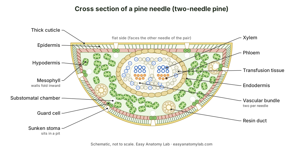
| Structure | What it does |
|---|---|
| Thick cuticle and epidermis | Waterproofing; the needle loses very little water even in frozen soil |
| Hypodermis | One or more layers of thick-walled fibre cells under the epidermis; stiffens the needle |
| Sunken stomata | Guard cells sit below the surface in a pit, where still, humid air reduces water loss |
| Mesophyll with infolded walls | Folds increase the wall area for chloroplasts; not divided into palisade and spongy |
| Resin ducts | Canals lined with secretory (epithelial) cells that make resin, which seals wounds and deters insects |
| Endodermis | A ring of cells around the central tissue, as in roots |
| Transfusion tissue | Short tracheids and parenchyma that carry water from the xylem out to the mesophyll |
| Vascular bundles | Two in two-needle and three-needle pines (one in some soft pines); xylem towards the flat side |
The half-moon outline of the section is a clue to the species group: pines with two needles per bundle have semicircular needles, pines with three have triangular ones. Sources describe the mesophyll differently: some call it spongy parenchyma, others arm-palisade cells, because each cell has ridges of wall folding into it. The number of vascular bundles is characteristic of the species.
Cross section of an aloe vera leaf (succulent)
An aloe vera leaf cross section shows a thin green rind around a thick, clear core. From the outside in: a thick cuticle and epidermis with sunken stomata, a band of green chlorenchyma (photosynthetic cells), a ring of vascular bundles at the edge of the core, and a large central mass of colourless, water-storing (aquiferous) parenchyma, which is the clear gel people use.
Aloe vera is a CAM plant, and its leaf shows the typical CAM traits: thick succulent tissue, large cells with big vacuoles that store water and, overnight, malic acid, and a thick waxy surface. The bitter yellow sap that seeps from a cut aloe leaf is stored near the vascular bundles, not in the gel. Unlike lilac, an aloe leaf has no palisade layer: in Aloe vera the chlorenchyma cells are rounded.
What a leaf cross section looks like under the microscope
Under a light microscope, a leaf cross section shows a clear epidermis on each side, a dark green row of upright palisade cells under the upper epidermis, a paler, loose spongy layer with white air spaces, and veins of thick-walled, empty-looking xylem cells above a patch of small phloem cells.
- Low power (×40): find the midrib bulge and the thin blade on either side. In a lilac slide the midrib shows a crescent of xylem and phloem in a mass of colourless cells.
- Medium power (×100): move into the blade and count the layers from top to bottom. The palisade row is easy to spot because the cells stand on end like a fence.
- High power (×400): look for chloroplasts lining the walls of the palisade cells, a guard cell pair in the lower epidermis, and the substomatal chamber above it.
Colours depend on how the slide was made. In a fresh hand section, the only colour is the natural green of the chloroplasts. Commercial prepared slides are usually double-stained with safranin and fast green: safranin stains lignified walls (xylem, fibres) and the cutin of the cuticle red, and fast green stains ordinary cellulose walls green. So in a typical lilac slide the xylem and cuticle look red and the mesophyll, epidermis, phloem and collenchyma look green or blue-green. Chloroplasts in old, stained slides often look like dark dots rather than green discs.
How to make a leaf cross section (hand sectioning)
You can make your own leaf cross section with a new razor blade and a piece of carrot or potato to hold the leaf. The aim is a slice thin enough to be almost transparent; you will need to cut many sections and keep only the best.
- Cut a strip of leaf about 5 mm wide across the midrib. Soft leaves such as privet, lilac, spinach or a geranium work well.
- Cut a small block of carrot or potato, split it in half, and cut a shallow groove in one half. Lay the leaf strip in the groove and press the other half on top, so the leaf is held upright in a sandwich.
- Keep the blade, the tissue and your fingers wet with water. Rest your forearms on the bench, use your forefinger as a guide, and cut across the top of the sandwich in one smooth pull or push. Do not saw.
- Cut many thin slices and float them all in water in a watch glass or Petri dish.
- Look at them against a dark background or under a dissecting microscope, and pick the thinnest. They should be almost transparent.
- Move the best sections with a fine brush into a small drop of water on a slide. Lower a coverslip slowly from one side with forceps to avoid air bubbles.
- Examine under low power first. For waxy leaves that repel water, a very small amount of detergent in the water helps the section sink and wet.
Safety: razor blades are very sharp. Use a single-edged blade or a blade holder, always cut away from your fingers, and follow your teacher's instructions. Change the blade as soon as it starts to drag, because a blunt blade crushes the cells.
Sun leaves and shade leaves: the palisade layer changes with light
The number of palisade layers is not fixed for a species. A leaf that grows in strong light develops a palisade tissue several cells deep, while a leaf that grows in shade keeps a single palisade layer. You can see this on one tree: leaves from the sunny outside of the crown are thicker than leaves from the shaded inside. (Botany Online (University of Hamburg): the mesophyll, main assimilation tissue of leaves.)
The two mesophyll layers also differ in how much they photosynthesise. Palisade cells contain three to five times as many chloroplasts as spongy cells, and the amount of air space in the mesophyll varies widely between species and habitats, from about 70 to 700 parts per thousand of the leaf volume. (Botany Online (University of Hamburg): the mesophyll, main assimilation tissue of leaves.)
Leaf cross sections from wet and dry habitats
Lilac is called a mesomorphic leaf because it grows where soil moisture is adequate: a broad, flat blade, a one-cell-thick epidermis, palisade and spongy mesophyll, and veins with xylem above phloem. The same lab compares it with oleander (Nerium), a xeromorphic leaf from dry places, whose epidermis is several cells thick and whose stomata sit in pits called stomatal crypts on the underside, and with the water lily (Nymphaea), whose spongy tissue is full of large air spaces (aerenchyma), whose stomata are on the upper surface only, and whose veins have little xylem. (University of Wisconsin–Madison, Botany 130: leaf anatomy (lilac, oleander, water lily, maize).)
Leaves from harsh, dry or salty places show the same trends. An Acacia leaf has a thick cuticle, a mangrove (Rhizophora) leaf has more closely packed cells and a thicker cuticle, and a bottlebrush (Callistemon) leaf contains a lot of thick-walled sclerenchyma. In the floating water lily leaf the cuticle is much thinner, and the aerenchyma lets the leaf float. (Bellairs, Plant Anatomy and Physiology (LibreTexts), 3.2 Leaves.)
How Kranz anatomy divides the work in a maize leaf
In maize, each vein is wrapped in a ring of bundle sheath cells, and one or more rings of mesophyll cells sit outside that. A proteomics study of the two cell types showed how the work is split: the Rubisco large subunit accumulates only in bundle sheath chloroplasts, while mesophyll chloroplasts hold photosystem II and the enzyme PPDK that regenerates the carbon dioxide acceptor. Bundle sheath cells take in malate and break it down to pyruvate, releasing carbon dioxide for the Calvin cycle. (Majeran et al. (2005): bundle sheath and mesophyll chloroplasts of maize, The Plant Cell.)
The two kinds of chloroplast also look different. Bundle sheath chloroplasts in maize sit towards the outer wall of the cell and contain large starch grains; mesophyll chloroplasts are arranged randomly and have stacked thylakoids (grana). (Majeran et al. (2005): bundle sheath and mesophyll chloroplasts of maize, The Plant Cell.)
Kranz means wreath. The bundle sheath cells of C4 plants have many chloroplasts, thick walls that gases pass through poorly, and no air spaces between them, which keeps the concentrated carbon dioxide inside. The first carbon dioxide-fixing enzyme in the mesophyll is PEP carboxylase, and the mesophyll cells lack Rubisco. (NCERT Biology Class XI, Chapter 11: Photosynthesis in Higher Plants (PDF).)
Bulliform cells: how a grass leaf rolls up in drought
Bulliform cells are enlarged, colourless epidermal cells found only on the upper side of grass leaves, usually in strips two to five cells wide that run along the leaf. They are also called hinge or motor cells, and they are linked to the way grass leaves roll inward when the plant is short of water. Exactly how they work is still being debated. (Matschi et al. (2020): the maize bulliform cell cuticle and leaf rolling, Plant Direct.)
A 2020 study of maize found that bulliform cells shrink more than other epidermal cells when a leaf dries out, and that their cuticle is about four times thicker than the cuticle of ordinary epidermal cells but lets water through more easily. The authors suggest this uneven shrinking helps the leaf fold. (Matschi et al. (2020): the maize bulliform cell cuticle and leaf rolling, Plant Direct.)
Where the bitter aloin is stored in an aloe leaf
A study of six Aloe species found that every leaf had the same basic layers: epidermis, chlorenchyma (the green photosynthetic layer), aquiferous (water-storing) tissue, and vascular bundles. All six showed xeromorphic features: thick epidermal walls, a thick cuticle, sunken stomata and well-developed water-storage tissue. In Aloe vera the chlorenchyma cells are rounded, not palisade-like as in Aloe arborescens. (Li et al. (2003): leaf structure and aloin content in six Aloe species, Journal of Integrative Plant Biology.)
Fluorescence microscopy showed that the bitter yellow compound aloin is stored in the large parenchyma cells of the vascular bundles, the bundle sheath and the sheath around the water-storage tissue, not in the green chlorenchyma. Aloe vera had less aloin than Aloe arborescens. (Li et al. (2003): leaf structure and aloin content in six Aloe species, Journal of Integrative Plant Biology.)
Label the cross section of a leaf: worksheet and answer key
Test yourself: name each numbered structure on this unlabeled leaf cross section diagram. You can print it or copy the numbers onto paper. The answer key is below.
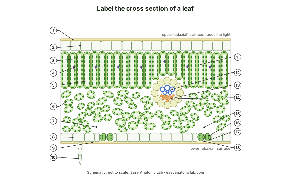
Show the answer key
- Waxy cuticle
- Upper epidermis
- Palisade mesophyll
- Nucleus
- Chloroplast
- Spongy mesophyll
- Air space
- Lower epidermis
- Lower cuticle
- Trichome (leaf hair)
- Vacuole
- Xylem
- Phloem
- Bundle sheath
- Substomatal chamber
- Guard cell chloroplast
- Guard cell
- Stoma (pore)
Extra questions:
- Which two leaf cell types contain chloroplasts, and which layer has the most?
- Why are most stomata on the lower surface of a lilac leaf?
- In a vein, which tissue is on the upper side and which on the lower side?
- Give two differences between a dicot and a monocot leaf cross section.
- What is Kranz anatomy, and in which plant would you see it?
- Name three features of a pine needle that reduce water loss.
Show answers to the extra questions
- Mesophyll cells (palisade and spongy) and guard cells. The palisade layer has the most.
- The lower surface is shaded and cooler, so less water evaporates through the stomata there.
- Xylem on the upper (adaxial) side, phloem on the lower (abaxial) side.
- Any two: monocot mesophyll is not divided into palisade and spongy; monocots have stomata on both surfaces; monocot veins are parallel and similar in size; grasses have bulliform cells.
- A wreath of large, chloroplast-rich bundle sheath cells around each vein, surrounded by mesophyll cells. It is found in C4 plants such as maize (corn), sorghum and sugarcane.
- Any three: thick cuticle, sunken stomata, thick-walled hypodermis, small surface area (needle shape).
Frequently asked questions
What are the layers of a leaf cross section in order?
From top to bottom: waxy cuticle, upper epidermis, palisade mesophyll, spongy mesophyll (with the veins between the two mesophyll layers), lower epidermis with stomata and guard cells, and the lower cuticle. Each vein has xylem on the upper side and phloem on the lower side, inside a bundle sheath.
Why is the palisade layer near the top of the leaf?
Because that is where the light arrives. Palisade cells are tall, tightly packed and full of chloroplasts, so they absorb most of the light for photosynthesis. The upper epidermis above them is transparent and has no chloroplasts, so it does not shade them.
Why are most stomata on the bottom of the leaf?
The lower surface is cooler and out of direct sun, so less water evaporates from stomata there. In most dicot leaves, such as lilac, the lower epidermis has many more stomata than the upper, which may have none. Floating leaves such as water lilies are the exception: their stomata are on top.
Is xylem on the top or the bottom of a leaf vein?
Xylem is on the top (adaxial side) and phloem is on the bottom (abaxial side). This matches the stem, where xylem is towards the centre: when the vascular bundle bends out into the leaf, the side that faced the centre of the stem becomes the upper side of the leaf.
What is the difference between a monocot and a dicot leaf cross section?
A dicot leaf (lilac) is dorsiventral: palisade on top, spongy below, and most stomata underneath, with a net of veins of different sizes. A monocot leaf (maize, grasses) is isobilateral: the mesophyll is not divided into palisade and spongy layers, stomata are on both surfaces, the parallel veins look similar in size, and grasses have large bulliform cells in the upper epidermis.
What is Kranz anatomy?
Kranz anatomy is the leaf structure of C4 plants such as maize and sorghum. Each vein is surrounded by a ring of large, chloroplast-rich bundle sheath cells, with mesophyll cells arranged in a ring outside them. Kranz is German for wreath. The mesophyll fixes carbon dioxide into a 4-carbon acid, and the bundle sheath releases it for the Calvin cycle.
What does a leaf cross section look like under a microscope?
On a fresh section you see a clear epidermis, a dark green palisade row of upright cells, a paler green spongy layer with white air spaces, and veins of clear, empty-looking xylem cells. On a commercial safranin and fast green slide, the cuticle and lignified xylem are red and the cellulose walls of the other cells are green or blue-green.
Why is a pine needle cross section different?
A pine needle is built to save water in cold, dry winters. It has a very thick cuticle, a thick-walled hypodermis, stomata sunken in pits, mesophyll cells with inward folds of the wall, resin ducts, and an endodermis surrounding transfusion tissue and one or two vascular bundles in the centre.
Which leaf cells contain chloroplasts?
Palisade and spongy mesophyll cells, and the guard cells. Ordinary epidermal cells, xylem, phloem and the colourless midrib parenchyma have none (or very few). In C4 plants, the bundle sheath cells also contain many chloroplasts.
References
- Clark, M. A., Douglas, M. & Choi, J. (2018). Biology 2e, section 30.4: Leaves. OpenStax, Rice University.
- NCERT (2026 reprint). Biology, Textbook for Class XI. Chapter 6: Anatomy of Flowering Plants (dorsiventral and isobilateral leaves).
- NCERT (2026 reprint). Biology, Textbook for Class XI. Chapter 11: Photosynthesis in Higher Plants (Kranz anatomy and the C4 pathway).
- University of Wisconsin–Madison, Department of Botany. Botany 130 lab: leaf anatomy of Syringa, Nerium, Nymphaea and Zea.
- von Sengbusch, P. Botany Online: the mesophyll, main assimilation tissue of leaves. University of Hamburg (archived).
- von Sengbusch, P. Botany Online: leaves of conifers, cross section of a pine needle. University of Hamburg (archived).
- University of British Columbia. BIOL/APBI 210 lab information: Pinus leaf anatomy.
- Matschi, S. et al. (2020). Structure-function analysis of the maize bulliform cell cuticle and its potential role in dehydration and leaf rolling. Plant Direct 4(10), e00282.
- Majeran, W., Cai, Y., Sun, Q. & van Wijk, K. J. (2005). Functional differentiation of bundle sheath and mesophyll maize chloroplasts determined by comparative proteomics. The Plant Cell 17, 3111–3140.
- Borland, A. M., Leverett, A., Hurtado-Castano, N., Hu, R. & Yang, X. (2018). Functional anatomical traits of the photosynthetic organs of plants with crassulacean acid metabolism. In: The Leaf: A Platform for Performing Photosynthesis, Springer, 281–305.
- Li, J.-Y., Wang, T.-X., Shen, Z.-G. & Hu, Z.-H. (2003). Relationship between leaf structure and aloin content in six species of Aloe L. Journal of Integrative Plant Biology 45(5), 594–600.
- White, R. Using other plant tissues as support tissue to make hand sections. Prometheus protocols.
- Megías, M., Molist, P. & Pombal, M. A. Atlas of Plant and Animal Histology: safranin–alcian blue/fast green staining. University of Vigo.
- Bellairs, S. Plant Anatomy and Physiology, 3.2 Leaves. Biology LibreTexts, Charles Darwin University.
More interactive anatomy models
- Flower anatomyParts of a flower in 3D: petals, sepals, stamens, pistil, ovary and ovules of a lily.
- Stomata diagramGuard cells, stomatal pore and how stomata open and close, in 3D.
- Seed anatomyBean and corn seed anatomy: seed coat, cotyledons, endosperm and embryo.
- Chloroplast structureEnvelope, stroma, thylakoids, grana and lumen of a chloroplast, in 3D.
- Human anatomy1,000+ bones, muscles, organs, vessels and nerves from real anatomical data.
- Octopus anatomyThree hearts, a ring-shaped brain, eight arms and a beak, in 3D.