

Cellular respiration at a glance
Cellular respiration is the set of reactions that break down glucose (and other fuels) with oxygen to make ATP, the energy molecule every cell uses. The overall equation is C6H12O6 + 6 O2 → 6 CO2 + 6 H2O + energy, and that energy is captured as about 30–32 ATP per glucose, with the rest given off as heat.
It happens in four stages. Glycolysis splits glucose into two pyruvate in the cytosol. Pyruvate oxidation turns pyruvate into acetyl CoA in the mitochondrial matrix. The Krebs cycle (citric acid cycle) strips the remaining carbons off as CO2, also in the matrix. Finally oxidative phosphorylation on the inner mitochondrial membrane, made up of the electron transport chain and chemiosmosis through ATP synthase, uses the electrons collected in the first three stages to make most of the ATP. Oxygen is needed only at the very end, as the final electron acceptor.
Two small molecules carry those electrons: NAD+, which becomes NADH, and FAD, which becomes FADH2. One glucose loads 10 NADH and 2 FADH2. Think of them as charged batteries that are emptied at the electron transport chain and sent back to be recharged.
Cellular respiration diagram: step by step
This interactive cellular respiration diagram shows where each stage happens and what goes in and comes out. Click a stage, use Back and Next, or press the arrow keys. The table under it adds up the ATP, NADH, FADH2 and CO2 from every stage. Switch between modern and older textbook ATP values, and between the two NADH shuttles, to see where the different totals in different books come from.
Cellular respiration: the whole pathway
- Where
- Cytosol, then the mitochondrion (matrix and inner membrane)
- Oxygen?
- Yes, overall (aerobic respiration)
Inputs
- 1 glucose (C6H12O6)
- 6 O2
- ~30–32 ADP + Pi
Outputs
- 6 CO2
- 6 H2O
- ~30–32 ATP
- Heat
Glucose is split in the cytosol, its carbon is stripped off as CO2 in the mitochondrial matrix, and its electrons are passed down the electron transport chain to oxygen. The energy released pumps protons, and the protons flowing back through ATP synthase make most of the ATP. Step through the four stages with the buttons or the arrow keys.
| Per glucose | ATP made directly | NADH | FADH₂ | CO₂ | ATP via ATP synthase |
|---|---|---|---|---|---|
| Glycolysis | 2 | 2 | 0 | 0 | 5 |
| Pyruvate oxidation | 0 | 2 | 0 | 2 | 5 |
| Krebs cycle | 2 | 6 | 2 | 4 | 18 |
| Oxidative phosphorylation | 0 | −10 (used) | −2 (used) | 0 | 28 |
| Total | 4 | 10 | 2 | 6 | 28 → 32 ATP in all |
Using 2.5 ATP per NADH and 1.5 per FADH₂ (measured P/O ratios). The malate–aspartate shuttle passes glycolysis NADH electrons to NAD⁺ in the matrix, so they count as full NADH. Real cells usually make less than this maximum, because some protons leak back across the membrane.
Cellular respiration diagram labeled
A labeled cellular respiration diagram shows glycolysis in the cytoplasm, pyruvate oxidation and the Krebs cycle in the mitochondrial matrix, and the electron transport chain and ATP synthase in the inner mitochondrial membrane, with glucose and oxygen going in and carbon dioxide, water and ATP coming out.
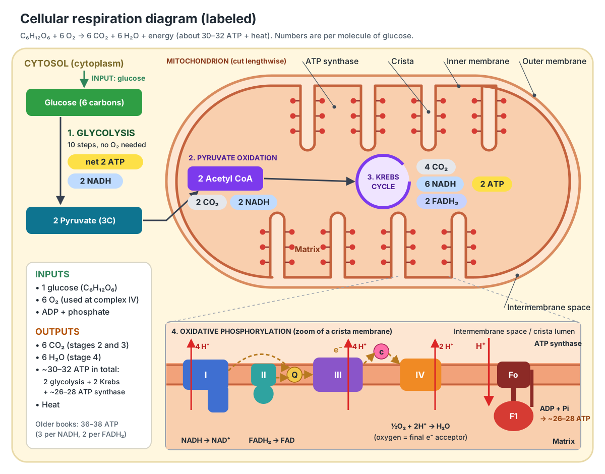
Read the diagram from top left to bottom right. Glucose enters the cell and is split by glycolysis. The two pyruvate cross both mitochondrial membranes into the matrix. There each loses a carbon as CO2 to become acetyl CoA, and the acetyl groups are burned in the Krebs cycle. The NADH and FADH2 made along the way deliver their electrons to the chain of protein complexes in the cristae, and the energy released ends up in ATP.
Simple diagram of cellular respiration
The simplest cellular respiration diagram is one line: glucose + oxygen → carbon dioxide + water + ATP. For a basic diagram, draw three boxes in a row, glycolysis → Krebs cycle → electron transport chain, write the place under each (cytoplasm, matrix, inner membrane) and the ATP made by each (2, 2 and about 26–28).
| Stage | Where | Main products per glucose | ATP |
|---|---|---|---|
| 1. Glycolysis | Cytosol | 2 pyruvate, 2 NADH | 2 (net) |
| 2. Pyruvate oxidation | Mitochondrial matrix | 2 acetyl CoA, 2 NADH, 2 CO2 | 0 |
| 3. Krebs cycle | Mitochondrial matrix | 6 NADH, 2 FADH2, 4 CO2 | 2 |
| 4. Oxidative phosphorylation | Inner mitochondrial membrane | 6 H2O; NAD+ and FAD recycled | ~26–28 |
| Total | 6 CO2, 6 H2O | ~30–32 |
A memory aid for the order: Get Pyruvate Ready, Krebs Cycles, Electrons Transport, or simply follow the carbon: 6 carbons in glucose, 3 + 3 in pyruvate, 2 + 2 in acetyl CoA, and 0 left after two turns of the Krebs cycle.
Cellular respiration diagram with inputs and outputs
The inputs of cellular respiration are glucose, oxygen, ADP and phosphate; the outputs are carbon dioxide, water and ATP. Inside the cell, each stage passes its products on as the next stage's inputs.
| Stage | Inputs (per glucose) | Outputs (per glucose) |
|---|---|---|
| Glycolysis | 1 glucose, 2 ATP, 2 NAD+, 4 ADP + 4 Pi | 2 pyruvate, 4 ATP (2 net), 2 NADH, 2 H2O |
| Pyruvate oxidation | 2 pyruvate, 2 coenzyme A, 2 NAD+ | 2 acetyl CoA, 2 CO2, 2 NADH |
| Krebs cycle (2 turns) | 2 acetyl CoA, 6 NAD+, 2 FAD, 2 ADP (or GDP) + 2 Pi, water | 4 CO2, 6 NADH, 2 FADH2, 2 ATP (or GTP), 2 coenzyme A |
| Electron transport chain | 10 NADH, 2 FADH2, 6 O2 | 10 NAD+, 2 FAD, 6 H2O, proton gradient |
| Chemiosmosis (ATP synthase) | Proton gradient, ADP + Pi | ~26–28 ATP |
Two points trip students up. First, the oxygen you breathe in ends up in water, not in CO2; the carbon dioxide comes from the carbons of glucose in stages 2 and 3. Second, glycolysis does not need oxygen, but stages 2–4 stop without it, because the NADH they make can only be recycled to NAD+ by the electron transport chain.
Glycolysis diagram: stage 1
Glycolysis is the ten-step breakdown of one glucose (6 carbons) into two pyruvate (3 carbons each) in the cytosol. It makes a net 2 ATP and 2 NADH and does not use oxygen, so it runs in almost every living cell, including bacteria.
The first half is the energy-investment phase (steps 1–5). Hexokinase adds a phosphate from ATP to glucose, an isomerase rearranges it into fructose-6-phosphate, and phosphofructokinase adds a second phosphate from another ATP. Aldolase then splits the 6-carbon sugar into two 3-carbon sugars, and an isomerase turns both into glyceraldehyde-3-phosphate (G3P).
The second half is the energy-payoff phase (steps 6–10), and it happens twice per glucose. G3P dehydrogenase oxidises each G3P and passes two electrons to NAD+, making NADH. Phosphoglycerate kinase moves a phosphate onto ADP, making ATP. After two rearrangements, by phosphoglycerate mutase and enolase, pyruvate kinase makes a second ATP and releases pyruvate. That is 4 ATP made minus 2 invested, so 2 net. ATP made this way, by an enzyme moving a phosphate directly from a substrate to ADP, is called substrate-level phosphorylation.
- Cytosol (site of glycolysis): Where glycolysis happens: glucose is split into two pyruvate, making a net 2 ATP and 2 NADH. No oxygen is needed.
- Glycolytic enzymes: Carry out the ten steps of glycolysis: 2 ATP invested in steps 1–5, then 4 ATP and 2 NADH made in steps 6–10.
- Glucose (C6H12O6): The fuel. Its carbon ends as CO2 and its electrons end on oxygen, forming water.
- Pyruvate: With oxygen, it enters the mitochondrion and is oxidised to acetyl CoA. Without oxygen, it is turned into lactate or ethanol (fermentation).
Pyruvate oxidation: stage 2 (the link reaction)
Pyruvate oxidation converts each pyruvate into acetyl CoA in the mitochondrial matrix, releasing one CO2 and making one NADH. It links glycolysis to the Krebs cycle, so it is often called the link reaction or transition step.
Pyruvate crosses the outer membrane through porins and the inner membrane through the mitochondrial pyruvate carrier. In the matrix, the very large pyruvate dehydrogenase complex removes the carboxyl group as CO2, oxidises the remaining two-carbon unit and attaches it to coenzyme A. Per glucose: 2 acetyl CoA, 2 CO2 and 2 NADH. Some textbooks count this step as part of the Krebs cycle and so describe three stages, not four.
- Porin (VDAC): Let small water-soluble molecules (pyruvate, ADP, ATP, phosphate, ions) cross the outer membrane, so the intermembrane space has almost the same small-molecule make-up as the cytosol.
- Mitochondrial pyruvate carrier: Moves pyruvate made by glycolysis in the cytosol across the inner membrane, so it can be oxidised in the matrix.
- Pyruvate dehydrogenase complex: Converts each pyruvate into acetyl CoA, releasing one CO2 and making one NADH (pyruvate oxidation, the link reaction).
Krebs cycle diagram: stage 3
The Krebs cycle (citric acid cycle, TCA cycle) is an eight-step loop in the mitochondrial matrix that oxidises the acetyl group of acetyl CoA to two CO2. Each turn makes 3 NADH, 1 FADH2 and 1 ATP (or GTP), and the cycle turns twice per glucose.
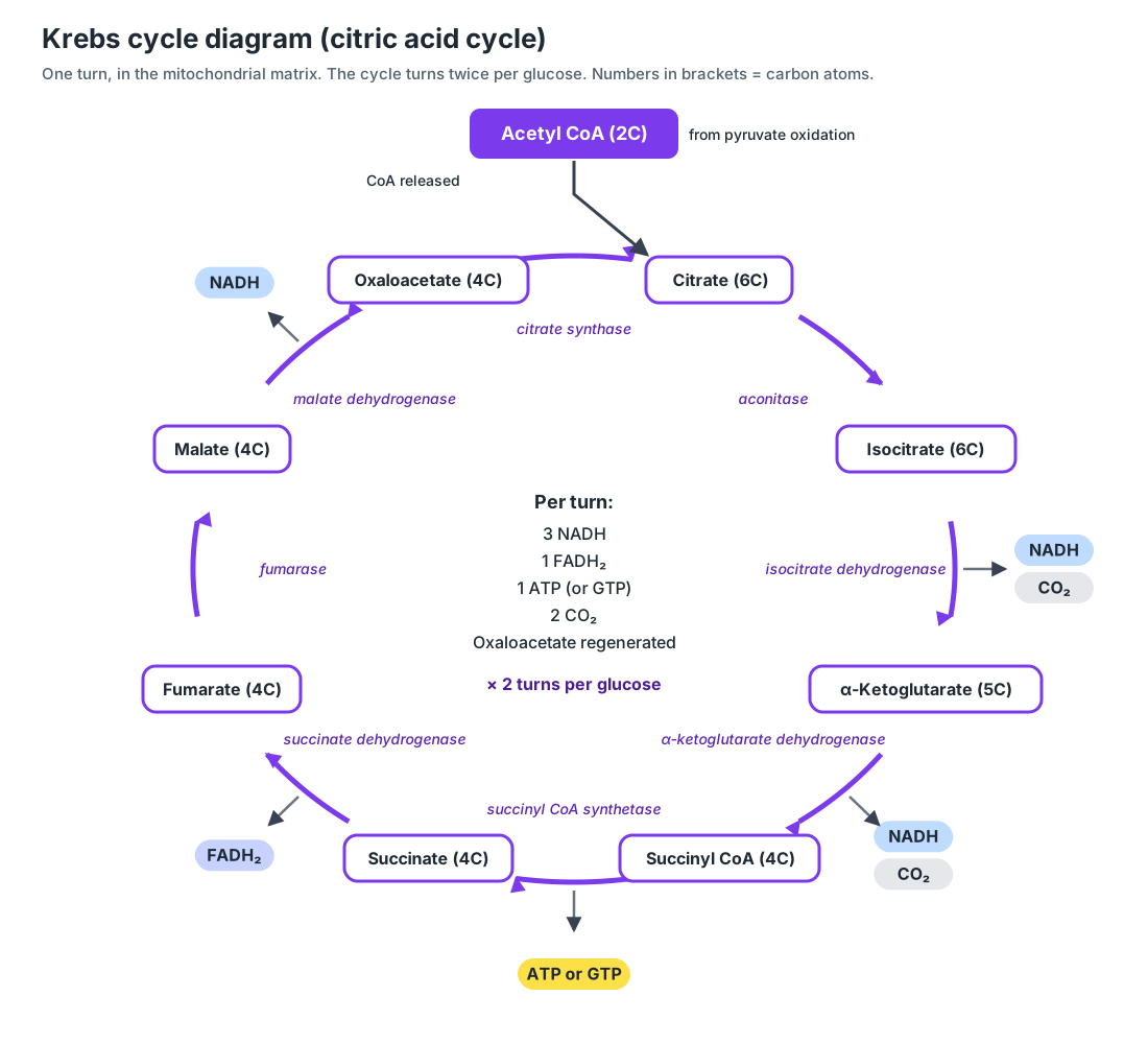
The cycle starts when the 2-carbon acetyl group joins the 4-carbon oxaloacetate to make 6-carbon citrate. Citrate is rearranged to isocitrate. Two oxidations in a row then each remove one carbon as CO2 and make one NADH: isocitrate becomes 5-carbon α-ketoglutarate, which becomes 4-carbon succinyl CoA. Succinyl CoA synthetase makes one ATP or GTP by substrate-level phosphorylation. Succinate dehydrogenase then oxidises succinate to fumarate and makes FADH2; this enzyme is also complex II of the electron transport chain and is the only cycle enzyme bound to the inner membrane. Fumarate becomes malate, and malate dehydrogenase regenerates oxaloacetate and makes the third NADH.
Because oxaloacetate is remade at the end, the same molecules are used over and over: the cycle is a processing wheel, not a straight line. Its intermediates are also starting materials for amino acids, fats and heme, so the Krebs cycle is a hub of the whole of metabolism, not only of energy production.
- Krebs cycle enzymes: Oxidise acetyl groups to CO2. Each turn makes 3 NADH, 1 FADH2, 1 ATP (or GTP) and 2 CO2; the cycle turns twice per glucose.
- Mitochondrial matrix: Site of pyruvate oxidation and the Krebs (citric acid) cycle, which make NADH, FADH2, CO2 and a little ATP. It also holds the mitochondrion's own DNA and ribosomes.
Electron transport chain diagram: stage 4
The electron transport chain is a series of four protein complexes (I–IV) and two mobile carriers (ubiquinone and cytochrome c) in the inner mitochondrial membrane. It passes electrons from NADH and FADH2 to oxygen and uses the energy released to pump protons out of the matrix.
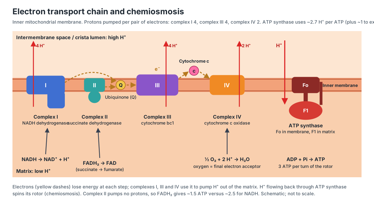
- Complex I (NADH dehydrogenase): Takes two electrons from NADH, passes them to ubiquinone and uses the energy to pump four protons out of the matrix.
- Complex II (succinate dehydrogenase): Oxidises succinate to fumarate in the Krebs cycle, and passes the electrons through FADH2 straight to ubiquinone. It pumps no protons.
- Ubiquinone (coenzyme Q): Carries pairs of electrons from complexes I and II (and other enzymes) to complex III.
- Complex III (cytochrome bc1): Takes electrons from ubiquinol, passes them one at a time to cytochrome c, and moves protons across the membrane (4 per electron pair, via the Q cycle).
- Cytochrome c: Shuttles one electron at a time from complex III to complex IV.
- Complex IV (cytochrome c oxidase): Hands electrons to oxygen, the final electron acceptor, which joins with protons to form water. It also pumps protons (2 per electron pair).
At each step the electrons move to a carrier that holds them more tightly, so they lose a little energy each time, like water running down a series of waterwheels. Per pair of electrons from NADH, complexes I, III and IV pump about 4, 4 and 2 protons into the intermembrane space (and the crista lumen): about 10 in all. Electrons from FADH2 enter at complex II, which pumps none, so they move only about 6.
Oxygen's job is to accept the electrons at the end of the chain. Each O2 takes 4 electrons and 4 protons and becomes 2 water molecules. If oxygen runs out, or if a poison such as cyanide blocks complex IV, electrons back up along the whole chain, NADH stops being recycled, and the Krebs cycle and pyruvate oxidation halt too.
Chemiosmosis and ATP synthase
Chemiosmosis is the use of a proton gradient across a membrane to make ATP: protons pumped out by the electron transport chain flow back into the matrix through ATP synthase, and that flow drives ATP formation. It makes about 90% of the ATP from aerobic respiration.
- Hydrogen ions (H⁺, protons): Their concentration and charge difference across the inner membrane, the proton-motive force, is the energy store that ATP synthase taps (chemiosmosis).
- ATP synthase Fo rotor and stalk: Protons flowing from the intermembrane space back into the matrix through Fo spin the c-ring and stalk, which drives ATP formation in F1.
- ATP synthase F1 head: Joins ADP and phosphate into ATP. Each full turn of the rotor makes 3 ATP.
- ADP/ATP translocase (ANT): Swaps ATP made in the matrix for ADP from the cytosol, one for one, so new ATP can reach the rest of the cell.
- Phosphate carrier: Brings inorganic phosphate (Pi) into the matrix together with a proton, supplying ATP synthase.
The inner membrane is almost impermeable to protons, so the only easy way back is through the Fo channel of ATP synthase. The proton-motive force has two parts: a concentration difference (the intermembrane space is more acidic) and an electrical charge difference (the matrix side is negative). Together they push protons back in, spinning the rotor. Peter Mitchell proposed this mechanism in 1961; it was so controversial that it took years to be accepted, and it won him the 1978 Nobel Prize in Chemistry.
Making ATP from ADP plus phosphate is the process called oxidative phosphorylation("oxidative" because the energy comes from oxidising NADH and FADH2). Contrast this with the substrate-level phosphorylation in glycolysis and the Krebs cycle, which makes only 4 of the 30–32 ATP.
How many ATP are made in cellular respiration?
Aerobic respiration makes about 30–32 ATP per glucose by modern accounting: 2 from glycolysis, 2 from the Krebs cycle and about 26–28 from oxidative phosphorylation. Older textbooks give 36–38; the difference comes from the number of ATP credited to each NADH and FADH2.
| Source | Count per glucose | Modern value (ATP) | Older value (ATP) |
|---|---|---|---|
| Glycolysis, substrate-level | 2 ATP | 2 | 2 |
| Krebs cycle, substrate-level | 2 ATP (or GTP) | 2 | 2 |
| NADH from pyruvate oxidation and Krebs | 8 NADH | 8 × 2.5 = 20 | 8 × 3 = 24 |
| FADH2 from Krebs | 2 FADH2 | 2 × 1.5 = 3 | 2 × 2 = 4 |
| NADH from glycolysis | 2 NADH | 5 (malate–aspartate) or 3 (glycerol phosphate) | 6 or 4 |
| Total | 32 or 30 | 38 or 36 |
Why fractions? The ATP made per NADH depends on how many protons are pumped (about 10 per NADH) divided by how many are used per ATP. In animal mitochondria the ATP synthase rotor needs about 2.7 protons per ATP, and moving phosphate in and ATP out costs about 1 more, so roughly 3.7–4 protons buy one ATP in the cytosol. 10 ÷ 4 ≈ 2.5, and 6 ÷ 4 ≈ 1.5. Measured P/O ratios agree.
Why two shuttles? NADH made in the cytosol cannot cross the inner membrane. The malate–aspartate shuttle (important in liver and heart) passes its electrons to NAD+ inside the matrix, so they are worth a full 2.5 ATP. The glycerol phosphate shuttle (important in skeletal muscle and brain) passes them to FAD, so they are worth only 1.5. In real cells some protons also leak back without making ATP, so the actual yield is usually below the maximum. Overall, respiration captures roughly a third of the energy in glucose; OpenStax gives about 34%, with the rest released as heat.
For AP Biology and most exams, write "about 30–32 ATP" and explain that the number is approximate. If your textbook or teacher uses 36 or 38, the method is the same; only the per-NADH value differs.
Mitochondria and cellular respiration: parts of the 3D diagram
The mitochondrion is the organelle where stages 2, 3 and 4 of cellular respiration happen. It has an outer membrane, a narrow intermembrane space, a heavily folded inner membrane whose folds are called cristae, and the protein-rich matrix inside. Its parts map directly onto the stages: the matrix holds the Krebs cycle, and the inner membrane holds the electron transport chain and ATP synthase.
Typical mitochondria are roughly 1 µm wide and 2 µm long, about the size of a bacterium, but in many cells they fuse into networks tens of micrometres long. Yeast cells have around 10 mitochondria and mammalian cells around 1,000–10,000. Muscle cells, which need a lot of ATP, are packed with them.
This is the text version of the 3D model: every labeled structure, grouped by system, with what it does. Use it to check a mitochondria labeling worksheet.
Membranes & spaces
- Outer mitochondrial membrane (Outer membrane, OMM)
- The smooth outer boundary of the mitochondrion: one phospholipid bilayer studded with proteins. Separates the mitochondrion from the cytosol but lets ions and small molecules such as pyruvate, ADP and ATP pass freely through its porin channels.
- Porin (VDAC) (Voltage-dependent anion channel)
- Barrel-shaped channel proteins that make holes through the outer membrane. Let small water-soluble molecules (pyruvate, ADP, ATP, phosphate, ions) cross the outer membrane, so the intermembrane space has almost the same small-molecule make-up as the cytosol.
- Intermembrane space (IMS)
- The narrow gap between the outer and inner membranes. Its width is exaggerated in the model. Collects the hydrogen ions (protons) pumped out of the matrix by the electron transport chain. The resulting proton gradient powers ATP synthase.
- Inner mitochondrial membrane (Inner boundary membrane, IMM)
- The second, inner bilayer. The part that runs parallel to the outer membrane is the inner boundary membrane. Holds the electron transport chain and ATP synthase, and is almost impermeable to ions, so it can keep the proton gradient that drives ATP synthesis.
- Cristae (Singular: crista)
- Folds of the inner membrane that reach into the matrix. Here they are drawn as flat plates (lamellar cristae). Hugely increase the area of inner membrane, so a mitochondrion can hold many more electron transport chains and ATP synthases.
- Crista junction
- The narrow neck where a crista joins the inner boundary membrane. Connects the space inside the crista to the rest of the intermembrane space while partly separating them, so proteins and protons are not spread evenly.
- Intracristal space (Cristae lumen)
- The thin space inside each crista, between its two membranes. Shown as the dark stripe in each cut crista. Receives most of the protons pumped by the respiratory complexes in the cristae, so it is where the proton gradient is used.
- Mitochondrial matrix (Matrix)
- The dense, protein-rich fluid enclosed by the inner membrane. Site of pyruvate oxidation and the Krebs (citric acid) cycle, which make NADH, FADH2, CO2 and a little ATP. It also holds the mitochondrion's own DNA and ribosomes.
Electron transport chain
- Complex I (NADH dehydrogenase) (NADH:ubiquinone oxidoreductase)
- A large L-shaped enzyme: one arm lies in the membrane, the other sticks into the matrix. Takes two electrons from NADH, passes them to ubiquinone and uses the energy to pump four protons out of the matrix.
- Complex II (succinate dehydrogenase)
- A small complex that is both a Krebs cycle enzyme and an electron transport chain complex. Oxidises succinate to fumarate in the Krebs cycle, and passes the electrons through FADH2 straight to ubiquinone. It pumps no protons.
- Ubiquinone (coenzyme Q) (Q, CoQ; reduced form ubiquinol)
- A small, fat-soluble electron carrier that moves within the membrane. Shown as yellow dots. Carries pairs of electrons from complexes I and II (and other enzymes) to complex III.
- Complex III (cytochrome bc1) (Ubiquinol:cytochrome c oxidoreductase)
- A dimeric complex containing cytochrome b, cytochrome c1 and a Rieske iron–sulfur protein. Takes electrons from ubiquinol, passes them one at a time to cytochrome c, and moves protons across the membrane (4 per electron pair, via the Q cycle).
- Cytochrome c
- A small, water-soluble protein with a heme group. Shown as small pink beads. Shuttles one electron at a time from complex III to complex IV.
- Complex IV (cytochrome c oxidase)
- The last complex of the chain, containing cytochromes a and a3 and copper ions. Hands electrons to oxygen, the final electron acceptor, which joins with protons to form water. It also pumps protons (2 per electron pair).
ATP synthesis & transport
- ATP synthase F1 head (Complex V, F1 portion)
- The knob-like catalytic head of ATP synthase. It sticks out into the matrix. Joins ADP and phosphate into ATP. Each full turn of the rotor makes 3 ATP.
- ATP synthase Fo rotor and stalk (Fo portion, c-ring)
- The membrane part of ATP synthase: a ring of c-subunits joined to a central stalk. Protons flowing from the intermembrane space back into the matrix through Fo spin the c-ring and stalk, which drives ATP formation in F1.
- ADP/ATP translocase (ANT) (Adenine nucleotide translocator)
- A carrier protein in the inner membrane. Shown in green. Swaps ATP made in the matrix for ADP from the cytosol, one for one, so new ATP can reach the rest of the cell.
- Phosphate carrier
- A carrier protein in the inner membrane. Shown in pale blue. Brings inorganic phosphate (Pi) into the matrix together with a proton, supplying ATP synthase.
- Hydrogen ions (H⁺, protons)
- Protons pumped out of the matrix. Drawn as tiny red beads in the intermembrane space. Their concentration and charge difference across the inner membrane, the proton-motive force, is the energy store that ATP synthase taps (chemiosmosis).
Matrix contents
- Mitochondrial pyruvate carrier (MPC)
- The carrier protein that lets pyruvate into the matrix. Shown in orange. Moves pyruvate made by glycolysis in the cytosol across the inner membrane, so it can be oxidised in the matrix.
- Pyruvate dehydrogenase complex (PDC, PDH complex)
- A very large multi-enzyme complex. Shown as faceted purple balls. Converts each pyruvate into acetyl CoA, releasing one CO2 and making one NADH (pyruvate oxidation, the link reaction).
- Krebs cycle enzymes (Citric acid cycle, TCA cycle enzymes)
- The soluble enzymes of the citric acid cycle. Shown as small lilac beads. Oxidise acetyl groups to CO2. Each turn makes 3 NADH, 1 FADH2, 1 ATP (or GTP) and 2 CO2; the cycle turns twice per glucose.
- Mitochondrial DNA (nucleoid) (mtDNA)
- Small circular chromosomes packed into nucleoids. Shown as purple loops. Encodes 13 proteins of the electron transport chain and ATP synthase, plus the 2 rRNAs and 22 tRNAs used to make them inside the mitochondrion.
- Mitochondrial ribosomes (Mitoribosomes)
- The mitochondrion's own ribosomes. Shown as small two-part beads, some attached to the inner membrane. Translate the 13 mtDNA-encoded proteins, all of them parts of the oxidative phosphorylation complexes.
- Matrix granules
- Small dense granules seen in electron micrographs of the matrix. Shown as dark beads. Thought to be deposits of calcium and other ions; mitochondria help buffer calcium in the cell.
Cytoplasm & molecules
- Cytosol (site of glycolysis) (Cytoplasm fluid)
- The fluid part of the cytoplasm around the mitochondrion. Shown as the pale floor of the scene. Where glycolysis happens: glucose is split into two pyruvate, making a net 2 ATP and 2 NADH. No oxygen is needed.
- Glycolytic enzymes
- The ten enzymes of glycolysis, from hexokinase to pyruvate kinase. Shown as green beads. Carry out the ten steps of glycolysis: 2 ATP invested in steps 1–5, then 4 ATP and 2 NADH made in steps 6–10.
- Glucose (C6H12O6)
- The six-carbon sugar that respiration breaks down. Shown as hexagonal rings. The fuel. Its carbon ends as CO2 and its electrons end on oxygen, forming water.
- Pyruvate (Pyruvic acid)
- The three-carbon end product of glycolysis. Two are made from each glucose. With oxygen, it enters the mitochondrion and is oxidised to acetyl CoA. Without oxygen, it is turned into lactate or ethanol (fermentation).
- Oxygen (O2)
- Shown as pairs of red balls diffusing toward the mitochondrion. The final electron acceptor at complex IV. Each O2 takes 4 electrons and 4 protons and becomes 2 H2O.
- Carbon dioxide (CO2)
- Shown as a dark carbon between two red oxygens, leaving the mitochondrion. Waste product: 2 CO2 come from pyruvate oxidation and 4 from the Krebs cycle, 6 per glucose.
- ATP (adenosine triphosphate)
- The cell's energy currency. Shown as an adenosine bead with a tail of three phosphates. Powers muscle contraction, active transport, biosynthesis and nearly every other energy-using process in the cell.
Aerobic vs anaerobic respiration and fermentation
Aerobic respiration uses oxygen as the final electron acceptor and makes about 30–32 ATP per glucose. Anaerobic respiration uses an electron transport chain with a different inorganic final acceptor, such as sulfate or carbon dioxide. Fermentation uses no electron transport chain at all: it only recycles NADH to NAD+ so that glycolysis can keep making its 2 ATP.
| Aerobic respiration | Anaerobic respiration | Fermentation | |
|---|---|---|---|
| Final electron acceptor | Oxygen (O2) | Inorganic molecule other than O2 (e.g. sulfate, CO2) | Organic molecule (pyruvate or acetaldehyde) |
| Electron transport chain | Yes | Yes | No |
| Krebs cycle | Yes | Often | No |
| ATP per glucose | ~30–32 | Fewer than aerobic; varies | 2 (from glycolysis only) |
| End products | CO2 + H2O | e.g. H2S (sulfate reducers), methane (methanogens) | Lactate, or ethanol + CO2 |
| Who | Most eukaryotes and many bacteria | Some bacteria and archaea | Yeast, muscle cells short of O2, red blood cells, many bacteria |
Lactic acid fermentation: lactate dehydrogenase uses NADH to reduce pyruvate to lactate (lactic acid), regenerating NAD+. It happens in red blood cells, which have no mitochondria, in skeletal muscle working faster than its oxygen supply, and in bacteria used to make yogurt. Lactic acid was once blamed for muscle stiffness, but newer research disputes this.
Alcohol fermentation: in yeast, pyruvate decarboxylase removes CO2 from pyruvate to give acetaldehyde, and alcohol dehydrogenase uses NADH to reduce acetaldehyde to ethanol. The CO2 makes bread rise and the ethanol is the alcohol in beer and wine.
Organisms that can switch between aerobic respiration and fermentation, like yeast and E. coli, are facultative anaerobes. Obligate anaerobes such as Clostridium cannot use oxygen and are poisoned by it. For bacteria, which have no mitochondria, the electron transport chain sits in the plasma membrane; see our bacterial cell diagram.
Cellular respiration in plants vs animal cells
Plant cells carry out cellular respiration exactly as animal cells do, in their mitochondria, day and night. The difference is where the glucose comes from: plants make their own by photosynthesis in chloroplasts, while animals get it from food.
| Plant cell | Animal cell | |
|---|---|---|
| Mitochondria | Yes, in every living cell | Yes, in nearly every cell (not mature red blood cells) |
| Chloroplasts | Yes, in green tissues | No |
| Source of glucose | Own photosynthesis; sugars moved from leaves; starch stores | Digested food; glycogen stores |
| When it respires | All the time, light and dark | All the time |
| Extra pathways | Alternative oxidase and other bypasses that make less ATP | Mostly the standard chain |
| Fermentation product | Ethanol (e.g. flooded roots) | Lactate |
In bright light a leaf photosynthesises much faster than it respires, so it gives out oxygen overall. At night it only respires and gives out CO2. Roots, seeds, flowers and fruits, which have no or few chloroplasts, rely entirely on respiration, which is why waterlogged soil, which cuts off oxygen to roots, can kill plants. To see where photosynthesis happens, explore our chloroplast structure and leaf cross-section pages.
Photosynthesis and cellular respiration diagram
Photosynthesis and cellular respiration are opposite processes that form a cycle: photosynthesis uses light, CO2 and water to make glucose and oxygen, and cellular respiration uses glucose and oxygen to make CO2, water and ATP. The products of each are the reactants of the other.
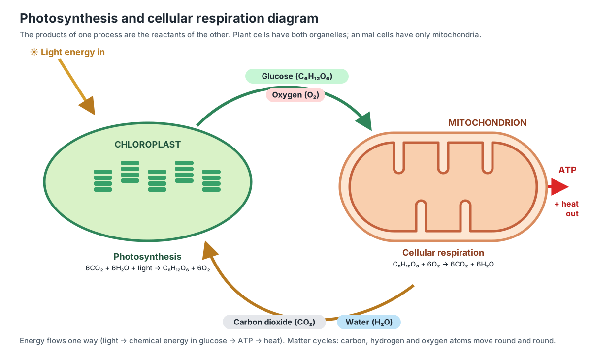
The equations are reverses of each other: photosynthesis is 6 CO2 + 6 H2O + light → C6H12O6 + 6 O2, and respiration is C6H12O6 + 6 O2 → 6 CO2 + 6 H2O + energy. But they are not the same reactions run backwards; they use different enzymes, different electron carriers (NADPH in photosynthesis, NADH and FADH2 in respiration) and different organelles.
The diagram also shows a key idea: matter cycles, energy flows. Carbon, hydrogen and oxygen atoms go round and round between the two processes, and between plants, animals and the air. Energy does not cycle. It comes in as sunlight, is stored in glucose, transferred to ATP, and is finally lost as heat, which is why life needs a constant input of light.
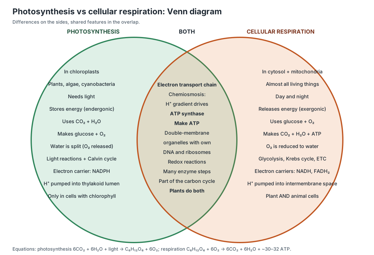
Photosynthesis vs cellular respiration: Venn diagram and comparison
Photosynthesis stores energy and happens only in chloroplasts of plants and algae (and in some bacteria); cellular respiration releases energy and happens in almost every living cell. Both use an electron transport chain and a proton gradient to drive ATP synthase.
| Feature | Photosynthesis | Cellular respiration |
|---|---|---|
| Organelle | Chloroplast | Cytosol + mitochondrion |
| Organisms | Plants, algae, cyanobacteria | Nearly all organisms, including plants |
| Equation | 6 CO2 + 6 H2O + light → C6H12O6 + 6 O2 | C6H12O6 + 6 O2 → 6 CO2 + 6 H2O + ATP |
| Energy | Light energy stored in glucose (endergonic) | Energy released from glucose (exergonic) |
| Stages | Light-dependent reactions (thylakoids) and Calvin cycle (stroma) | Glycolysis, pyruvate oxidation, Krebs cycle, oxidative phosphorylation |
| Electron source → acceptor | Water → NADP+ (makes NADPH) | Glucose (via NADH, FADH2) → O2 |
| Gas used / released | Uses CO2, releases O2 | Uses O2, releases CO2 |
| Proton gradient | Into the thylakoid lumen | Into the intermembrane space |
| ATP synthase | Yes (thylakoid membrane) | Yes (inner mitochondrial membrane) |
| When | Only in light | All the time |
For a Venn diagram, the middle should hold what both share: both are chains of redox reactions run by enzymes; both use an electron transport chain, chemiosmosis and ATP synthase; both happen in double-membrane organelles that have their own DNA and ribosomes; both are part of the carbon cycle; and plant cells do both. Chloroplasts and mitochondria are thought to descend from free-living bacteria, which is why they share so many features.
Cellular respiration diagram for AP Biology
For AP Biology, a cellular respiration diagram should show the location of each stage, the role of NAD+ and FAD as electron carriers, the proton gradient across the inner mitochondrial membrane, and oxygen as the final electron acceptor. Exam questions usually test reasoning about the system, not memorised numbers.
- Follow the electrons. Glucose is oxidised (loses electrons); oxygen is reduced (gains them). NADH and FADH2 carry the electrons in between.
- Predict the effect of a block. A drug that makes the inner membrane leaky to protons (an uncoupler) lets electron transport continue but stops ATP synthesis, so the energy becomes heat. Blocking complex IV stops electron flow, proton pumping and ATP synthesis. Blocking ATP synthase builds up the gradient until pumping slows.
- Link structure to function. Cristae increase membrane area for more chains and ATP synthases; a membrane impermeable to protons is what makes the gradient possible.
- Compare with photosynthesis. Both use chemiosmosis; in chloroplasts protons are pumped into the thylakoid lumen and ATP synthase faces the stroma.
- Know the exceptions. Prokaryotes do all four stages without mitochondria, using their plasma membrane; fermentation regenerates NAD+ without oxygen.
ATP synthase: the rotary motor of cellular respiration
ATP synthase is a molecular motor with two parts. The F1 head, which makes ATP, sticks out into the matrix. It is held to the membrane part, Fo, by a central stalk and a peripheral stalk. Protons crossing the membrane through Fo turn a ring of c-subunits and the central stalk at about 100 revolutions per second, and every full turn makes three ATP molecules. (Watt et al. (2010): Bioenergetic cost of making an adenosine triphosphate molecule in animal mitochondria, PNAS.)
The size of the c-ring sets the cost of each ATP, because each c-subunit carries one proton per turn. When the structure of the cow enzyme was solved, its ring had only 8 c-subunits, so animal mitochondria need about 8 ÷ 3 ≈ 2.7 protons per ATP. Bacteria, fungi and chloroplasts have rings of 10 to 15 subunits and pay 3.3 to 5 protons per ATP. Because c-subunit sequences are almost identical across vertebrates, the authors concluded that about 50,000 vertebrate species probably share the 8-subunit ring. (Watt et al. (2010): Bioenergetic cost of making an adenosine triphosphate molecule in animal mitochondria, PNAS.)
How the cristae organise the electron transport chain
The parts of the inner membrane are not random. Electron cryotomography of whole mitochondria from cow heart, potato and three fungi showed long rows of ATP synthase dimers only along the tightly curved edges of the cristae, with the two heads of each dimer at an angle of 70° or more. Complex I was found in the flat membrane regions. The authors proposed that proton pumps on the flat faces and proton-using ATP synthase rows on the edges together make ATP synthesis efficient. The 3D model on this page follows that layout. (Davies et al. (2011): Macromolecular organization of ATP synthase and complex I in whole mitochondria, PNAS.)
Cristae are not fixed shapes either. They change with physiological conditions, and their shape affects how the respiratory complexes are organised and how well oxidative phosphorylation works. Proteins that shape cristae have become candidate controls of cellular energy production. (Cogliati, Enriquez & Scorrano (2016): Mitochondrial cristae: where beauty meets functionality, Trends in Biochemical Sciences.)
Complexes I, III and IV often bind together into larger units called respiratory supercomplexes, with ubiquinone and cytochrome c shuttling electrons between them. Supercomplexes are thought to make electron transfer more efficient and to reduce the formation of reactive oxygen species, although how much they matter is still debated. (Guan, Zhao & Peng (2022): Mitochondrial respiratory chain supercomplexes, International Journal of Molecular Sciences.)
Why textbooks changed from 36–38 ATP to about 30–32
The ATP yield of respiration depends on the P/O ratio: how many ATP are made for each pair of electrons (each oxygen atom) passed to O2. A review of the main measurements since 1937 concluded that values of about 2.5 for NADH-linked substrates and 1.5 for succinate (FADH2) fit most of the data once errors are corrected. These fractional values come straight from the proton counts: about 10 protons are pumped per NADH, and making and exporting one ATP costs about 4. (Hinkle (2005): P/O ratios of mitochondrial oxidative phosphorylation, Biochimica et Biophysica Acta.)
Older textbooks rounded these to 3 and 2. With 10 NADH and 2 FADH2 per glucose, that gives 30 + 4 = 34 ATP from oxidative phosphorylation plus 4 ATP from glycolysis and the Krebs cycle, or 38 in total; 36 if the NADH from glycolysis enters by the glycerol phosphate shuttle. With 2.5 and 1.5, the same sum gives 32 (or 30). The same review noted that if ATP synthase needs 10/3 protons per ATP, as early structures suggested, the ratios could be as low as 2.3 and 1.4, so the true maximum may be a little under 30. (Hinkle (2005): P/O ratios of mitochondrial oxidative phosphorylation, Biochimica et Biophysica Acta.)
Mitochondrial DNA and mitochondrial ribosomes
Mitochondria carry their own small chromosome. The human mitochondrial genome is a circle of 16,569 base pairs that encodes the 12S and 16S ribosomal RNAs, 22 tRNAs and 13 proteins. It is extremely compact: genes have few or no spaces between them, and some stop codons are only completed after the RNA is copied. (Anderson et al. (1981): Sequence and organization of the human mitochondrial genome, Nature.)
All 13 proteins are parts of the oxidative phosphorylation machinery: subunits of complexes I, III and IV and of ATP synthase. Each cell holds hundreds to thousands of mitochondria, and each mitochondrion has one or more copies of the DNA. Faults in these genes cause mitochondrial diseases. (MedlinePlus Genetics: Mitochondrial DNA.)
The 13 proteins are made on the mitochondrion's own ribosomes. The human mitoribosome contains 80 proteins, 36 of them found only in mitochondria, plus three RNA molecules, and it has many features not seen in bacterial or cytosolic ribosomes. (Amunts et al. (2015): The structure of the human mitochondrial ribosome, Science.)
Respiration in plants: day, night and the alternative oxidase
Plants respire all the time. Respiration runs both day and night to supply energy for every energy-requiring process, and estimates indicate that plants release almost half of the CO2 they assimilate back to the atmosphere by respiration, with large differences between species and conditions. (Wendering & Nikoloski (2023): Toward mechanistic modeling and rational engineering of plant respiration, Plant Physiology.)
Plant mitochondria have an extra electron path that animals lack. Alternative oxidase (AOX) takes electrons from ubiquinone straight to oxygen, skipping complexes III and IV, so it pumps fewer protons and makes less ATP. This lets plants keep respiring and keep their metabolism balanced under stresses such as cold, drought, nutrient shortage and infection. (Vanlerberghe (2013): Alternative oxidase, a mitochondrial respiratory pathway in plants, International Journal of Molecular Sciences.)
Label it yourself: blank cellular respiration diagram
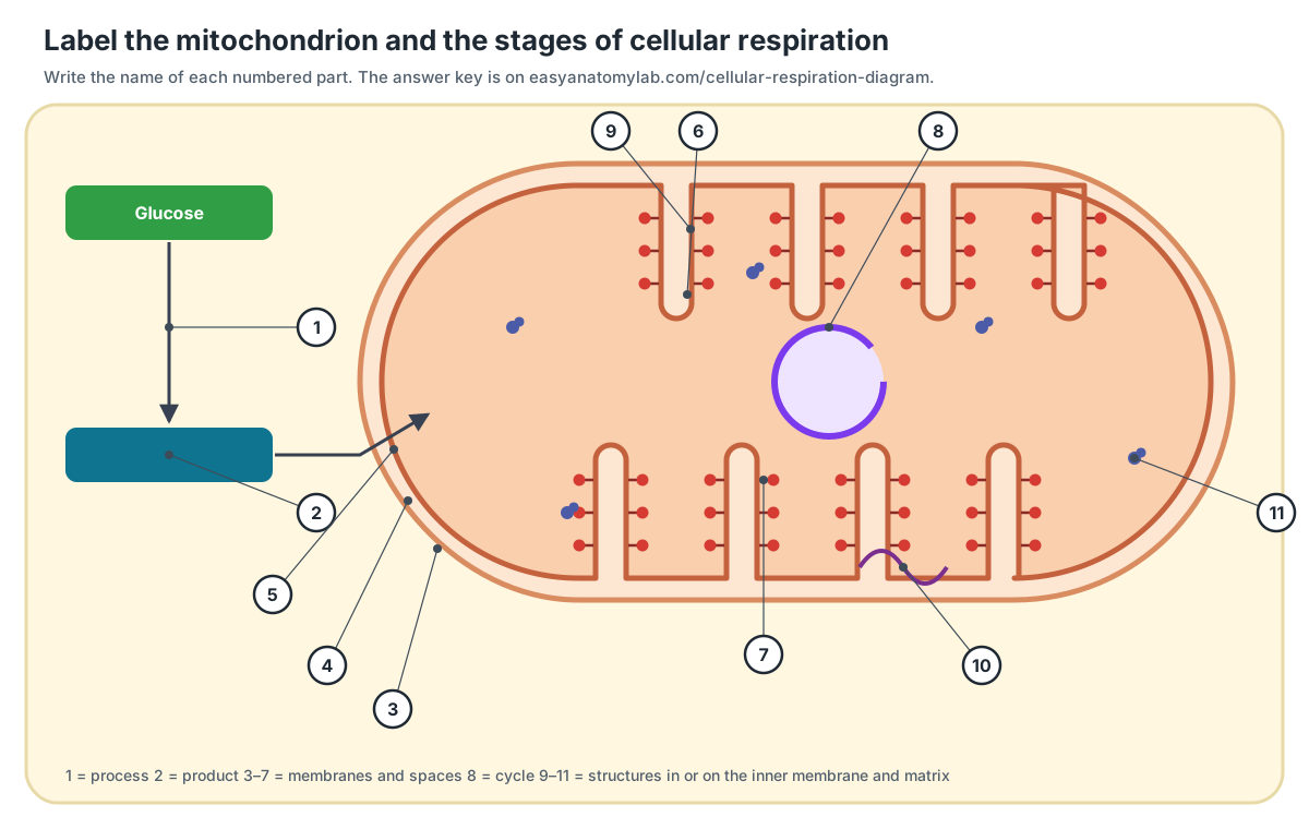
Test yourself with this unlabeled cellular respiration diagram. Write the name of each numbered part, then check the answer key. You can also turn on "All labels" in the 3D viewer and try to name each part before reading it.
Answer key
- Glycolysis (in the cytosol)
- Pyruvate (2 per glucose)
- Outer mitochondrial membrane
- Intermembrane space
- Inner mitochondrial membrane
- Crista (fold of the inner membrane)
- Matrix
- Krebs cycle (citric acid cycle)
- ATP synthase
- Mitochondrial DNA
- Mitochondrial ribosome
Quick quiz
- Which stage of cellular respiration does not need oxygen, and where does it happen?
- How many CO2 molecules are released per glucose, and in which stages?
- What is the final electron acceptor of the electron transport chain, and what does it become?
- Why do FADH2 electrons make fewer ATP than NADH electrons?
- Name the two ways ATP is made in cellular respiration.
- Why do muscle cells make lactate during a sprint?
Quiz answers
- Glycolysis, in the cytosol.
- Six: 2 in pyruvate oxidation and 4 in the Krebs cycle.
- Oxygen; it becomes water.
- They enter at complex II, which pumps no protons, so they skip complex I's 4 protons: ~6 instead of ~10 protons, ~1.5 instead of ~2.5 ATP.
- Substrate-level phosphorylation (glycolysis, Krebs cycle) and oxidative phosphorylation (ATP synthase).
- Oxygen supply cannot keep up, so NADH builds up; reducing pyruvate to lactate recycles NAD+ so glycolysis can keep making ATP.
Frequently asked questions
What are the 4 stages of cellular respiration?
Glycolysis in the cytosol, pyruvate oxidation (the link reaction) in the mitochondrial matrix, the Krebs (citric acid) cycle in the matrix, and oxidative phosphorylation (the electron transport chain plus chemiosmosis) on the inner mitochondrial membrane. Some textbooks merge pyruvate oxidation into the Krebs cycle and list three stages.
What is the equation for cellular respiration?
C6H12O6 + 6 O2 → 6 CO2 + 6 H2O + energy. In words: glucose plus oxygen gives carbon dioxide plus water, and the energy released is stored as about 30–32 ATP, with the rest lost as heat.
How many ATP are made in cellular respiration?
About 30–32 ATP per glucose with modern values of 2.5 ATP per NADH and 1.5 ATP per FADH2. Older textbooks gave 36–38 because they assumed whole numbers, 3 and 2, and ignored the cost of moving ATP, ADP and phosphate across the inner membrane. Only 4 ATP are made directly (2 in glycolysis, 2 in the Krebs cycle); the rest come from ATP synthase.
Where does cellular respiration take place?
Glycolysis happens in the cytosol. Pyruvate oxidation and the Krebs cycle happen in the mitochondrial matrix. The electron transport chain and ATP synthase sit in the inner mitochondrial membrane, especially the cristae. In bacteria, which have no mitochondria, the chain is in the plasma membrane.
Do plants do cellular respiration?
Yes. Every living plant cell has mitochondria and respires day and night, using sugars made by photosynthesis. Plants release almost half of the CO2 they fix back to the air by respiration. In the light, a leaf's photosynthesis is usually faster than its respiration, so it shows net O2 release.
What is the difference between photosynthesis and cellular respiration?
Photosynthesis stores light energy in glucose: 6 CO2 + 6 H2O → C6H12O6 + 6 O2, in chloroplasts, and only in plants, algae and some bacteria. Cellular respiration releases that energy as ATP: C6H12O6 + 6 O2 → 6 CO2 + 6 H2O, in mitochondria, in almost all eukaryotes. Both use an electron transport chain and ATP synthase driven by a proton gradient.
What is the difference between aerobic and anaerobic respiration?
Aerobic respiration uses oxygen as the final electron acceptor and makes about 30–32 ATP per glucose. Anaerobic respiration, found in some bacteria and archaea, still uses an electron transport chain but with another inorganic acceptor, such as sulfate or nitrate. Fermentation is different again: it uses no electron transport chain and makes only the 2 ATP from glycolysis.
Why is oxygen needed for cellular respiration?
Oxygen is the final electron acceptor at complex IV. It takes the electrons that have passed down the chain and combines with protons to form water. Without it, electrons back up, NADH cannot be recycled to NAD+, and the Krebs cycle and electron transport chain stop.
What is the role of NADH and FADH2?
They are electron carriers. Glycolysis, pyruvate oxidation and the Krebs cycle load electrons onto NAD+ and FAD, making NADH and FADH2. These carry the electrons to the electron transport chain, where their energy pumps protons. One glucose gives 10 NADH and 2 FADH2.
References
- OpenStax Biology 2e (2018), section 7.2 Glycolysis. Rice University.
- OpenStax Biology 2e (2018), section 7.3 Oxidation of pyruvate and the citric acid cycle.
- OpenStax Biology 2e (2018), section 7.4 Oxidative phosphorylation.
- OpenStax Biology 2e (2018), section 7.5 Metabolism without oxygen.
- OpenStax Biology 2e (2018), section 8.1 Overview of photosynthesis.
- OpenStax Biology 2e (2018), section 4.3 Eukaryotic cells.
- Milo, R. & Phillips, R. Cell Biology by the Numbers: How big are mitochondria? (BioNumbers).
- MedlinePlus Genetics. Mitochondrial DNA. U.S. National Library of Medicine.
- Anderson, S. et al. (1981). Sequence and organization of the human mitochondrial genome. Nature 290, 457–465.
- Amunts, A., Brown, A., Toots, J., Scheres, S. H. W. & Ramakrishnan, V. (2015). The structure of the human mitochondrial ribosome. Science 348, 95–98.
- Watt, I. N., Montgomery, M. G., Runswick, M. J., Leslie, A. G. W. & Walker, J. E. (2010). Bioenergetic cost of making an adenosine triphosphate molecule in animal mitochondria. PNAS 107, 16823–16827.
- Hinkle, P. C. (2005). P/O ratios of mitochondrial oxidative phosphorylation. Biochimica et Biophysica Acta 1706, 1–11.
- Davies, K. M. et al. (2011). Macromolecular organization of ATP synthase and complex I in whole mitochondria. PNAS 108, 14121–14126.
- Cogliati, S., Enriquez, J. A. & Scorrano, L. (2016). Mitochondrial cristae: where beauty meets functionality. Trends in Biochemical Sciences 41, 261–273.
- Guan, S., Zhao, L. & Peng, R. (2022). Mitochondrial respiratory chain supercomplexes: from structure to function. International Journal of Molecular Sciences 23, 13880.
- Sazanov, L. A. (2015). A giant molecular proton pump: structure and mechanism of respiratory complex I. Nature Reviews Molecular Cell Biology 16, 375–388.
- Vanlerberghe, G. C. (2013). Alternative oxidase: a mitochondrial respiratory pathway to maintain metabolic and signaling homeostasis during abiotic and biotic stress in plants. International Journal of Molecular Sciences 14, 6805–6847.
- Wendering, P. & Nikoloski, Z. (2023). Toward mechanistic modeling and rational engineering of plant respiration. Plant Physiology 191, 2150–2166.
More interactive anatomy models
- Human anatomy1,000+ bones, muscles, organs, vessels and nerves from real anatomical data.
- Octopus anatomyThree hearts, a ring-shaped brain, eight arms and a beak, in 3D.
- Squid anatomyPen, eight arms, two tentacles, giant axons and three hearts of the longfin squid.
- Squid dissectionStep-by-step guided squid dissection with every structure labeled.
- Fish anatomyExternal and internal anatomy of a bony fish: fins, gills, swim bladder, heart and brain.
- Perch dissectionGuided yellow perch (fish) dissection, from scales to the swim bladder.