

Heart anatomy and blood flow at a glance
The heart is two pumps side by side. The right side receives oxygen-poor blood from the body and pumps it to the lungs. The left side receives oxygen-rich blood from the lungs and pumps it to the body. Each side has a thin-walled receiving chamber (an atrium) above a muscular pumping chamber (a ventricle), and each ventricle has a one-way valve at its inlet and its outlet.
An adult heart is about the size of a fist: roughly 12 cm long, 8 cm wide and 6 cm thick, and about 250–350 g. It sits in the mediastinum between the lungs, with its base at about the third costal cartilage and its apex pointing down and to the left. (OpenStax Anatomy & Physiology 2e, 19.1 Heart Anatomy.)
Every drop of blood passes through the heart twice per full circuit: once through the right side on its way to the lungs (the pulmonary circuit) and once through the left side on its way to the tissues (the systemic circuit). Both sides beat at the same time, so the right and left ventricles eject the same volume with each beat, about 70–80 mL at rest. (OpenStax Anatomy & Physiology 2e, 19.3 Cardiac Cycle.)
Order of blood flow through the heart, step by step
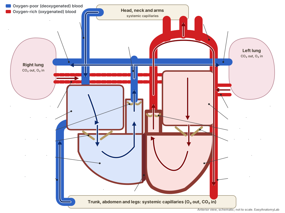
Blood flows through the heart in this order: venae cavae → right atrium → tricuspid valve → right ventricle → pulmonary valve → pulmonary arteries → lungs → pulmonary veins → left atrium → mitral valve → left ventricle → aortic valve → aorta → body. In full:
- Venae cavae and coronary sinus. Oxygen-poor blood from the body returns through the superior vena cava (upper body), the inferior vena cava (lower body) and the coronary sinus (heart muscle).
- Right atrium. The right atrium fills while the ventricles contract. Then the tricuspid valve opens and most blood falls passively into the right ventricle.
- Tricuspid valve. Blood passes through the three-leaflet tricuspid valve. The atrium contracts last, topping up the ventricle.
- Right ventricle. The right ventricle contracts. The tricuspid valve snaps shut (part of 'lub') and pressure rises until the pulmonary valve opens.
- Pulmonary valve. Blood is pushed through the pulmonary semilunar valve. When the ventricle relaxes the valve closes (part of 'dub').
- Pulmonary trunk and pulmonary arteries. The pulmonary trunk divides into the right and left pulmonary arteries, the only arteries that carry oxygen-poor blood.
- Lung capillaries: gas exchange. In the capillaries around the alveoli, blood unloads carbon dioxide and picks up oxygen. It leaves the lungs oxygen-rich.
- Pulmonary veins. Four pulmonary veins, two from each lung, carry oxygen-rich blood back to the heart.
- Left atrium. The left atrium collects the oxygen-rich blood and passes it on when the mitral valve opens.
- Mitral (bicuspid) valve. Blood flows through the two-leaflet mitral valve into the left ventricle.
- Left ventricle. The thick left ventricle contracts. The mitral valve closes ('lub') and pressure climbs to about 120 mmHg.
- Aortic valve. Blood is ejected through the aortic semilunar valve, which closes ('dub') as the ventricle relaxes.
- Aorta and its branches. The aorta rises, arches and descends. The coronary arteries, brachiocephalic trunk, left common carotid and left subclavian arteries branch off it.
- Body tissues and back to the venae cavae. In the systemic capillaries blood gives oxygen to the tissues and collects carbon dioxide, then returns through veins to the venae cavae.
A quick way to remember it: the right side is the "lung pump" and the left side the "body pump"; on each side blood goes atrium → atrioventricular valve → ventricle → semilunar valve → great artery. Try the interactive pathway below. It shares its step with the 3D model above.
Heart anatomy blood flow diagram
This labeled diagram of blood flow through the heart shows the chambers as a schematic frontal section, seen from the front. Arrows in blue trace oxygen-poor blood; arrows in red trace oxygen-rich blood. The right pulmonary veins are dashed because they pass behind the superior vena cava and aorta to reach the left atrium.
| Chamber | Receives blood from | Inlet valve | Outlet valve | Sends blood to | Blood |
|---|---|---|---|---|---|
| Right atrium | Superior and inferior venae cavae, coronary sinus | None | Tricuspid | Right ventricle | Oxygen-poor |
| Right ventricle | Right atrium | Tricuspid | Pulmonary | Pulmonary trunk → lungs | Oxygen-poor |
| Left atrium | Four pulmonary veins | None | Mitral | Left ventricle | Oxygen-rich |
| Left ventricle | Left atrium | Mitral | Aortic | Aorta → body | Oxygen-rich |
Anatomy of the heart: the parts that direct blood flow
The parts of the heart that matter for blood flow are the four chambers, the two septa, the four valves with their supporting cords and muscles, and the great vessels. The interatrial septum separates the two atria and the thicker interventricular septum separates the two ventricles, so oxygen-poor and oxygen-rich blood never mix in a healthy adult heart. (OpenStax Anatomy & Physiology 2e, 19.1 Heart Anatomy.)
The heart wall has three layers. The epicardium is the thin outer layer (the visceral layer of the serous pericardium). The myocardium is the thick middle layer of cardiac muscle that does the pumping. The endocardium is the smooth inner lining, continuous with the lining of the blood vessels. The whole heart sits in the pericardial sac, whose serous fluid lets it beat without friction. (OpenStax Anatomy & Physiology 2e, 19.1 Heart Anatomy.)
The two ventricles differ most in their walls. The left ventricle must push blood through the long, high-resistance systemic circuit, so its wall is far thicker than the right ventricle's. In a catheter study, normal left ventricular systolic pressure is about 90–140 mmHg, while pulmonary artery systolic pressure, which the right ventricle has to beat, is only about 15–30 mmHg. (Davis's Lab & Diagnostic Tests, Cardiac catheterization (normal pressures).)
Inside the ventricles, the atrioventricular valves are held by chordae tendineae, fibrous cords that run to papillary muscles on the ventricle wall. OpenStax describes three papillary muscles in the right ventricle (anterior, posterior and septal) and two in the left. The right ventricle also has the moderator band, a muscular bridge from the septum to the anterior papillary muscle that carries part of the right bundle branch. The semilunar valves have no chordae or papillary muscles. (OpenStax Anatomy & Physiology 2e, 19.1 Heart Anatomy.)
Pulmonary vs systemic circulation
Pulmonary circulation carries blood from the right ventricle to the lungs and back to the left atrium; systemic circulation carries blood from the left ventricle to every other tissue and back to the right atrium. The pulmonary circuit exists only to exchange gases: in the lung capillaries blood unloads carbon dioxide and loads oxygen. The systemic circuit delivers oxygen and nutrients and collects wastes. (OpenStax Anatomy & Physiology 2e, 20.5 Circulatory Pathways.)
The two circuits are in series, so blood must pass through one to reach the other. This is why the pulmonary arteries are the only arteries that carry oxygen-poor blood and the pulmonary veins the only veins (in the adult) that carry oxygen-rich blood. An artery is any vessel carrying blood away from the heart; a vein is any vessel carrying it back.
Typical anatomy has four pulmonary veins, a superior and an inferior vein from each lung, and Radiopaedia reports this pattern in about 70% of people. The right superior vein also drains the right middle lobe. Common variants include a single common trunk for the two left veins and an extra right middle-lobe vein (about 10%). (Radiopaedia, Pulmonary veins.)
On the systemic side, the aortic arch normally gives off three branches: the brachiocephalic trunk (which splits into the right subclavian and right common carotid arteries), the left common carotid artery and the left subclavian artery. This standard pattern is found in about 75% of people. In about 15% the brachiocephalic trunk and left common carotid share an origin, an arrangement often called a bovine arch. (Radiopaedia, Aortic arch.)
| Pulmonary circulation | Systemic circulation | |
|---|---|---|
| Pumped by | Right ventricle | Left ventricle |
| Leaves the heart in | Pulmonary trunk → right and left pulmonary arteries | Aorta |
| Returns in | Four pulmonary veins → left atrium | Superior and inferior venae cavae (and coronary sinus) → right atrium |
| Arteries carry | Oxygen-poor blood | Oxygen-rich blood |
| Veins carry | Oxygen-rich blood | Oxygen-poor blood |
| Capillaries | Around the alveoli of the lungs | In every other tissue |
| Purpose | Unload CO₂, load O₂ | Deliver O₂ and nutrients, collect CO₂ and wastes |
| Typical systolic pressure | About 15–30 mmHg (pulmonary artery) | About 90–140 mmHg (left ventricle) |
Heart valves and heart sounds (lub-dub)
The heart has four valves that keep blood moving one way: the tricuspid and mitral (bicuspid) atrioventricular valves between the atria and ventricles, and the pulmonary and aortic semilunar valves at the exits of the ventricles. Valves are passive. They open and close only because of pressure differences on their two sides. (OpenStax Anatomy & Physiology 2e, 19.1 Heart Anatomy.)
The first heart sound, 'lub' (S1), is made when the tricuspid and mitral valves close at the start of ventricular contraction. The second heart sound, 'dub' (S2), is made when the aortic and pulmonary valves close as the ventricles relax. A third sound (S3) can be normal in children, some athletes and pregnancy but may signal heart failure later in life; a fourth (S4) comes from the atria pushing blood into a stiff ventricle. Murmurs are caused by turbulent flow, for example through a narrowed or leaking valve. (OpenStax Anatomy & Physiology 2e, 19.3 Cardiac Cycle.)
The atrioventricular valves are supported from below. When a ventricle contracts, its papillary muscles contract too and tighten the chordae tendineae, so the leaflets meet edge to edge instead of being blown back into the atrium. If a chorda breaks, part of the valve flails and blood leaks backward (regurgitation). (OpenStax Anatomy & Physiology 2e, 19.1 Heart Anatomy.)
| Valve | Type | Leaflets / cusps | Between | Closes | Sound |
|---|---|---|---|---|---|
| Tricuspid | Atrioventricular | 3, with chordae | Right atrium → right ventricle | Start of ventricular systole | S1 'lub' |
| Mitral (bicuspid) | Atrioventricular | 2, with chordae | Left atrium → left ventricle | Start of ventricular systole | S1 'lub' |
| Pulmonary | Semilunar | 3, no chordae | Right ventricle → pulmonary trunk | Start of ventricular diastole | S2 'dub' |
| Aortic | Semilunar | 3, no chordae | Left ventricle → aorta | Start of ventricular diastole | S2 'dub' |
The cardiac cycle: systole, diastole and how valves open and close
The cardiac cycle is one heartbeat: a period of contraction (systole) and relaxation (diastole) of the atria and then the ventricles. At about 75 beats per minute one cycle lasts about 0.8 s. OpenStax gives atrial systole about 100 ms, ventricular systole about 270 ms and ventricular diastole about 430 ms. (OpenStax Anatomy & Physiology 2e, 19.3 Cardiac Cycle.)
Most ventricular filling is passive. While the atria and ventricles are both relaxed, blood flows from the veins through the atria and open atrioventricular valves; this supplies about 70–80% of the filling. Atrial systole then adds the last 20–30%, the 'atrial kick'. The ventricle ends diastole holding about 130 mL (the end-diastolic volume). (OpenStax Anatomy & Physiology 2e, 19.3 Cardiac Cycle.)
Ventricular systole has two phases. In isovolumic contraction, ventricular pressure rises above atrial pressure, which slams the atrioventricular valves shut (S1); the semilunar valves are still closed, so volume does not change. When ventricular pressure exceeds the pressure in the aorta and pulmonary trunk, the semilunar valves open and blood is ejected: about 70–80 mL leaves each ventricle and 50–60 mL stays behind (the end-systolic volume). (OpenStax Anatomy & Physiology 2e, 19.3 Cardiac Cycle.)
Ventricular diastole also has two phases. In isovolumic relaxation, ventricular pressure falls below arterial pressure, blood briefly flows back and fills the semilunar cusps, and the valves close (S2). This shows on the aortic pressure trace as the dicrotic notch. When ventricular pressure falls below atrial pressure, the atrioventricular valves open and passive filling starts again. (OpenStax Anatomy & Physiology 2e, 19.3 Cardiac Cycle.)
| Phase | AV valves | Semilunar valves | What happens |
|---|---|---|---|
| Atrial systole (~100 ms) | Open | Closed | Atria contract and top up the ventricles (last 20–30% of filling) |
| Isovolumic contraction | Close (S1) | Closed | Ventricular pressure rises; volume stays about 130 mL |
| Ventricular ejection | Closed | Open | About 70–80 mL ejected from each ventricle |
| Isovolumic relaxation | Closed | Close (S2) | Ventricular pressure falls; dicrotic notch on the aortic trace |
| Ventricular filling | Open | Closed | Passive filling from the atria (70–80% of filling) |
Conduction system of the heart and the ECG
Each heartbeat starts in the sinoatrial (SA) node, a patch of pacemaker cells in the right atrial wall near the opening of the superior vena cava. Its cells depolarise on their own; without nervous or hormonal input the SA node would fire about 80–100 times a minute. The impulse spreads across both atria (Bachmann's bundle carries it to the left atrium) and reaches the atrioventricular (AV) node in about 50 ms. (OpenStax Anatomy & Physiology 2e, 19.2 Cardiac Muscle and Electrical Activity.)
The AV node, in the lower interatrial septum, delays the impulse by about 100 ms. That pause lets the atria finish emptying before the ventricles contract. The impulse then runs down the atrioventricular bundle (bundle of His), which is the only normal electrical bridge through the insulating fibrous skeleton, and splits into the right and left bundle branches in the interventricular septum. (OpenStax Anatomy & Physiology 2e, 19.2 Cardiac Muscle and Electrical Activity.)
The bundle branches run toward the apex and hand the impulse to the Purkinje fibres, which spread it through the ventricular muscle in about 75 ms. Because activation starts at the apex and moves toward the base, the ventricles squeeze blood upward toward the pulmonary and aortic valves, like squeezing a tube of toothpaste from the bottom. (OpenStax Anatomy & Physiology 2e, 19.2 Cardiac Muscle and Electrical Activity.)
The ECG records this sequence from the skin. The P wave is atrial depolarisation, and atrial contraction follows about 25 ms after it starts. The PR interval covers the spread through the atria and the AV node delay; it lengthens in first-degree heart block. The QRS complex is ventricular depolarisation (large because the ventricles are large), and ventricular contraction begins near the R peak. The T wave is ventricular repolarisation. Atrial repolarisation happens during the QRS and is hidden by it. (OpenStax Anatomy & Physiology 2e, 19.2 Cardiac Muscle and Electrical Activity.)
| Step | Structure | Timing (OpenStax) | ECG |
|---|---|---|---|
| 1 | SA node fires; impulse spreads over both atria | Reaches AV node in ~50 ms | P wave |
| 2 | AV node delay | ~100 ms | PR segment |
| 3 | Bundle of His → right and left bundle branches | ~25 ms to the Purkinje fibres | Start of QRS |
| 4 | Purkinje fibres → ventricular muscle, apex first | ~75 ms | QRS complex |
| 5 | Ventricles repolarise | T wave |
Coronary circulation: blood flow to the heart muscle
The heart muscle is fed by the right and left coronary arteries, the first branches of the aorta. They arise from the aortic sinuses just above the aortic valve cusps. The left coronary artery divides into the left anterior descending (anterior interventricular) artery and the circumflex artery, which together supply most of the left ventricle, the left atrium and the interventricular septum. The right coronary artery supplies the right atrium, much of the right ventricle and, usually, the conduction system; its branches include the marginal artery and the posterior interventricular (posterior descending) artery. (OpenStax Anatomy & Physiology 2e, 19.1 Heart Anatomy.)
Coronary dominance describes which artery gives rise to the posterior descending artery. Radiopaedia reports right dominance in about 85% of hearts, left dominance (from the circumflex) in about 8% and codominance in about 7%; individual studies vary. The BodyParts3D heart in the viewer is right-dominant. (Radiopaedia, Coronary arterial dominance.)
After passing through the myocardial capillaries, blood drains into cardiac veins. The great cardiac vein (beside the LAD), the middle cardiac vein (in the posterior groove) and the small cardiac vein (with the right coronary artery) empty into the coronary sinus, a wide vein on the back of the heart that opens into the right atrium. Anterior cardiac veins drain the front of the right ventricle straight into the right atrium. (OpenStax Anatomy & Physiology 2e, 19.1 Heart Anatomy.)
Fetal circulation in brief: foramen ovale and ductus arteriosus
Before birth the lungs are not used for gas exchange, so the fetal heart has two shunts that send most blood around them. The foramen ovale, an opening in the interatrial septum, lets blood pass from the right atrium straight into the left atrium. The ductus arteriosus, a short vessel between the pulmonary trunk and the aorta, carries most of the right ventricle's output into the aorta. A third shunt, the ductus venosus, lets much of the oxygen-rich blood from the umbilical vein bypass the liver. (OpenStax Anatomy & Physiology 2e, 20.6 Development of Blood Vessels and Fetal Circulation.)
At birth the first breaths expand the lungs and lower their resistance, and pressure in the left atrium rises above that in the right. This pushes the flap of the foramen ovale shut; it leaves the fossa ovalis. Rising oxygen levels make the ductus arteriosus constrict; it becomes the ligamentum arteriosum. The ductus venosus becomes the ligamentum venosum. The foramen ovale stays probe-patent in up to about a third of adults, usually without symptoms. (Radiopaedia, Patent foramen ovale.)
Liver blood flow anatomy
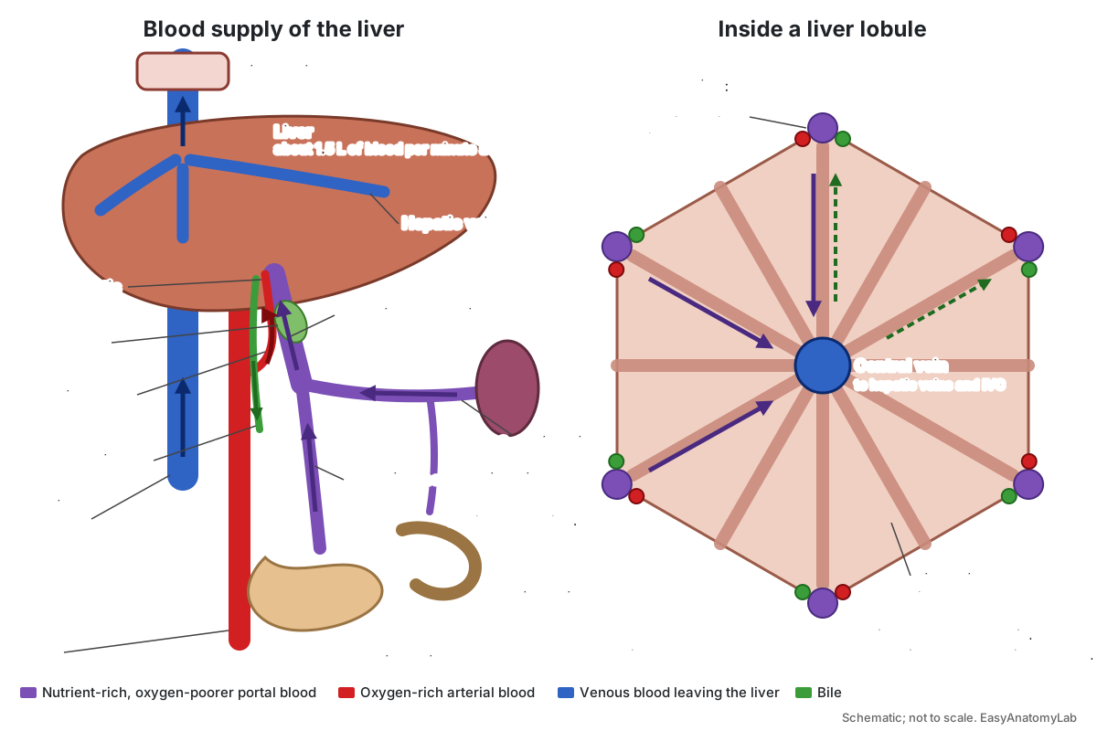
The liver has a dual blood supply: about 75% of its blood arrives through the hepatic portal vein and about 25% through the hepatic artery, yet each supplies roughly half of the liver's oxygen, because portal blood has already passed through the gut and is partly deoxygenated. (LibreTexts Medicine, Blood Supply to the Liver (Boundless Anatomy and Physiology).)
The hepatic portal vein forms behind the pancreas where the superior mesenteric vein (from the small intestine and much of the large intestine) joins the splenic vein (from the spleen, pancreas and part of the stomach, and usually also receiving the inferior mesenteric vein). The hepatic artery proper, a branch of the common hepatic artery from the coeliac trunk, brings oxygen-rich systemic blood. Both enter the liver at the porta hepatis, the 'gate' on its lower surface. (OpenStax Anatomy & Physiology 2e, 20.5 Circulatory Pathways.)
Inside the liver both vessels branch alongside bile ducts in portal triads at the corners of each hepatic lobule. Their blood mixes and flows inward through sinusoids, wide leaky capillaries lined by plates of hepatocytes and patrolled by Kupffer cells. Hepatocytes take up nutrients and toxins, store and release glucose, and make plasma proteins. The sinusoids drain into the central vein of the lobule; central veins join into the right, middle and left hepatic veins, which empty into the inferior vena cava just below the right atrium. At rest the liver receives about 1,500 mL of blood per minute. (OpenStax Anatomy & Physiology 2e, 23.6 Accessory Organs in Digestion.)
Bile flows the opposite way to blood: hepatocytes secrete it into bile canaliculi, which carry it outward to the bile ductules of the portal triads, then to the hepatic ducts and common bile duct. (OpenStax Anatomy & Physiology 2e, 23.6 Accessory Organs in Digestion.)
- Hepatic portal vein (~75% of the blood) and hepatic artery proper (~25%) → porta hepatis
- Interlobular branches in the portal triads
- Hepatic sinusoids, between plates of hepatocytes
- Central vein of each lobule
- Right, middle and left hepatic veins
- Inferior vena cava → right atrium
Cerebral blood flow anatomy in brief: the circle of Willis
The brain is supplied by two pairs of arteries: the internal carotid arteries (from the common carotids) and the vertebral arteries (from the subclavians). The two vertebral arteries join to form the basilar artery. The anterior cerebral artery supplies mainly the frontal lobe, and the middle cerebral artery the temporal and parietal lobes. (OpenStax Anatomy & Physiology 2e, 20.5 Circulatory Pathways.)
At the base of the brain these vessels connect in the circle of Willis (arterial circle), around the optic chiasm and pituitary stalk. It is made of the two internal carotid arteries, the first (A1) segments of both anterior cerebral arteries joined by the anterior communicating artery, the two posterior communicating arteries, and the first (P1) segments of both posterior cerebral arteries from the tip of the basilar artery. It can even out flow between the two sides and provide a detour if one feeding artery is blocked. A complete circle with no missing or very thin segment is found in only about 20–25% of people. (Radiopaedia, Circle of Willis.)
Blood flow through the heart: anatomy and physiology course summary
For an anatomy and physiology exam, these are the points most often tested on heart anatomy and blood flow:
- Order of flow: venae cavae → RA → tricuspid → RV → pulmonary valve → pulmonary arteries → lungs → pulmonary veins → LA → mitral → LV → aortic valve → aorta.
- The right side handles oxygen-poor blood and the pulmonary circuit; the left side handles oxygen-rich blood and the systemic circuit.
- Pulmonary arteries carry oxygen-poor blood and pulmonary veins oxygen-rich blood. Vessels are named by direction, not oxygen.
- Valves open and close because of pressure differences. AV valves close at the start of systole (S1, lub); semilunar valves close at the start of diastole (S2, dub).
- Chordae tendineae and papillary muscles stop AV valve prolapse; semilunar valves have neither.
- The left ventricle wall is thickest because systemic pressure is much higher than pulmonary pressure.
- Stroke volume ≈ 70–80 mL = end-diastolic volume (~130 mL) − end-systolic volume (~50–60 mL).
- Conduction: SA node → atria → AV node (delay) → bundle of His → bundle branches → Purkinje fibres. P wave, QRS complex and T wave match atrial depolarisation, ventricular depolarisation and ventricular repolarisation.
- Coronary arteries leave the aortic sinuses; the coronary sinus returns cardiac venous blood to the right atrium.
- Fetal shunts: foramen ovale (→ fossa ovalis), ductus arteriosus (→ ligamentum arteriosum), ductus venosus (→ ligamentum venosum).
To see the same structures on a real specimen, work through the sheep heart dissection, which compares sheep, pig and human hearts. The heart's place in the whole body, with every other organ system, is on the 3D human anatomy page, built from the same BodyParts3D data. Blood carries oxygen to cells for cellular respiration, and the kidney's nephrons filter the blood that the renal arteries bring from the aorta.
Label the heart blood flow diagram yourself
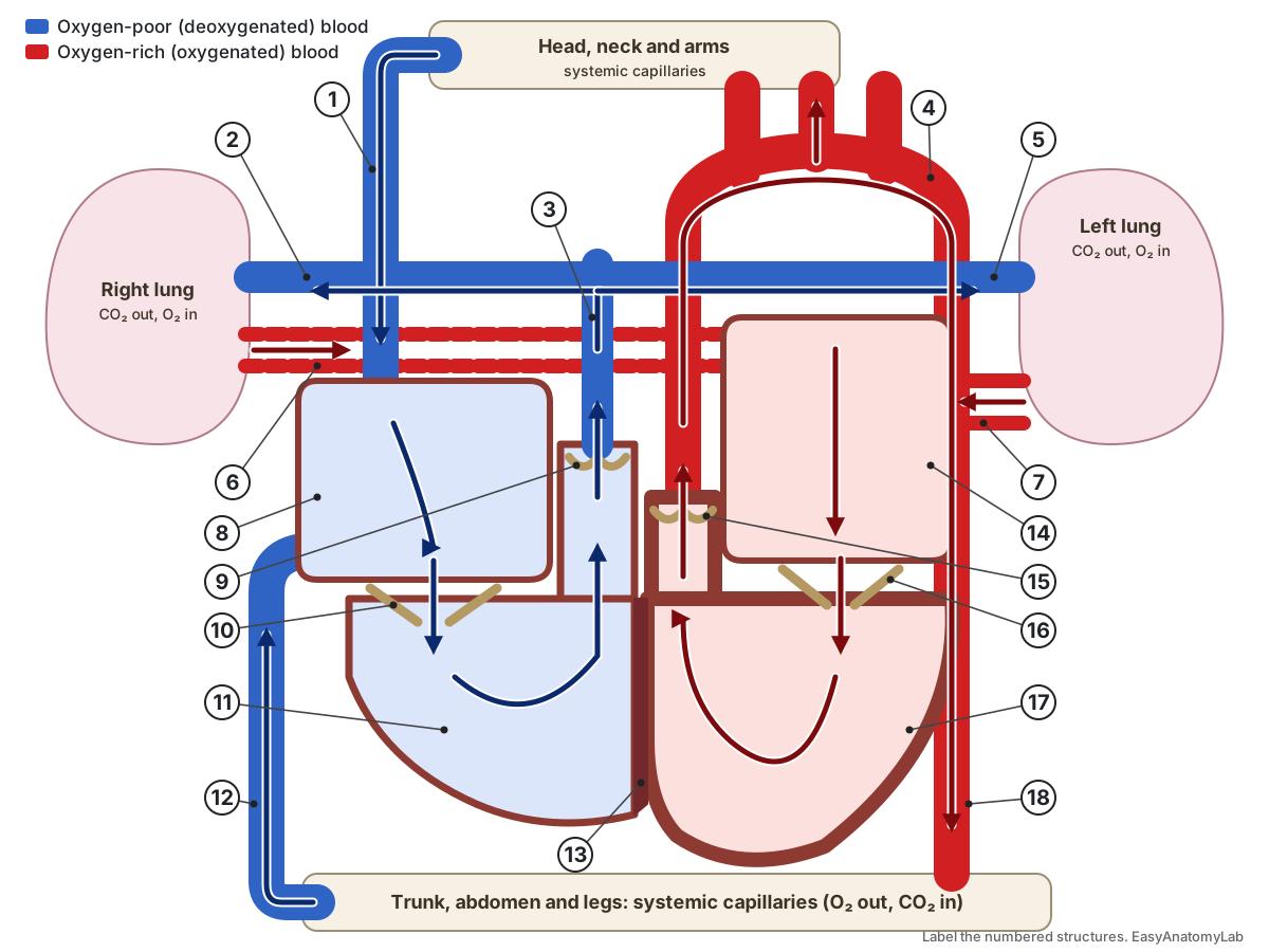
Test yourself: write the name of each numbered structure, then trace the path of a red blood cell from the superior vena cava to the aorta. Open the answer key when you are done.
Answer key
- Superior vena cava
- Right pulmonary artery
- Pulmonary trunk
- Aorta (arch)
- Left pulmonary artery
- Right pulmonary veins
- Left pulmonary veins
- Right atrium
- Pulmonary valve
- Tricuspid valve
- Right ventricle
- Inferior vena cava
- Interventricular septum
- Left atrium
- Aortic valve
- Mitral (bicuspid) valve
- Left ventricle
- Descending aorta
Heart structures: labeled reference
Every structure in the 3D heart, grouped as in the viewer. The coloured groups follow the blood they carry.
Chambers & septa
- Right atrium
- The thin-walled upper right chamber. It receives oxygen-poor blood from the body and from the heart muscle itself. Collects blood from the superior vena cava, inferior vena cava and coronary sinus, then passes it through the tricuspid valve into the right ventricle.
- Right ventricle
- The lower right chamber. It pumps oxygen-poor blood through the pulmonary valve to the lungs. Contracts to push blood into the pulmonary trunk, starting the pulmonary circuit.
- Left atrium
- The upper left chamber. It receives oxygen-rich blood from the lungs through the pulmonary veins. Passes oxygen-rich blood through the mitral valve into the left ventricle.
- Left ventricle
- The lower left chamber and the strongest pump. It sends oxygen-rich blood through the aortic valve to the whole body. Generates the high pressure (normally about 90–140 mmHg systolic) that drives blood around the systemic circuit.
- Apex of the heart (Cardiac apex)
- The blunt tip of the heart, formed by the left ventricle. It points down, forward and to the left. A landmark: the apex beat can be felt here, and ventricular contraction spreads from the apex toward the base.
- Interventricular septum
- The thick muscular wall between the right and left ventricles. Keeps oxygen-poor and oxygen-rich blood apart and carries the bundle of His and the bundle branches.
- Interatrial septum
- The thin wall between the right and left atria. Separates the two atria after birth.
Valves & supports
- Tricuspid valve (Right atrioventricular valve)
- The valve between the right atrium and right ventricle, with three leaflets. Opens while the ventricle fills and closes when the right ventricle contracts, so blood cannot flow back into the atrium.
- Pulmonary valve (Pulmonary semilunar valve)
- The semilunar valve between the right ventricle and the pulmonary trunk, with three pocket-shaped cusps. Opens when right ventricular pressure exceeds pulmonary trunk pressure, and closes when the ventricle relaxes.
- Mitral valve (Bicuspid valve, left atrioventricular valve)
- The valve between the left atrium and left ventricle, with two leaflets. Lets oxygen-rich blood into the left ventricle and seals when the ventricle contracts.
- Aortic valve (Aortic semilunar valve)
- The semilunar valve between the left ventricle and the aorta, normally with three cusps. Opens during ventricular ejection and closes at the start of ventricular diastole, producing the dicrotic notch on the aortic pressure trace.
- Chordae tendineae (Heart strings)
- Fibrous cords that tie the free edges of the tricuspid and mitral leaflets to the papillary muscles. Stop the leaflets from turning inside out (prolapsing) into the atria when the ventricles contract.
- Anterior papillary muscle (right ventricle)
- The largest papillary muscle of the right ventricle. Contracts with the ventricle and pulls on chordae to the anterior and posterior tricuspid leaflets.
- Posterior papillary muscle (right ventricle) (Inferior papillary muscle)
- A smaller papillary muscle on the lower (inferior) wall of the right ventricle. Anchors chordae to the posterior and septal tricuspid leaflets.
- Septal papillary muscle (right ventricle)
- A small papillary muscle on the septal wall of the right ventricle. Anchors chordae to the septal and anterior tricuspid leaflets.
- Papillary muscles (left ventricle) (Anterolateral and posteromedial papillary muscles)
- The muscle columns of the left ventricle that anchor the mitral valve chordae. Tense the chordae so the mitral leaflets meet and do not prolapse during systole.
- Moderator band (Septomarginal trabecula)
- A muscular band that crosses the right ventricle from the septum to the anterior papillary muscle. Carries part of the right bundle branch, so the anterior papillary muscle contracts on time.
Oxygen-poor vessels
- Superior vena cava (SVC)
- The large vein that returns oxygen-poor blood from the head, neck, arms and chest to the right atrium. Step 1 of blood flow: delivers blood from the upper body into the right atrium.
- Inferior vena cava (IVC)
- The largest vein, returning oxygen-poor blood from the abdomen, pelvis and legs to the right atrium. Step 1 of blood flow: delivers blood from the lower body, including the liver through the hepatic veins.
- Brachiocephalic veins (Innominate veins)
- The two veins that join to form the superior vena cava. Drain the internal jugular and subclavian veins of each side.
- Pulmonary trunk
- The large artery leaving the right ventricle. It carries oxygen-poor blood toward the lungs. Carries the right ventricle's output to the right and left pulmonary arteries.
- Right pulmonary artery
- The branch of the pulmonary trunk to the right lung. Carries oxygen-poor blood to the right lung's capillaries for gas exchange.
- Left pulmonary artery
- The branch of the pulmonary trunk to the left lung. Carries oxygen-poor blood to the left lung.
Oxygen-rich vessels
- Right superior pulmonary vein
- Carries oxygen-rich blood from the upper and middle lobes of the right lung to the left atrium. Step 8 of blood flow: returns oxygenated blood from the lungs to the heart.
- Right inferior pulmonary vein
- Carries oxygen-rich blood from the lower lobe of the right lung to the left atrium. Returns oxygenated blood from the right lower lobe.
- Left superior pulmonary vein
- Carries oxygen-rich blood from the upper lobe (including the lingula) of the left lung. Returns oxygenated blood from the left upper lobe.
- Left inferior pulmonary vein
- Carries oxygen-rich blood from the lower lobe of the left lung. Returns oxygenated blood from the left lower lobe.
- Ascending aorta
- The first part of the aorta, rising from the left ventricle. Carries the left ventricle's output upward. Its only branches are the two coronary arteries.
- Aortic arch (Arch of the aorta)
- The curved part of the aorta that turns from rising to falling and gives off the three great branches. Sends blood to the head, neck and arms through the brachiocephalic trunk, left common carotid and left subclavian arteries.
- Descending (thoracic) aorta
- The continuation of the aorta down through the chest, behind the heart. Carries oxygen-rich blood to the chest wall, abdomen, pelvis and legs.
- Brachiocephalic trunk (Brachiocephalic artery, innominate artery)
- The first and largest branch of the aortic arch. Divides into the right common carotid and right subclavian arteries (OpenStax).
- Left common carotid artery
- The second branch of the aortic arch, supplying the left side of the head and neck. Divides in the neck into the internal carotid (brain) and external carotid (face, scalp) arteries.
- Left subclavian artery
- The third branch of the aortic arch, supplying the left arm. Supplies the left arm and, through the left vertebral artery, part of the brain.
- Right common carotid artery
- A branch of the brachiocephalic trunk to the right side of the head and neck. Supplies the right side of the head and brain through its internal and external carotid branches.
- Right subclavian artery
- A branch of the brachiocephalic trunk to the right arm. Supplies the right arm and, through the right vertebral artery, part of the brain.
Coronary vessels
- Left main coronary artery (Left coronary artery (LCA))
- The short trunk of the left coronary artery, arising from the aorta just above the aortic valve. Divides into the left anterior descending and circumflex arteries, which supply most of the left ventricle and septum.
- Left anterior descending artery (LAD, anterior interventricular artery)
- The branch of the left coronary artery that runs down the front groove between the ventricles. Supplies the front of the left ventricle, the front two-thirds of the septum and part of the right ventricle.
- Circumflex artery (Left circumflex (LCx))
- The branch of the left coronary artery that curves around the left side in the coronary groove. Supplies the left atrium and the lateral and back walls of the left ventricle.
- Right coronary artery (RCA)
- The coronary artery that runs in the right atrioventricular groove. Supplies the right atrium, most of the right ventricle and, in most people, the SA and AV nodes.
- Posterior descending artery (PDA, posterior interventricular artery)
- The artery in the posterior groove between the ventricles. In this right-dominant model it comes from the right coronary artery. Supplies the back third of the septum and the lower walls of both ventricles.
- Coronary sinus
- A wide, thin-walled vein on the back of the heart that collects most of the blood from the heart muscle. Returns oxygen-poor blood from the myocardium to the right atrium.
- Great cardiac vein
- The main vein on the front of the heart, beside the LAD. Drains the area supplied by the left coronary artery into the coronary sinus.
- Middle cardiac vein (Posterior interventricular vein)
- The vein in the posterior interventricular groove, beside the posterior descending artery. Drains the back of both ventricles into the coronary sinus.
- Small cardiac vein
- A small vein that runs with the right coronary artery. Drains the right atrium and right ventricle into the coronary sinus.
- Posterior vein of the left ventricle
- A vein on the back of the left ventricle. Drains the back wall of the left ventricle into the coronary sinus.
- Anterior cardiac veins
- Small veins on the front of the right ventricle. Drain the front of the right ventricle straight into the right atrium, bypassing the coronary sinus (OpenStax).
Conduction system
- Sinoatrial (SA) node (Sinus node, pacemaker)
- The natural pacemaker: a small patch of specialised muscle that starts each heartbeat. Fires spontaneously; on its own it would set a rate of about 80–100 beats per minute, slowed at rest by the vagus nerve.
- Atrioventricular (AV) node
- A small node in the floor of the right atrium that delays the impulse before it reaches the ventricles. Holds the impulse for about 100 ms so the atria can finish filling the ventricles (OpenStax).
- Bundle of His (Atrioventricular bundle)
- The only normal electrical connection between the atria and the ventricles. Carries the impulse from the AV node into the septum, where it splits into the bundle branches.
- Right bundle branch
- The branch of the bundle of His that runs down the right side of the septum. Carries the impulse to the right ventricle and its anterior papillary muscle.
- Left bundle branch
- The branch of the bundle of His that runs down the left side of the septum and divides into fascicles. Carries the impulse to the thick left ventricle through its anterior and posterior fascicles (OpenStax).
- Purkinje fibres (Subendocardial branches)
- A network of fast-conducting fibres under the endocardium of both ventricles. Spread the impulse through the ventricles in about 75 ms, activating the apex first so blood is squeezed toward the outflow valves.
Fetal remnants
- Fossa ovalis
- An oval dip on the right atrial side of the interatrial septum. A remnant of the foramen ovale, the fetal opening that let blood bypass the lungs.
- Ligamentum arteriosum
- A short fibrous cord joining the pulmonary trunk to the underside of the aortic arch. The remnant of the ductus arteriosus, which carried blood from the pulmonary trunk to the aorta before birth.
Frequently asked questions
What is the correct order of blood flow through the heart?
Superior and inferior vena cava → right atrium → tricuspid valve → right ventricle → pulmonary valve → pulmonary trunk and arteries → lungs → pulmonary veins → left atrium → mitral (bicuspid) valve → left ventricle → aortic valve → aorta → body.
Which side of the heart carries oxygenated blood?
The left side. The left atrium and left ventricle carry oxygen-rich blood from the lungs to the body. The right atrium and right ventricle carry oxygen-poor blood from the body to the lungs.
Why is the left ventricle wall thicker than the right?
The left ventricle pumps blood around the whole body against high resistance, reaching about 90–140 mmHg. The right ventricle only pumps to the nearby lungs, at about 15–30 mmHg, so it needs less muscle.
Do pulmonary arteries carry oxygenated blood?
No. The pulmonary trunk and pulmonary arteries carry oxygen-poor blood from the right ventricle to the lungs. The pulmonary veins carry oxygen-rich blood back to the left atrium. Arteries and veins are named by direction of flow (away from or toward the heart), not by oxygen content.
What causes the lub-dub heart sounds?
'Lub' (S1) is the closing of the tricuspid and mitral valves at the start of ventricular contraction. 'Dub' (S2) is the closing of the aortic and pulmonary valves when the ventricles relax.
How many valves does the heart have?
Four: two atrioventricular valves (tricuspid on the right, mitral on the left) and two semilunar valves (pulmonary and aortic). They keep blood moving in one direction.
Where does the heart get its own blood supply?
From the right and left coronary arteries, which leave the aorta just above the aortic valve. The left coronary artery divides into the left anterior descending and circumflex arteries. Blood returns through the cardiac veins to the coronary sinus and the right atrium.
What is the pathway of electrical conduction in the heart?
SA node → atrial muscle → AV node (about 100 ms delay) → bundle of His → right and left bundle branches → Purkinje fibres → ventricular muscle, starting at the apex.
How does blood flow through the liver?
The hepatic portal vein (about 75% of the supply) and hepatic artery (about 25%) enter at the porta hepatis. Their branches empty into sinusoids, which drain into central veins, then hepatic veins, then the inferior vena cava.
References
- OpenStax. Anatomy and Physiology 2e, section 19.1: Heart Anatomy. Rice University, 2022.
- OpenStax. Anatomy and Physiology 2e, section 19.2: Cardiac Muscle and Electrical Activity.
- OpenStax. Anatomy and Physiology 2e, section 19.3: Cardiac Cycle.
- OpenStax. Anatomy and Physiology 2e, section 20.5: Circulatory Pathways.
- OpenStax. Anatomy and Physiology 2e, section 20.6: Development of Blood Vessels and Fetal Circulation.
- OpenStax. Anatomy and Physiology 2e, section 23.6: Accessory Organs in Digestion: the Liver, Pancreas, and Gallbladder.
- LibreTexts Medicine. Blood Supply to the Liver (Boundless Anatomy and Physiology, 22.7D).
- Radiopaedia. Coronary arterial dominance.
- Radiopaedia. Pulmonary veins.
- Radiopaedia. Aortic arch.
- Radiopaedia. Circle of Willis.
- Radiopaedia. Patent foramen ovale.
- Van Leeuwen, A. M. & Bladh, M. L. Davis's Lab & Diagnostic Tests: Cardiac catheterization (normal pressures).
- Mitsuhashi, N. et al. BodyParts3D: 3D structure database for anatomical concepts. Nucleic Acids Research (published online October 2008); data © The Database Center for Life Science, CC BY 4.0.
More interactive anatomy models
- Action potential diagramDepolarization, repolarization and ion channels of a neuron action potential.
- Skin layersEpidermis, dermis and hypodermis with hair follicles, glands and receptors, in 3D.
- Sarcomere diagramZ discs, A and I bands, H zone, actin and myosin of a sarcomere, in 3D.
- Nephron anatomyGlomerulus, Bowman's capsule, tubules, loop of Henle and collecting duct, in 3D.
- Human anatomy1,000+ bones, muscles, organs, vessels and nerves from real anatomical data.
- Octopus anatomyThree hearts, a ring-shaped brain, eight arms and a beak, in 3D.