

Bacterial cell diagram: the essentials
A bacterial cell is a single prokaryotic cell with no nucleus and no membrane-bound organelles. Every bacterium has a plasma membrane, cytoplasm, ribosomes and DNA in a region called the nucleoid, and almost all have a cell wall made of peptidoglycan. Many also have a capsule, plasmids, storage granules, fimbriae, a sex pilus and one or more flagella.
This page uses Escherichia coli as the reference cell. It is a rod-shaped (bacillus), Gram-negative bacterium that lives in the gut of humans and other warm-blooded animals and is the standard organism of bacterial genetics. As a Gram-negative cell it has two membranes, an inner plasma membrane and an outer membrane, with a thin peptidoglycan wall in the gel-filled space between them, the periplasm. Where Gram-positive bacteria such as Staphylococcus or Bacillus differ, the text says so.
Bacteria are small. Most prokaryotic cells are 0.1–5 µm across, while typical plant and animal cells are 10–100 µm. Small size gives a large surface area for the volume, so nutrients and wastes can move in and out by diffusion fast enough to serve the whole cell. (OpenStax Biology 2e, 4.2 Prokaryotic cells.)
Labeled bacterial cell diagram
A labeled diagram of a bacterial cell shows, from the outside in: the capsule, the cell wall (in a Gram-negative cell the outer membrane, periplasm and peptidoglycan layer), the plasma membrane, and the cytoplasm with its nucleoid, plasmids, ribosomes and granules. Attached to the surface are fimbriae, a sex pilus and flagella, each flagellum with a hook and a basal body.
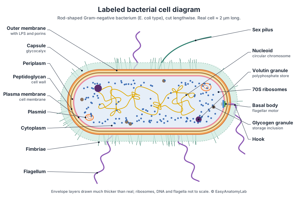
In the 3D model above, switch on All labels to see every part named at once, or click a part to zoom to it. The right-hand pole of the model shows the motor of one flagellum, with its rings set into each layer of the envelope.
Bacterial cell structure and functions
Each part of a bacterial cell has one main job. The table lists every structure on a typical exam diagram, what it does, and whether all bacteria have it.
| Structure | Function | Found in |
|---|---|---|
| Capsule (slime layer) | Sticks the cell to surfaces, stops it drying out, helps pathogens avoid white blood cells | Some bacteria |
| Cell wall (peptidoglycan) | Keeps the shape and stops the cell bursting by osmosis | Almost all bacteria |
| Outer membrane with LPS | Extra barrier against antibiotics and detergents; LPS contains the endotoxin | Gram-negative bacteria only |
| Periplasm | Holds enzymes that process nutrients and build the wall | Gram-negative bacteria |
| Plasma membrane | Controls what enters and leaves; site of respiration and ATP synthesis | All bacteria |
| Cytoplasm | Where the cell's chemical reactions take place | All bacteria |
| Nucleoid (chromosome) | One circular DNA molecule carrying the essential genes | All bacteria |
| Plasmids | Small DNA rings with extra genes, e.g. antibiotic resistance | Many bacteria |
| 70S ribosomes | Make proteins | All bacteria |
| Storage granules (inclusions) | Store glycogen, phosphate, sulfur or other reserves | Many bacteria |
| Flagellum | Rotates to move the cell | Some bacteria |
| Fimbriae | Attach the cell to surfaces and host cells | Many bacteria |
| Sex pilus | Joins two cells so plasmid DNA can be transferred (conjugation) | Donor (F+) cells |
| Endospore | Tough, dormant survival form | Some Gram-positives (Bacillus, Clostridium); not E. coli |
Parts of a bacterial cell: every structure in the model
This is the text version of the 3D model: every labeled structure of the E. coli cell, grouped by region, with what it does. Use it as a study list or to check a bacterial cell labeling worksheet.
Capsule & pili
- Capsule (Glycocalyx; a loose version is called a slime layer)
- A sticky layer of polysaccharide (sugar polymer) or protein that coats the outside of many bacteria. Helps the cell stick to surfaces and to other cells, form biofilms, resist drying out and, in pathogens, avoid being engulfed by white blood cells.
- Fimbriae (Attachment pili; singular fimbria)
- Many short, thin, bristle-like protein fibres that cover the cell surface. Attach the bacterium to surfaces and to host cells, for example the lining of the urinary tract or gut.
- Sex pilus (F pilus, conjugation pilus)
- A single long, hollow protein tube made by cells that carry the F (fertility) plasmid. Reaches out to a recipient cell, then retracts to pull the two cells together so a copy of a plasmid can pass between them (conjugation).
Cell envelope
- O antigen (O polysaccharide, O side chain)
- The long, outer sugar chain of each lipopolysaccharide (LPS) molecule. Forms a hydrophilic (water-loving) coat over the cell. Its exact sugars differ between strains and are recognised by antibodies, so it is used to type strains.
- Core oligosaccharide (LPS core polysaccharide)
- A short, non-repeating chain of sugars that joins lipid A to the O antigen. Links the membrane anchor (lipid A) to the O antigen and helps make the outer membrane a tight barrier.
- Lipid A (endotoxin) (Outer leaflet of the outer membrane; LPS anchor)
- The fatty part of lipopolysaccharide. Lipid A molecules form the outer half (leaflet) of the outer membrane. Anchors LPS in the membrane and makes the outer membrane a strong barrier. It is the endotoxin of Gram-negative bacteria: the part that triggers fever and, in severe infections, septic shock.
- Outer membrane (OM)
- A second lipid bilayer outside the cell wall, found only in Gram-negative bacteria. Its inner leaflet is phospholipid and its outer leaflet is mostly LPS. A protective barrier that keeps out many detergents, bile salts and antibiotics, while porins let small nutrients through.
- Porins (OmpF and OmpC channels)
- Barrel-shaped protein channels that cross the outer membrane, each a trimer of three barrels. Let water, ions, sugars, amino acids and other small hydrophilic molecules diffuse through the outer membrane.
- Periplasm (Periplasmic space)
- A gel-filled compartment between the outer membrane and the plasma membrane, which contains the peptidoglycan layer. Holds proteins that bind and break down nutrients, fold and check new envelope proteins, and destroy some antibiotics (for example β-lactamases).
- Peptidoglycan cell wall (Murein, cell wall)
- A mesh-like bag made of sugar chains (alternating NAG and NAM) cross-linked by short peptides. In E. coli it is only one to a few layers thick. Gives the cell its rod shape and stops it bursting when water rushes in by osmosis. It is the target of penicillin and other β-lactam antibiotics, and of lysozyme.
- Plasma membrane (Cell membrane, cytoplasmic membrane, inner membrane)
- A phospholipid bilayer with embedded proteins that encloses the cytoplasm. Controls what enters and leaves the cell, and does the jobs of mitochondria in our cells: it holds the respiratory chain and ATP synthase that make ATP. It also anchors the flagellar motor.
Cytoplasm & DNA
- Cytoplasm (Cytosol)
- The crowded, gel-like interior of the cell: water, enzymes, ions, nutrients, RNA and ribosomes. Where nearly all of the cell's chemistry happens, including glycolysis and protein synthesis.
- Nucleoid (chromosome) (Bacterial chromosome, circular DNA)
- The region of cytoplasm that holds the cell's single circular chromosome, folded into loops. It has no membrane around it. Carries the cell's genes. The DNA is copied before the cell divides, and genes are transcribed into mRNA right in the cytoplasm.
- Plasmids
- Small, circular, double-stranded DNA molecules separate from the chromosome. Carry extra genes that are useful but not essential, such as antibiotic resistance, toxins or the genes for a sex pilus. They copy themselves independently and can be passed to other cells.
- 70S ribosomes (Ribosomes)
- Small particles of RNA and protein that make proteins. Each has a small 30S and a large 50S subunit. Translate messenger RNA into proteins.
- Glycogen granules (Storage granules, inclusion bodies)
- Small granules of glycogen, a branched glucose polymer, stored in the cytoplasm. A store of carbon and energy that the cell can break down when food runs short.
- Volutin granules (Polyphosphate granules, metachromatic granules)
- Dense granules of polymerised inorganic phosphate. Store phosphate, and energy in its phosphate bonds, for making DNA, RNA, ATP and membranes.
Flagellum
- Flagellum (filament) (Flagella; made of flagellin)
- A long, rigid, helical protein whip. E. coli has several, scattered over its surface (peritrichous). Rotates like a propeller to push the cell through liquid.
- Hook (Flagellar hook)
- A short, curved, flexible protein coupling at the base of each filament. Acts as a universal joint, passing the motor's rotation to the filament at an angle.
- Rod (drive shaft) (Flagellar rod)
- The central shaft of the motor, running through the whole envelope. Transmits torque from the rotor in the plasma membrane out to the hook.
- L ring
- The outer ring of the basal body, named for the LPS layer. Acts as a bushing (bearing) that lets the rod spin through the outer membrane.
- P ring
- The second ring of the basal body, named for the peptidoglycan layer. A second bearing for the rotating rod.
- MS ring (Membrane and supramembrane ring)
- A ring of protein in the plasma membrane that is part of the rotor. The foundation of the motor; it turns with the rod and carries the C ring.
- C ring (Switch complex (FliG, FliM, FliN))
- A bell-shaped ring hanging from the MS ring into the cytoplasm. Part of the rotor and the switch: when the signalling protein CheY-P binds, the motor flips from counter-clockwise to clockwise rotation.
- Stator units (MotA/MotB) (Mot proteins)
- Ion channels arranged around the rotor, each made of MotA and MotB proteins. The motor's engine: protons flowing into the cell through the stators (the proton motive force) push on the rotor and turn it.
Bacterial cell wall diagram: Gram-positive vs Gram-negative
Bacterial cell walls come in two main types. A Gram-positive wall is a thick mesh of peptidoglycan, 30–100 nm and many layers deep, threaded with teichoic acids and lying directly on the plasma membrane. A Gram-negative wall is a peptidoglycan layer only a few nanometres thick, sitting in the periplasm under a second, outer membrane. (Silhavy, Kahne & Walker (2010): The bacterial cell envelope, Cold Spring Harbor Perspectives in Biology.)
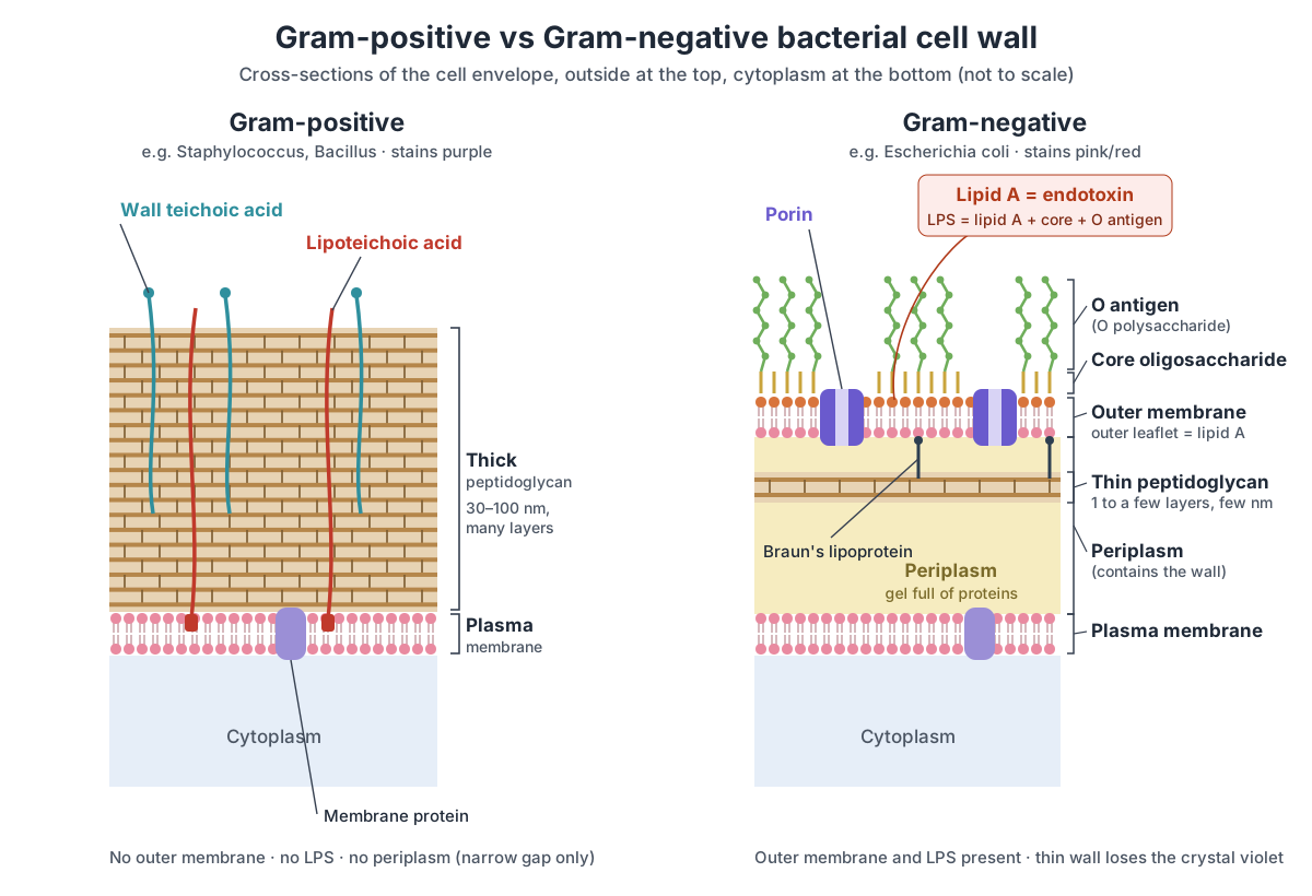
| Feature | Gram-positive | Gram-negative |
|---|---|---|
| Gram stain result | Purple: keeps the crystal violet | Pink to red: takes up the safranin counterstain |
| Peptidoglycan | Thick, 30–100 nm, many layers | Thin, a few nm, one to a few layers |
| Teichoic acids | Wall teichoic acids and lipoteichoic acids | None |
| Outer membrane | None | Present; outer leaflet is LPS |
| Lipopolysaccharide (endotoxin) | None | Lipid A + core + O antigen |
| Periplasm | None, or only a narrow gap | Present, a gel full of proteins |
| Porins | None | Present (e.g. OmpF, OmpC) |
| Peptide cross-links | Often via pentaglycine bridges (e.g. Staphylococcus) | Direct tetrapeptide cross-links |
| Flagellar basal body | No L or P ring | L ring (outer membrane) and P ring (peptidoglycan) |
| Examples | Staphylococcus, Streptococcus, Bacillus, Clostridium | E. coli, Salmonella, Pseudomonas, Neisseria |
Peptidoglycan, also called murein, is made of long sugar chains of alternating NAG (N-acetylglucosamine) and NAM (N-acetylmuramic acid), cross-linked by short peptides into one giant bag-shaped molecule around the cell. Teichoic acids can make up over 60% of the mass of a Gram-positive wall; wall teichoic acids are bonded to the peptidoglycan, and lipoteichoic acids are anchored in the membrane. (OpenStax Microbiology, 3.3 Unique characteristics of prokaryotic cells; Silhavy et al., 2010.)
How the Gram stain works. Crystal violet stains every cell purple; Gram's iodine locks it in as a crystal violet–iodine complex; alcohol or acetone then washes the dye out of cells with a thin wall but not from cells with a thick one; and the red counterstain safranin colors the cells that lost the purple. So Gram-positive cells end purple and Gram-negative cells end pink. Mycobacteria, such as the tuberculosis bacterium, are a third case: a waxy outer layer of mycolic acids makes them hard to Gram stain, so they are identified with an acid-fast stain instead. (OpenStax Microbiology, 2.4 Staining microscopic specimens.)
Endotoxins in the bacterial cell diagram
On a bacterial cell diagram, the endotoxin is found in the outer membrane of Gram-negative bacteria: it is lipid A, the fatty anchor of lipopolysaccharide (LPS) that forms the outer leaflet of that membrane. In the 3D model it is the thin dark-orange skin over the outer membrane; the green fuzz above it is the O antigen, and the short gold stalks between are the core oligosaccharide.
- Lipid A (endotoxin): Anchors LPS in the membrane and makes the outer membrane a strong barrier. It is the endotoxin of Gram-negative bacteria: the part that triggers fever and, in severe infections, septic shock.
- Core oligosaccharide: Links the membrane anchor (lipid A) to the O antigen and helps make the outer membrane a tight barrier.
- O antigen: Forms a hydrophilic (water-loving) coat over the cell. Its exact sugars differ between strains and are recognised by antibodies, so it is used to type strains.
- Porins: Let water, ions, sugars, amino acids and other small hydrophilic molecules diffuse through the outer membrane.
Endotoxins differ from exotoxins. An exotoxin is a protein that a living bacterium secretes, such as tetanus or cholera toxin, and it can come from Gram-positive or Gram-negative species. An endotoxin is a structural part of the Gram-negative wall and is released mostly when the cell dies or is broken apart. Gram-positive bacteria have no LPS and so no endotoxin. The research section below explains how lipid A triggers fever and septic shock.
Bacterial cell membrane and cytoplasm
The bacterial cell membrane (plasma membrane) is a phospholipid bilayer studded with proteins that wraps the cytoplasm. It controls what enters and leaves the cell, and because bacteria have no mitochondria, it also carries the respiratory chain and ATP synthase that make ATP. It anchors the flagellar motors, and it is where the cell builds new membrane and wall.
Small molecules cross it by simple diffusion, facilitated diffusion through channel and carrier proteins, active transport using ATP, or group translocation, in which the molecule is chemically changed as it crosses, as in the phosphotransferase system that brings in sugars. (OpenStax Microbiology, 3.3 Unique characteristics of prokaryotic cells.) To see how a membrane drives ATP synthesis, see the cellular respiration diagram.
Inside the membrane, the cytoplasm is a crowded gel of water, ions, enzymes, RNA and ribosomes. With no nucleus, transcription and translation happen side by side in the same space.
Nucleoid, plasmids, ribosomes and storage granules
Inside a bacterial cell, the DNA is in the nucleoid, a region of cytoplasm with no membrane around it. It holds one circular chromosome. Extra genes may sit on small DNA rings called plasmids. Proteins are made on 70S ribosomes, and reserves are kept in inclusions (storage granules).
- Nucleoid (chromosome): Carries the cell's genes. The DNA is copied before the cell divides, and genes are transcribed into mRNA right in the cytoplasm.
- Plasmids: Carry extra genes that are useful but not essential, such as antibiotic resistance, toxins or the genes for a sex pilus. They copy themselves independently and can be passed to other cells.
- 70S ribosomes: Translate messenger RNA into proteins.
- Glycogen granules: A store of carbon and energy that the cell can break down when food runs short.
- Volutin granules: Store phosphate, and energy in its phosphate bonds, for making DNA, RNA, ATP and membranes.
Inclusions vary between species. Besides glycogen and volutin (polyphosphate), bacteria store polyhydroxybutyrate (PHB) in Bacillus and Pseudomonas and sulfur in Thiobacillus. Some water bacteria have gas vacuoles for buoyancy, magnetotactic bacteria carry chains of magnetic crystals (magnetosomes), and photosynthetic bacteria pack the CO₂-fixing enzyme RuBisCO into carboxysomes. (OpenStax Microbiology, 3.3 Unique characteristics of prokaryotic cells.)
Flagella, pili and fimbriae
Bacteria have three kinds of surface appendage. Flagella are long helical filaments turned by a motor and used for swimming. Fimbriae are many short, thin bristles used to stick to surfaces. The sex pilus (F pilus) is a single long tube used to pull another cell close for DNA transfer.
- Flagellum (filament): Rotates like a propeller to push the cell through liquid.
- Hook: Acts as a universal joint, passing the motor's rotation to the filament at an angle.
- Rod (drive shaft): Transmits torque from the rotor in the plasma membrane out to the hook.
- L ring: Acts as a bushing (bearing) that lets the rod spin through the outer membrane.
- P ring: A second bearing for the rotating rod.
- MS ring: The foundation of the motor; it turns with the rod and carries the C ring.
- C ring: Part of the rotor and the switch: when the signalling protein CheY-P binds, the motor flips from counter-clockwise to clockwise rotation.
- Stator units (MotA/MotB): The motor's engine: protons flowing into the cell through the stators (the proton motive force) push on the rotor and turn it.
- Fimbriae: Attach the bacterium to surfaces and to host cells, for example the lining of the urinary tract or gut.
- Sex pilus: Reaches out to a recipient cell, then retracts to pull the two cells together so a copy of a plasmid can pass between them (conjugation).
Flagella are arranged in four patterns: monotrichous (one at one pole), amphitrichous (one at each pole), lophotrichous (a tuft at one pole) and peritrichous (scattered all over the cell). E. coli is peritrichous, with about three or four flagella per cell. (OpenStax Microbiology, 3.3 Unique characteristics of prokaryotic cells; Turner, Ryu & Berg, 2000.)
In conjugation, a donor cell carrying the F plasmid extends its sex pilus to a recipient, the pilus retracts to draw the cells together, and a single strand of the plasmid is copied across. R plasmids carrying several antibiotic-resistance genes spread the same way, even between species, which is one reason resistance spreads so fast. (OpenStax Microbiology, 11.6 How asexual prokaryotes achieve genetic diversity.)
Endospores (not found in E. coli)
An endospore is a tough, dormant structure that some Gram-positive bacteria, notably Bacillus and Clostridium, form inside the cell when conditions turn bad. E. coli does not make endospores, which is why there is none in the model. Inside the spore is a dehydrated copy of the chromosome, wrapped in a double membrane, a thick cortex rich in calcium and dipicolinic acid, a protein coat and, in some species, an outer exosporium. Endospores survive heat, radiation, drying and many disinfectants, and germinate when conditions improve. (OpenStax Microbiology, 3.3 Unique characteristics of prokaryotic cells.)
Endospores do not take up the Gram stain and look like clear holes in a Gram-stained cell. The Schaeffer-Fulton stain drives green malachite green into them with heat, so spores show green inside pink cells. Diseases caused by spore-formers include anthrax (Bacillus anthracis), tetanus and botulism (Clostridium). (OpenStax Microbiology, 2.4 Staining microscopic specimens.)
Bacterial cell shapes: coccus, bacillus and spirillum
Bacteria come in three basic shapes: round cocci, rod-shaped bacilli and spiral spirilla. In-between forms include the short, oval coccobacillus, the comma-shaped vibrio and the long, flexible spirochete. The cell wall, helped by internal cytoskeleton proteins, holds each shape. (OpenStax Microbiology, 3.3 Unique characteristics of prokaryotic cells.)
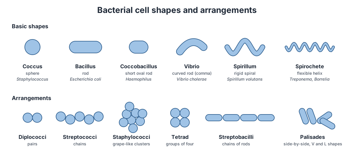
Cells that stay joined after dividing form typical arrangements: pairs (diplococci), chains (streptococci and streptobacilli), grape-like clusters (staphylococci) and squares of four (tetrads). The names of many bacteria come straight from this: Streptococcus grows in chains, Staphylococcus in clusters. A coccal bacterial cell has the same parts as the rod in the model, just packed into a sphere.
Prokaryotic vs eukaryotic cells
Bacteria are prokaryotic cells: they have no nucleus and no membrane-bound organelles, and their DNA is usually a single circular chromosome. Eukaryotic cells (animals, plants, fungi and protists) keep their linear chromosomes in a nucleus and divide their cytoplasm into organelles such as mitochondria and the endoplasmic reticulum.
| Feature | Prokaryotic cell (bacterium) | Eukaryotic cell |
|---|---|---|
| Typical size | 0.1–5 µm across; E. coli about 2 µm long | 10–100 µm across |
| Nucleus | None; DNA in the nucleoid | Yes, with a double nuclear envelope |
| Chromosomes | Usually one, circular | Several, linear |
| Histones | None in bacteria (other DNA-packing proteins); some archaea have them | Yes |
| Membrane-bound organelles | None | Mitochondria, ER, Golgi, lysosomes; chloroplasts in plants and algae |
| Ribosomes | 70S (30S + 50S) | 80S (40S + 60S) in the cytoplasm; 70S in mitochondria and chloroplasts |
| Cell wall | Usually peptidoglycan | Cellulose (plants), chitin (fungi), none (animals) |
| Flagella | Stiff helix of flagellin that rotates | Flexible 9+2 microtubule whip that bends, powered by dynein |
| Cell division | Binary fission | Mitosis (and meiosis for sex cells) |
| Examples | Bacteria and archaea | Animals, plants, fungi, protists |
The 70S ribosomes and bacterial-like DNA inside mitochondria and chloroplasts are key evidence that these organelles began as bacteria living inside an ancestral eukaryotic cell (the endosymbiotic theory). (OpenStax Microbiology, 3.4 Unique characteristics of eukaryotic cells.) Compare the double membrane of a chloroplast in our chloroplast structure model, and see eukaryotic single cells in the amoeba and Euglena diagrams.
Plant, animal and bacterial cells compared
Plant, animal and bacterial cells all have a cell membrane, cytoplasm, ribosomes and DNA. Plant and animal cells are eukaryotic and share a nucleus and mitochondria. Plant and bacterial cells both have a cell wall, but plant walls are cellulose and bacterial walls are peptidoglycan. Only bacterial cells lack a nucleus.
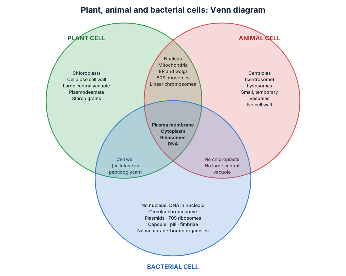
| Feature | Plant cell | Animal cell | Bacterial cell |
|---|---|---|---|
| Cell membrane | Yes | Yes | Yes |
| Cytoplasm | Yes | Yes | Yes |
| Nucleus | Yes | Yes | No; nucleoid |
| Genetic material | Linear chromosomes in the nucleus | Linear chromosomes in the nucleus | One circular chromosome, often plus plasmids |
| Cell wall | Yes, cellulose | No | Yes, peptidoglycan |
| Mitochondria | Yes | Yes | No; respiration on the plasma membrane |
| Chloroplasts | Yes, in green tissues | No | No (photosynthetic bacteria use membranes in the cytoplasm) |
| Vacuole | Large, permanent central vacuole | Small, temporary vacuoles if any | No |
| Ribosomes | 80S | 80S | 70S |
| Centrioles and lysosomes | Usually absent | Present | Absent |
| Typical size | 10–100 µm | 10–100 µm | About 1–5 µm |
Sources for the plant and animal columns: OpenStax Biology 2e, which notes that animal cells each have a centrosome and lysosomes whereas most plant cells do not, and that plant cells have a cellulose wall, chloroplasts and a large central vacuole. (OpenStax Biology 2e, 4.3 Eukaryotic cells.)
Bacterial cell division: binary fission
Bacteria divide by binary fission: the cell copies its chromosome, grows longer, builds a wall across its middle and splits into two identical daughter cells. Under ideal laboratory conditions E. coli can divide every 20 minutes. (OpenStax Microbiology, 9.1 How microbes grow.)
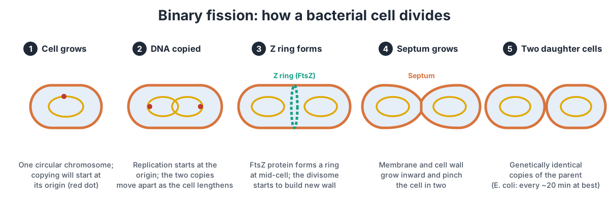
- Replication. Copying of the circular chromosome begins at a single site, the origin of replication, and runs both ways around the circle until the two forks meet at the terminus.
- Segregation and growth. The cell lengthens and the two copies of the chromosome move apart, so each half of the cell gets one complete copy.
- Z ring. The protein FtsZ assembles into a ring on the inside of the plasma membrane at mid-cell. It marks the plane of division and recruits other proteins to form the divisome.
- Septum. The divisome builds new peptidoglycan and membrane inward, forming a cross-wall (septum) that pinches the cell in two.
- Separation. Enzymes split the shared wall and two daughter cells of similar size separate, each with a copy of the parent's genome.
Binary fission is not mitosis. There is no nucleus to break down, no spindle and no condensed chromosomes. Each division doubles the population, which is why bacterial numbers rise exponentially when food is plentiful.
Simple bacterial cell diagram for GCSE and A-level
For a simple GCSE bacterial cell diagram, draw a rod or oval with a cell wall around a cell membrane, fill it with cytoplasm, and draw the DNA as one large loop lying free (no nucleus), one or two small plasmids, dots for ribosomes, and a tail-like flagellum. Label each part and state that a bacterial cell is prokaryotic and much smaller than a plant or animal cell.
- GCSE must-knows: cell membrane, cell wall, cytoplasm, chromosomal DNA as a single loop, plasmids, ribosomes, flagellum; no nucleus, no mitochondria, no chloroplasts. Most bacteria are a few micrometres long at most, about ten times or more smaller than a typical plant or animal cell.
- A-level additions: 70S ribosomes (vs 80S), a murein (peptidoglycan) cell wall, capsule, pili and plasmids; binary fission; the Gram-positive vs Gram-negative wall and its link to antibiotic action.
- Common mistakes: drawing a nucleus; calling the cell wall cellulose; drawing mitochondria; giving ribosomes a membrane; drawing the DNA as several linear chromosomes.
- Mesosomes: some older textbooks show folds of the membrane called mesosomes. They turned out to be artefacts of preparing cells for the electron microscope, so most courses no longer include them.
Bacteria are not viruses. A virus has no cytoplasm, ribosomes or cell membrane and cannot reproduce on its own. Compare the two in our virus structure diagram.
How the bacterial flagellum works: a rotary motor
A bacterial flagellum does not wave like a whip. It spins. At its base is a rotary motor of about 25 different proteins that spans the whole cell envelope. A shaft called the rod runs through the middle; the MS ring in the plasma membrane and the C ring below it form the rotor; the L ring in the outer membrane and the P ring in the peptidoglycan act as bushings; and a ring of stator units, each made of the proteins MotA and MotB, sits around the rotor and drives it. About 12 stators fit around the E. coli motor. (Guo & Liu (2022): The bacterial flagellar motor, Frontiers in Microbiology.)
The energy comes from protons, not ATP. The cell pumps protons out across its plasma membrane, and as they flow back in through the stator channels they push on the rotor. Rotation rates are on the order of 100 revolutions per second, which drives filaments about 20 nm thick and several micrometres long. (Turner, Ryu & Berg (2000): Real-time imaging of fluorescent flagellar filaments, Journal of Bacteriology.)
E. coli steers by switching the motor's direction. When the motors turn counter-clockwise, the left-handed filaments wrap into one bundle behind the cell and it swims straight, a 'run' that lasts about a second. When one or more motors reverse to clockwise, the bundle flies apart for about a tenth of a second, the cell 'tumbles', and it sets off in a new random direction. Filming fluorescently labelled filaments showed that a change in just one filament can cause a tumble. (Turner, Ryu & Berg (2000): Real-time imaging of fluorescent flagellar filaments, Journal of Bacteriology.)
By making runs longer when it moves towards food and shorter when it moves away, the cell drifts up a nutrient gradient. This is chemotaxis. The signal reaches the motor through the protein CheY: when it is phosphorylated (CheY-P), it binds the C ring and flips the motor to clockwise. (Guo & Liu (2022): The bacterial flagellar motor, Frontiers in Microbiology.)
Endotoxin in the bacterial cell: lipid A and septic shock
The endotoxin of a Gram-negative bacterium is not a secreted poison; it is part of the cell wall. Lipopolysaccharide (LPS) has three regions: lipid A, a hydrophobic anchor that forms the outer leaflet of the outer membrane; a non-repeating core oligosaccharide; and the long O-antigen polysaccharide on the outside. Lipid A is the part that carries the endotoxin activity. An E. coli cell carries about a million lipid A molecules. (Raetz & Whitfield (2002): Lipopolysaccharide endotoxins, Annual Review of Biochemistry.)
Our innate immune system detects lipid A through the receptor TLR4 and its helper protein MD2. A small amount of LPS is a useful alarm that switches on defences. Released in large amounts in the bloodstream in severe sepsis, it can drive the over-production of inflammatory mediators and clotting factors that leads to Gram-negative septic shock. Synthetic E. coli lipid A on its own reproduces these effects in animals, which is how scientists know lipid A is the active part. (Raetz & Whitfield (2002): Lipopolysaccharide endotoxins, Annual Review of Biochemistry.)
LPS also explains why the outer membrane is such a good barrier. The outer membrane is a lipid bilayer, but not a phospholipid bilayer: phospholipids are confined to its inner leaflet, while the outer leaflet is mostly LPS. Hydrophilic molecules can only cross through porins such as OmpF and OmpC, which let through molecules smaller than about 700 daltons. That is one reason many antibiotics that work on Gram-positive bacteria fail against Gram-negative ones. (Silhavy, Kahne & Walker (2010): The bacterial cell envelope, Cold Spring Harbor Perspectives in Biology.)
E. coli by the numbers
A useful rule of thumb is that an E. coli cell is about 1 µm across and 2 µm long, with a volume of about 1 µm³ (one femtolitre) and a mass of about 1 picogram. Treating the cell as a cylinder with rounded ends gives a slightly larger volume, about 1.3 µm³. Cells that grow faster are bigger: dry mass ranges from about 148 fg in cells that divide every 100 minutes to 865 fg in cells that divide every 24 minutes. (Milo & Phillips, Cell Biology by the Numbers: How big is an E. coli cell and what is its mass?.)
Ribosome numbers rise steeply with growth rate, because a fast-growing cell needs to make protein quickly. Measurements on E. coli B/r at 37 °C give about 6,800 ribosomes per cell at a 100-minute doubling time and about 72,000 at a 24-minute doubling time. The 3D model above shows only about 300, enlarged, on the cut faces of the cytoplasm. (BioNumbers 101441: ribosomes per E. coli cell at different growth rates (Bremer & Dennis).)
The chromosome of the standard laboratory strain, E. coli K-12, is 4,639,221 base pairs long and codes for 4,288 annotated proteins. At about 0.34 nm per base pair, that DNA would stretch to about 1.6 mm, roughly 800 times the length of the cell, which is why it must be folded into the compact nucleoid. (Blattner et al. (1997): The complete genome sequence of Escherichia coli K-12, Science (Europe PMC).)
E. coli is also the host of the best-studied bacterial viruses. See how one injects its DNA through the envelope in our bacteriophage T4 diagram.
Label the bacterial cell: unlabeled diagram and answer key
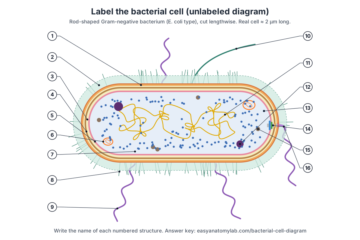
Test yourself with this blank bacterial cell diagram. Write the name of each of the 16 numbered parts, then open the answer key. For a quick quiz, turn off the labels in the 3D model, click parts at random and name them before you read the panel.
Show the answer key
- Outer membrane (with LPS and porins)
- Capsule (glycocalyx)
- Periplasm
- Peptidoglycan cell wall
- Plasma membrane (cell membrane)
- Plasmid
- Cytoplasm
- Fimbriae
- Flagellum (filament)
- Sex pilus (F pilus)
- Nucleoid (circular chromosome)
- Volutin (polyphosphate) granule
- 70S ribosomes
- Basal body (flagellar motor)
- Glycogen granule (storage inclusion)
- Hook
Short-answer questions:
- Name four structures found in every bacterial cell. (Plasma membrane, cytoplasm, ribosomes, DNA/nucleoid.)
- What are the two subunits of a bacterial ribosome? (30S and 50S, making a 70S ribosome.)
- Which layer of a Gram-negative cell contains the endotoxin? (The outer membrane: lipid A of LPS.)
- Why do Gram-positive cells stay purple in a Gram stain? (Their thick peptidoglycan keeps the crystal violet–iodine complex.)
- What is the difference between a fimbria and a sex pilus? (Fimbriae attach; the sex pilus joins cells for DNA transfer.)
- Name the protein that forms the ring at the division site. (FtsZ.)
Frequently asked questions
What are the main parts of a bacterial cell?
Every bacterial cell has a plasma membrane, cytoplasm, ribosomes and a nucleoid containing its circular chromosome. Almost all also have a peptidoglycan cell wall. Many add plasmids, storage granules, a capsule, fimbriae, a sex pilus and one or more flagella. Gram-negative bacteria such as E. coli also have an outer membrane and a periplasm.
Does a bacterial cell have a nucleus?
No. Bacteria are prokaryotes, which means 'before the nucleus'. Their DNA lies in a region of the cytoplasm called the nucleoid, with no membrane around it. They also lack mitochondria, chloroplasts, endoplasmic reticulum and other membrane-bound organelles.
What is the difference between Gram-positive and Gram-negative cell walls?
A Gram-positive wall is a thick peptidoglycan layer (30–100 nm) threaded with teichoic acids, sitting directly on the plasma membrane. A Gram-negative wall is a thin peptidoglycan layer (a few nm) inside a periplasm, covered by an outer membrane whose outer leaflet is lipopolysaccharide (LPS). In the Gram stain, Gram-positive cells stay purple and Gram-negative cells turn pink.
Where is the endotoxin in a bacterial cell?
In the outer membrane of Gram-negative bacteria. The endotoxin is lipid A, the fatty anchor of lipopolysaccharide, which forms the outer leaflet of the outer membrane. It is released mainly when bacteria die or break apart, and it triggers fever and inflammation. Gram-positive bacteria have no LPS.
What is the function of plasmids in a bacterial cell?
Plasmids are small circles of DNA that carry extra genes, for example for antibiotic resistance. They copy themselves independently of the chromosome and can be passed to other bacteria, which is one way antibiotic resistance spreads.
What is the difference between pili and flagella?
Flagella are long, helical filaments driven by a rotary motor in the cell envelope; they make the cell swim. Pili and fimbriae are thinner, straight protein fibres that do not rotate: fimbriae attach the cell to surfaces, and the sex pilus pulls two cells together so DNA can pass between them.
Why are bacterial ribosomes 70S?
The S is a svedberg unit, a measure of how fast a particle sinks in a centrifuge, which depends on its size and shape. A bacterial ribosome is made of a 30S and a 50S subunit and settles at 70S. Eukaryotic cytoplasmic ribosomes are larger, at 80S.
How big is a bacterial cell?
Most bacteria are 0.5–5 µm long. E. coli is about 2 µm long and about 1 µm wide, with a volume of about 1 µm³. A typical animal or plant cell is 10–100 µm across, so it could hold hundreds to thousands of E. coli cells.
Do all bacteria have a capsule and flagella?
No. Capsules, flagella, pili and plasmids are found in some bacteria and not others, and can differ between strains of the same species. Exam diagrams draw them all on one cell to show every possible part.
References
- Parker, N., Schneegurt, M., Thi Tu, A.-H., Lister, P. & Forster, B. M. (2016). Microbiology, section 3.3: Unique characteristics of prokaryotic cells. OpenStax.
- OpenStax Microbiology, section 3.4: Unique characteristics of eukaryotic cells.
- OpenStax Microbiology, section 2.4: Staining microscopic specimens.
- OpenStax Microbiology, section 9.1: How microbes grow.
- OpenStax Microbiology, section 11.6: How asexual prokaryotes achieve genetic diversity.
- Clark, M. A., Douglas, M. & Choi, J. (2018). Biology 2e, section 4.2: Prokaryotic cells. OpenStax.
- OpenStax Biology 2e, section 4.3: Eukaryotic cells.
- Silhavy, T. J., Kahne, D. & Walker, S. (2010). The bacterial cell envelope. Cold Spring Harbor Perspectives in Biology 2, a000414.
- Raetz, C. R. H. & Whitfield, C. (2002). Lipopolysaccharide endotoxins. Annual Review of Biochemistry 71, 635–700.
- Guo, S. & Liu, J. (2022). The bacterial flagellar motor: insights into torque generation, rotational switching, and mechanosensing. Frontiers in Microbiology 13, 911114.
- Turner, L., Ryu, W. S. & Berg, H. C. (2000). Real-time imaging of fluorescent flagellar filaments. Journal of Bacteriology 182, 2793–2801.
- Blattner, F. R. et al. (1997). The complete genome sequence of Escherichia coli K-12. Science 277, 1453–1462.
- Matias, V. R. F., Al-Amoudi, A., Dubochet, J. & Beveridge, T. J. (2003). Cryo-transmission electron microscopy of frozen-hydrated sections of Escherichia coli and Pseudomonas aeruginosa. Journal of Bacteriology 185, 6112–6118.
- Milo, R. & Phillips, R. Cell Biology by the Numbers: How big is an E. coli cell and what is its mass?
- BioNumbers BNID 101441: number of ribosomes per cell in E. coli B/r, from Bremer, H. & Dennis, P. P. (1996), in Escherichia coli and Salmonella, 2nd ed.
More interactive anatomy models
- Amoeba diagramLabeled Amoeba proteus: pseudopodia, nucleus, contractile and food vacuoles.
- Euglena diagramLabeled Euglena: flagellum, eyespot, chloroplasts, pellicle and contractile vacuole.
- Bacteriophage diagramT4 phage structure in 3D: head, collar, tail sheath, baseplate and tail fibres.
- Virus structureCapsid, envelope, spikes and genome of helical, icosahedral and enveloped viruses.
- Human anatomy1,000+ bones, muscles, organs, vessels and nerves from real anatomical data.
- Octopus anatomyThree hearts, a ring-shaped brain, eight arms and a beak, in 3D.