

What is chloroplast structure? The chloroplast at a glance
A chloroplast is the organelle where photosynthesis happens in plants and algae. Its structure has three membrane systems and three spaces: a double-membrane envelope (outer membrane, intermembrane space and inner membrane), a fluid-filled stroma, and inside the stroma a network of flattened sacs called thylakoids, stacked into grana and joined by stroma lamellae. The space inside each thylakoid is the thylakoid lumen.
Chloroplasts in flowering plants are lens-shaped, about 5 µm across and 2.5 µm thick, roughly a twentieth of the width of a human hair. A single leaf mesophyll cell of Arabidopsis holds about 110 of them. They are green because their thylakoid membranes are full of chlorophyll.
Each part does a different job. The thylakoids capture light and make ATP and NADPH (the light-dependent reactions); the stroma uses them to turn CO₂ into sugar (the Calvin cycle). The chloroplast also has its own circular DNA and its own small 70S ribosomes, clues that it evolved from a bacterium.
Structure of a chloroplast: labeled diagram
To describe the structure of a chloroplast, work from the outside in: envelope, stroma, thylakoid system, then the smaller contents of the stroma. The diagram shows a chloroplast cut lengthwise, the view you see in a transmission electron micrograph.
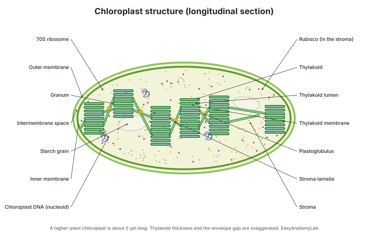
- Envelope. Two membranes. The outer membrane is leaky to small molecules; the inner membrane is selective and full of transporters. Between them is a narrow intermembrane space.
- Stroma. A thick, protein-rich fluid that fills the chloroplast. It holds the Calvin-cycle enzymes (above all Rubisco), the chloroplast DNA, 70S ribosomes, starch grains and plastoglobuli.
- Thylakoids. Flattened membrane sacs. Stacks of them are grana (singular granum); the single, unstacked sheets that link the grana are stroma lamellae, also called stroma thylakoids, intergranal lamellae or frets.
- Thylakoid lumen. The space inside the thylakoids. All the thylakoids are joined, so the lumen is one continuous compartment, separate from the stroma.
For where chloroplasts sit inside a leaf, see the leaf cross-section; for the chloroplast-filled guard cells that open and close the pores, see the stomata diagram.
Chloroplast structure labeled: every part and its function
This is the text version of the 3D model: every labeled structure of the chloroplast, grouped by region, with where it is and what it does. Use it as a study list or to check a labeling worksheet.
Envelope
- Chloroplast envelope (Double membrane)
- The two membranes that wrap the whole chloroplast: an outer membrane and an inner membrane with a narrow space between them. Separates the stroma from the cytosol of the plant cell and controls what goes in and out.
- Outer membrane (Outer envelope membrane)
- The outer of the two envelope membranes. It is fairly leaky to small molecules. Forms the outer boundary. Pore-forming proteins let small molecules and ions through, and the TOC complex recognises proteins that are to be imported.
- Intermembrane space
- The narrow gap between the outer and inner envelope membranes. A thin watery compartment that imported proteins cross on their way from the TOC to the TIC channel.
- Inner membrane (Inner envelope membrane)
- The inner envelope membrane. It is selective and is packed with transport proteins. Controls traffic between the stroma and the cytosol. Transporters move sugar phosphates, phosphate and other metabolites, and the TIC complex completes protein import.
Stroma and its contents
- Stroma
- The protein-rich fluid that fills the inside of the chloroplast around the thylakoids. Site of the Calvin cycle (the light-independent reactions), where CO₂ is fixed into sugar using ATP and NADPH from the thylakoids. It also holds the chloroplast DNA, ribosomes and starch.
- Rubisco (Ribulose-1,5-bisphosphate carboxylase/oxygenase)
- The enzyme that fixes CO₂ in the first step of the Calvin cycle. Drawn as many small orange particles. Joins CO₂ to the five-carbon sugar RuBP, making two molecules of 3-phosphoglycerate.
- 70S ribosomes (Chloroplast ribosomes)
- Small, bacterial-type ribosomes that make the proteins encoded by the chloroplast's own DNA. Translate chloroplast mRNAs, for example for the D1 protein of photosystem II and the large subunit of Rubisco.
- Chloroplast DNA (nucleoids) (cpDNA, ctDNA, plastome)
- The chloroplast's own genome, packed with proteins into small clumps called nucleoids. Carries about 120–130 genes for photosynthesis proteins, ribosomal RNAs, transfer RNAs and ribosomal proteins.
- Starch grains (Starch granules, transitory starch)
- White, flattened granules of starch made from the sugars of photosynthesis. Store carbohydrate by day and are broken down at night to maltose and glucose, which are exported to keep the plant supplied in the dark.
- Plastoglobuli (Plastoglobules, lipid droplets)
- Small yellow lipid droplets attached to the thylakoids. Store and make lipid molecules such as vitamin E (tocopherol), plastoquinone and carotenoids, and exchange them with the thylakoid membrane.
Thylakoid system
- Granum (grana) (Grana stacks)
- A stack of disc-shaped thylakoids, like a pile of coins. These are the 'disk-shaped structures inside chloroplasts'. Packs a large area of light-absorbing membrane into a small space. The stacked (appressed) membranes hold most of photosystem II and its light-harvesting antenna.
- Thylakoid (Granal thylakoid, thylakoid disc)
- One flattened, membrane-bound sac. Stacks of thylakoids form a granum. Carries out the light-dependent reactions. Its membrane holds the pigments and protein complexes; its inside (the lumen) collects protons.
- Thylakoid membrane
- The membrane of the thylakoids, holding chlorophyll and the light-reaction complexes. Absorbs light and runs the electron transport chain, which splits water, releases oxygen, pumps protons into the lumen and makes ATP and NADPH.
- Thylakoid lumen (Thylakoid space, intrathylakoid space)
- The narrow space inside each thylakoid. Collects the protons (H⁺) pumped in by the electron transport chain and released by water splitting. The resulting gradient drives ATP synthase.
- Stroma lamellae (Stroma thylakoids, intergranal lamellae, frets)
- Unstacked thylakoid sheets that link one granum to the next. Connect the grana into one network and carry most of the photosystem I and ATP synthase.
Thylakoid membrane complexes (close-up)
- Photosystem II (PSII, P680)
- The light-driven complex that takes electrons from water. Drawn as a dimer. Absorbs light at its reaction centre (P680), passes excited electrons to plastoquinone, and replaces them with electrons from water, releasing O₂ and protons into the lumen.
- Light-harvesting complex II (LHCII, antenna)
- Chlorophyll a/b–protein complexes that act as an antenna for photosystem II. Absorb light and pass the energy to the reaction centre. LHCII also helps hold grana membranes together.
- Oxygen-evolving complex (Water-splitting complex, Mn₄CaO₅ cluster)
- The part of photosystem II that splits water. Removes electrons from water for P680 and releases O₂ and H⁺ into the lumen.
- Plastoquinone (PQ / PQH₂)
- A small, fat-soluble electron carrier that moves within the membrane. Picks up two electrons from photosystem II and two protons from the stroma, then carries them to cytochrome b6f, where the protons are released into the lumen.
- Cytochrome b6f complex (Cyt b6f)
- The membrane complex that links the two photosystems and pumps extra protons. Takes electrons from plastoquinol and passes them to plastocyanin, moving more protons into the lumen as it does so.
- Plastocyanin (PC)
- A small copper protein that shuttles electrons through the lumen. Carries one electron at a time from cytochrome b6f to photosystem I.
- Photosystem I (PSI, P700, with its LHCI antenna)
- The second light-driven complex. It energises electrons a second time. Absorbs light at P700 and passes electrons to ferredoxin on the stroma side; its electron gap is filled by plastocyanin.
- Ferredoxin (Fd)
- A small iron–sulfur protein on the stroma side of photosystem I. Takes electrons from photosystem I and hands them to FNR (and to other stromal enzymes).
- Ferredoxin–NADP⁺ reductase (FNR)
- The enzyme that makes NADPH. Uses two electrons from ferredoxin to reduce NADP⁺ to NADPH in the stroma, ready for the Calvin cycle.
- ATP synthase (CF₀CF₁, chloroplast ATP synthase)
- A rotary motor that makes ATP as protons flow back from the lumen to the stroma. Protons flowing through the CF₀ ring turn it; the turning stalk makes the CF₁ head join ADP and phosphate into ATP.
Internal structure of a chloroplast: thylakoids, grana and stroma lamellae
The disk-shaped structures inside chloroplasts are thylakoids, and a stack of them is a granum. A typical granum is about 300–500 nm across and holds about 5 to 20 thylakoids. A mesophyll chloroplast contains many grana, scattered through the stroma with their discs parallel to the long axis of the organelle.
Grana are not separate piles. Single-layer sheets, the stroma lamellae, connect each granum to its neighbours, so the whole thylakoid system is one continuous membrane folded into stacks and sheets. Close to each granum, the lamellae wind around it as right-handed helical ramps and join every disc through a narrow slit. The 3D model shows these ramps as the lighter green spirals around each stack.
| Region | What it is | Main complexes |
|---|---|---|
| Grana (appressed membranes) | Tightly stacked thylakoids inside a granum | Photosystem II and LHCII; some cytochrome b6f |
| Grana end membranes and margins | Top and bottom discs, and the curved edges of each disc | Photosystem I and ATP synthase at the flat ends; curved margins |
| Stroma lamellae | Unstacked sheets that link grana | Photosystem I, ATP synthase, cytochrome b6f |
| Lumen | The space inside all thylakoids | Plastocyanin; the water-splitting site of PSII faces it |
The membrane itself is unusual. Most cell membranes are built from phospholipids, but about 52% of thylakoid lipid is the galactolipid MGDG and another 27% is DGDG. These sugar-headed lipids help the membrane form the sharp curves at the edges of every disc.
The thylakoid membrane: photosystems and ATP synthase
The thylakoid membrane holds four large protein complexes that work in a chain: photosystem II, the cytochrome b6f complex, photosystem I and ATP synthase. Two small mobile carriers link them: plastoquinone moves inside the membrane and plastocyanin moves through the lumen.
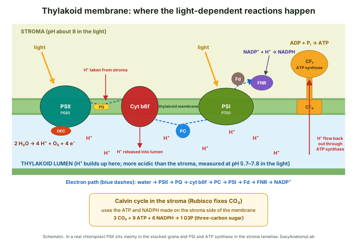
- Photosystem II absorbs light, and its reaction centre (P680) passes excited electrons to plastoquinone. Its oxygen-evolving complex replaces them by splitting water on the lumen side: two water molecules give one O₂, four protons and four electrons.
- Plastoquinone carries the electrons, plus protons picked up from the stroma, to the cytochrome b6f complex, which releases the protons into the lumen and passes the electrons to plastocyanin.
- Photosystem I (P700) absorbs light again and boosts the electrons to ferredoxin on the stroma side. The enzyme FNR uses them to make NADPH.
- Protons now crowd the lumen. They flow back to the stroma through ATP synthase, turning its CF₀ rotor; the CF₁ head, which sticks out into the stroma, makes ATP.
Note the direction. ATP and NADPH are both released into the stroma, exactly where the Calvin cycle needs them. This is why the CF₁ head always faces the stroma, never the lumen.
Chloroplast structure and function
Chloroplast structure and function go together: the thylakoids run the light-dependent reactions and the stroma runs the light-independent reactions (the Calvin cycle). The table summarises which part does what.
| Structure | Function |
|---|---|
| Outer membrane | Boundary with the cytosol; lets small molecules through; starts protein import (TOC) |
| Inner membrane | Controls traffic of sugars, phosphate and ions; completes protein import (TIC) |
| Stroma | Calvin cycle: fixes CO₂ into sugar using ATP and NADPH; makes starch, amino acids and fatty acids |
| Thylakoid membrane | Absorbs light; runs electron transport; splits water; pumps protons; makes ATP and NADPH |
| Thylakoid lumen | Stores the protons that drive ATP synthase |
| Grana | Pack a large membrane area into a small volume; hold most of photosystem II |
| Stroma lamellae | Join the grana; hold most of photosystem I and ATP synthase |
| Chloroplast DNA and 70S ribosomes | Make some of the chloroplast's own proteins, such as D1 of PSII and the large subunit of Rubisco |
| Starch grains | Store sugar made by day; broken down at night |
| Plastoglobuli | Store and make lipids such as vitamin E, plastoquinone and carotenoids |
In the Calvin cycle, Rubisco joins CO₂ to the five-carbon sugar RuBP. The cycle has three stages: fixation, reduction and regeneration. For every three CO₂ fixed it uses nine ATP and six NADPH and releases one three-carbon sugar, G3P, which the cell uses to build glucose, sucrose and starch. The full pathway is covered alongside its mirror image, respiration, on the cellular respiration diagram page.
How chloroplast structure relates to its function
Chloroplast structure relates to its function in four main ways: a huge membrane surface for capturing light, separate compartments for the two stages of photosynthesis, a sealed lumen that can hold a proton gradient, and its own genes and ribosomes. This is the classic AP Biology and A-level question “explain how the structure of a chloroplast is related to its function”. Each point below links a feature to what it achieves.
| Structural feature | How it helps photosynthesis |
|---|---|
| Many thylakoids stacked into grana | Large surface area for chlorophyll, photosystems and electron carriers, so more light is absorbed and more ATP and NADPH made |
| Photosystems grouped with antenna complexes | Hundreds of chlorophyll molecules funnel energy to each reaction centre, capturing light over a wide area |
| Thylakoid membrane impermeable to H⁺ except through ATP synthase | Lets a proton gradient build up between lumen and stroma; the only way back is through ATP synthase, which makes ATP (chemiosmosis) |
| Small lumen volume | A small number of protons makes a big change in concentration, so the gradient builds quickly |
| ATP synthase heads and FNR face the stroma | ATP and NADPH are released straight into the stroma for the Calvin cycle |
| Stroma holds all Calvin-cycle enzymes | Enzymes, substrates and products are concentrated together, so CO₂ fixation is fast |
| Double envelope with transporters | Keeps stroma conditions (pH about 8, high Mg²⁺ in the light) separate from the cytosol; exports sugar phosphates |
| Own DNA and 70S ribosomes | Can make key photosynthetic proteins, such as the quickly damaged D1 protein of PSII, on the spot |
| Starch grains | Store the products of photosynthesis as insoluble starch that does not upset osmotic balance |
Model answer (6 marks). The thylakoid membranes are folded and stacked into grana, giving a large surface area for the pigments, electron carriers and ATP synthase of the light-dependent reaction. The thylakoid membrane is impermeable to protons, so protons pumped into the small lumen form a concentration gradient; they can only return to the stroma through ATP synthase, which uses this flow to make ATP. The stroma contains the enzymes of the Calvin cycle, including Rubisco, close to where ATP and NADPH are released, so the light-independent reaction is efficient. The chloroplast contains DNA and ribosomes, so it can make some of its own proteins. Starch grains store the carbohydrate made.
Chloroplast DNA: the chloroplast genome
Chloroplasts have their own DNA (cpDNA), often called the plastome. In flowering plants it maps as a single circular molecule of roughly 120 to 160 thousand base pairs (kb) with about 120 to 130 genes. The smallest and largest known chloroplast genomes, at 107 kb and 218 kb, are outliers.
Most plastomes have a four-part (quadripartite) layout: two identical inverted repeats separate a large single-copy region and a small single-copy region. In Arabidopsis the genome is 154,478 bp long, with inverted repeats of 26,264 bp, a large single-copy region of 84,170 bp and a small single-copy region of 17,780 bp. It carries 87 potential protein-coding genes, 4 ribosomal RNA genes and 37 transfer RNA genes. Spinach's plastome is 150,725 bp.
The genes code mainly for parts of the photosystems, cytochrome b6f, ATP synthase, the large subunit of Rubisco (rbcL), and the chloroplast's own transcription and translation machinery. That is far too few for the whole organelle: an estimated 2,000–4,000 different chloroplast proteins are encoded in the nucleus, made in the cytosol and imported through the TOC and TIC channels of the envelope.
The DNA is packed with proteins into small bodies called nucleoids, several per chloroplast, attached to the thylakoids. Each chloroplast holds many copies of the genome, and a leaf cell may hold 10,000 or more. Although textbooks draw the genome as a single circle, inside the organelle it also exists as linear and branched molecules.
Endosymbiotic origin of chloroplasts
Chloroplasts descend from a cyanobacterium that was engulfed by a eukaryotic cell and kept alive inside it. This primary endosymbiosis is estimated to have happened more than 1.5 billion years ago, and it gave rise to the common ancestor of land plants, green algae, red algae and glaucophytes (together, the Archaeplastida).
The evidence that chloroplasts were once free-living bacteria:
- They have their own circular DNA, not wrapped around histones as in the nucleus.
- They have 70S ribosomes like bacteria, not the 80S ribosomes of the cytosol.
- They are about the size of a bacterium.
- They are surrounded by two membranes, and the outer membrane contains beta-barrel pore proteins like those of Gram-negative bacteria.
- They are never made from scratch; they divide in two from existing chloroplasts.
- Their genes and photosystems are most similar to those of cyanobacteria.
Over time most of the bacterium's genes moved to the host nucleus, which is why the chloroplast now imports most of its proteins. A second, much younger example exists: the amoeba Paulinella took up a different cyanobacterium about 90–140 million years ago, and its ‘chromatophores’ are still losing genes today.
Chloroplast vs mitochondrion
Chloroplasts and mitochondria are both double-membrane organelles with their own DNA and 70S-type ribosomes, and both make ATP with a proton gradient and ATP synthase. They differ in where the gradient is and what energy source they use.
| Feature | Chloroplast | Mitochondrion |
|---|---|---|
| Found in | Plants and algae (photosynthetic cells) | Almost all eukaryotic cells, including plant cells |
| Shape and size | Lens-shaped, about 5 µm long | Rod or oval, about 1–2 µm long (very variable) |
| Membranes | Outer, inner and a separate thylakoid membrane | Outer and inner; inner membrane folded into cristae |
| Inner fluid | Stroma | Matrix |
| Proton gradient | Across the thylakoid membrane; H⁺ pumped into the lumen | Across the inner membrane; H⁺ pumped into the intermembrane space |
| ATP synthase head faces | Stroma | Matrix |
| Energy source | Light | Chemical energy in food (oxidation of sugars and fats) |
| Main process | Photosynthesis: makes sugar, releases O₂ | Aerobic respiration: breaks down sugar, uses O₂ |
| Electron carriers made | NADPH | NADH and FADH₂ are used up |
| Pigments | Chlorophyll a and b, carotenoids | None (cytochromes only) |
| Genome | Circular, ~120–160 kb in flowering plants | Circular in animals (about 16.6 kb in humans); larger and more complex in plants |
| Ancestor | Cyanobacterium | Alphaproteobacterium |
Where chloroplasts are found in a plant cell
Chloroplasts are found mainly in the mesophyll cells of leaves, the palisade and spongy layers between the upper and lower epidermis. Inside a mesophyll cell they lie in the thin layer of cytoplasm pressed against the cell wall, around the large central vacuole, where they can move toward or away from light.
- Palisade and spongy mesophyll: the most chloroplasts per cell; about 110 per mesophyll cell in Arabidopsis.
- Guard cells: contain chloroplasts that are smaller and have fewer grana than mesophyll chloroplasts. They are photosynthetically active and help supply energy for stomatal opening.
- Epidermal pavement cells: textbooks often say they have no chloroplasts. In Arabidopsis they have about 10 small, underdeveloped ones per cell, so ‘few and small’ is more accurate. The epidermis is still largely transparent, letting light through to the mesophyll.
- Green stems and unripe fruit: have chloroplasts in their outer tissues.
- Roots, petals and storage tissue: have other kinds of plastids instead (colourless leucoplasts and amyloplasts, or coloured chromoplasts), which develop from the same kind of precursor.
Chlorophyll molecule structure
A chlorophyll molecule has two parts: a flat, ring-shaped ‘head’ with a magnesium ion at its centre, and a long hydrocarbon ‘tail’. The head is a chlorin, a ring built from four pyrrole rings (A to D) linked by =CH– bridges, with a fifth ring (E) fused to ring C. The magnesium sits in the middle, held by the four nitrogen atoms. The tail is phytol, a 20-carbon chain made of four isoprene units, joined by an ester bond to a side chain on ring D.
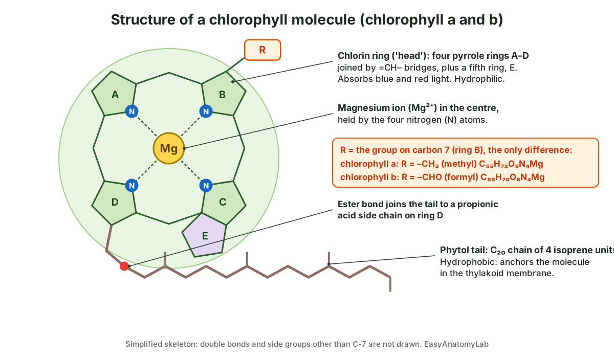
The head absorbs light, mainly blue and red, and reflects green. The hydrophobic tail anchors the molecule among the lipids of the thylakoid membrane, where the protein of a light-harvesting complex or photosystem holds it at a precise angle. Chlorophyll a (C₅₅H₇₂O₅N₄Mg) and chlorophyll b (C₅₅H₇₀O₆N₄Mg) differ only at carbon 7 on ring B: a methyl group (–CH₃) in chlorophyll a, a formyl group (–CHO) in chlorophyll b. That small change shifts which wavelengths each one absorbs, so together they capture more of the spectrum.
How grana and stroma lamellae fit together in 3D: the helical model
Flat textbook drawings show grana as separate piles of coins joined by straight bridges. Electron tomography, which builds a 3D image from many tilted electron micrographs, shows something more elegant. Each granum is wrapped by several stroma lamellae that wind around it like the ramps of a multi-storey car park. The ramps join each thylakoid disc of the granum through narrow, slit-like openings and then run off as flat sheets to neighbouring grana. The result is a single, continuous membrane that encloses a single, continuous lumen. (Pribil, Labs & Leister (2014): Structure and dynamics of thylakoids in land plants, Journal of Experimental Botany.)
This 'helical model' was first proposed by Paolillo and co-workers in the late 1960s. A detailed tomography study of lettuce chloroplasts in 2019 measured it precisely: grana averaged 278 nm across and 9 thylakoids high (range 3 to 19), about 4 to 5 lamellae wound around each granum as right-handed helices tilted on average about 16°, and the helices were linked to each other by smaller left-handed junctions. The authors explain that a sheet built only from same-handed helices would twist over long distances; the opposite-handed junctions cancel that twist, keep the network flat and tightly packed, and connect the lamellae to one another. (Bussi et al. (2019): Fundamental helical geometry consolidates the plant photosynthetic membrane, PNAS.)
An earlier tomography study had proposed instead that stroma lamellae run straight out from grana and split in two (a 'bifurcation' model). The 2019 work concluded that real thylakoids combine features of both models. The 3D model on this page shows the helical arrangement in simplified form: four right-handed ramps around each granum, joined by straight sheets. (Bussi et al. (2019): Fundamental helical geometry consolidates the plant photosynthetic membrane, PNAS.)
Why PSII sits in the grana and PSI in the stroma lamellae
The four big complexes of the light reactions are not spread evenly over the thylakoid membrane. Photosystem II and its antenna, LHCII, are packed into the stacked (appressed) membranes inside the grana. Photosystem I and ATP synthase sit in the unstacked membranes, the stroma lamellae and the top and bottom end membranes of each granum. Cytochrome b6f is found in both. Biologists call this uneven distribution lateral heterogeneity. (Pribil, Labs & Leister (2014): Structure and dynamics of thylakoids in land plants, Journal of Experimental Botany.)
The reason is partly simple geometry. Photosystem I and ATP synthase have large parts that stick out into the stroma: ATP synthase's catalytic head (CF₁) and the stromal ridge of PSI that docks ferredoxin. In stacked membranes, neighbouring thylakoids are pressed so close that there is no room for these bulky parts. ATP synthase is therefore found in the stroma lamellae and the flat end membranes of the grana, where the membrane faces open stroma. (Hahn et al. (2018): Structure, mechanism, and regulation of the chloroplast ATP synthase, Science.)
Because PSII and PSI are kept apart, the mobile carriers plastoquinone (in the membrane) and plastocyanin (in the lumen) have to carry electrons between them. That is one reason the width of the lumen matters: in dark-adapted Arabidopsis grana it is only about 4.7 nm, and it swells in the light, which may make it easier for plastocyanin to move. (Pribil, Labs & Leister (2014): Structure and dynamics of thylakoids in land plants, Journal of Experimental Botany.)
Chloroplast by the numbers
Size: higher-plant chloroplasts are lens-shaped, about 5 µm across and 2.5 µm thick. Measured values vary with species and growth: rice mesophyll chloroplasts, for example, measured about 4.2–5.6 µm long and 1.8–3.0 µm thick, and grew larger with more nitrogen. (BioNumbers 107012: size of a chloroplast, from Staehelin (2003), Photosynthesis Research 76:185–196.)
Number: a mesophyll cell of Arabidopsis holds about 110 chloroplasts, while an epidermal pavement cell holds about 10, each roughly half the size of a mesophyll chloroplast. (Barton et al. (2016): Epidermal pavement cells of Arabidopsis have chloroplasts, Plant Physiology.)
Genome: the Arabidopsis chloroplast genome has 154,478 base pairs, with two inverted repeats of 26,264 bp separating a large single-copy region of 84,170 bp and a small single-copy region of 17,780 bp. It carries 87 potential protein-coding genes (8 of them duplicated in the repeats), 4 ribosomal RNA genes and 37 transfer RNA genes. (Sato et al. (1999): Complete structure of the chloroplast genome of Arabidopsis thaliana, DNA Research (abstract, Europe PMC).)
Copies: a leaf cell can hold 10,000 or more copies of the chloroplast genome, and the number per plastid changes during development, from a few to a few hundred. (Morley & Nielsen (2016): Chloroplast DNA copy number changes during plant development, Frontiers in Plant Science.)
Proton gradient: in the light the stroma rises to about pH 7.8–8.0 and the lumen is measured at about pH 5.7–7.8. The chloroplast ATP synthase needs on average 4.67 protons for each ATP, because its rotor ring has 14 subunits and each full turn makes 3 ATP. (Höhner et al. (2016): Proton gradients and proton-dependent transport processes in the chloroplast, Frontiers in Plant Science.)
Rubisco: the global mass of Rubisco is estimated at about 0.7 billion tonnes, and its average effective rate in wild land plants is only about 1% of its maximum. (Bar-On & Milo (2019): The global mass and average rate of rubisco, PNAS.)
Chloroplast structure diagram: label it yourself (worksheet and answers)
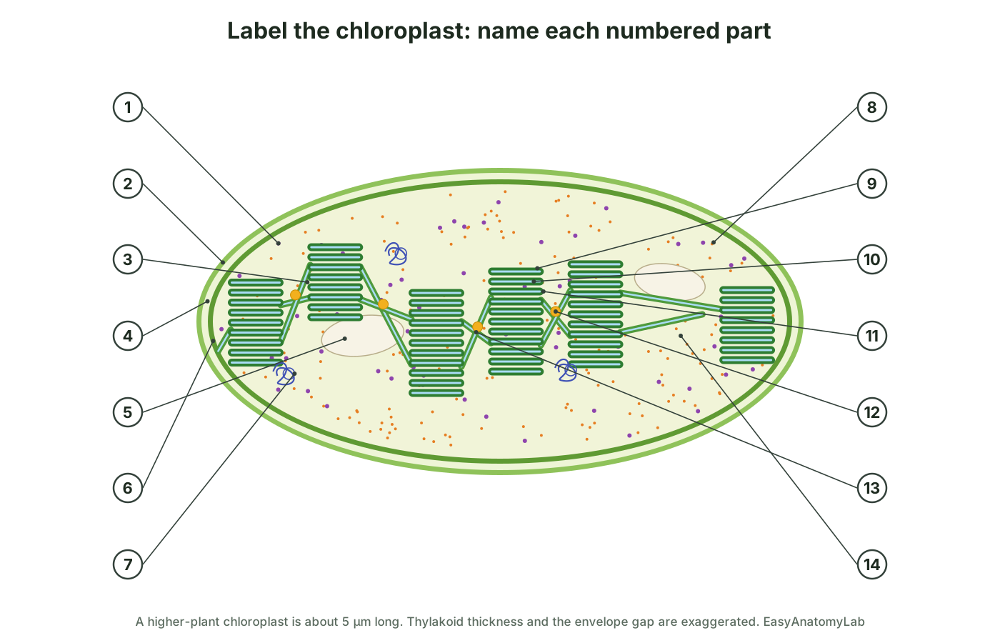
Test yourself with this chloroplast structure diagram with no labels. Match the label to the correct structure for each of the 14 numbers. It works as a coloring worksheet too: color the envelope light green, the grana dark green, the lumen blue, the starch white and the plastoglobuli yellow.
Answer key: chloroplast labeling worksheet
- 70S ribosome
- Outer membrane
- Granum
- Intermembrane space
- Starch grain
- Inner membrane
- Chloroplast DNA (nucleoid)
- Rubisco
- Thylakoid
- Thylakoid lumen
- Thylakoid membrane
- Plastoglobulus
- Stroma lamella
- Stroma
Chloroplast structure and function: practice questions
These questions cover what tests and online homework sets usually ask about chloroplast structure. Answers follow each question.
- Where in the chloroplast is water split? On the lumen side of photosystem II, in the thylakoid membrane. The protons stay in the lumen and the O₂ diffuses out of the leaf.
- Which compartment has the lowest pH in the light? The thylakoid lumen, because protons are pumped into it.
- Where is Rubisco found? In the stroma.
- Which way do protons flow through ATP synthase? From the lumen to the stroma.
- Name the membranes a CO₂ molecule crosses from the cytosol to reach Rubisco. The outer and inner envelope membranes (not the thylakoid membrane).
- How many membranes does an O₂ molecule made by PSII cross to leave the chloroplast? Three. O₂ is released into the lumen, so it crosses the thylakoid membrane and then the inner and outer envelope membranes.
- What is a granum? A stack of thylakoids.
- Why does the CF₁ part of ATP synthase not occur in the middle of a granum? The stacked membranes are pressed together with no stroma between them, so there is no room for the bulky head.
Frequently asked questions
What is the structure of a chloroplast?
A chloroplast is a lens-shaped organelle about 5 µm across with a double-membrane envelope (outer membrane, intermembrane space and inner membrane). Inside is a fluid, the stroma, containing stacks of disc-shaped thylakoids called grana, joined by stroma lamellae. Each thylakoid encloses a space called the lumen. The stroma also holds the chloroplast's circular DNA, 70S ribosomes, Rubisco, starch grains and lipid droplets called plastoglobuli.
What are the disk-shaped structures inside chloroplasts called?
They are thylakoids. A stack of thylakoids is called a granum (plural grana). The thylakoid membranes hold chlorophyll and the protein complexes of the light-dependent reactions.
How does the structure of a chloroplast relate to its function?
Stacked thylakoids give a huge membrane area for pigments and electron carriers, so a lot of light can be captured. The thylakoid membrane separates the small lumen from the stroma, so protons pumped into the lumen build a gradient that drives ATP synthase. The stroma keeps the Calvin-cycle enzymes together, right next to the ATP and NADPH made on the stroma side of the membrane. Its own DNA and ribosomes let it make some of its own proteins quickly.
Where does each stage of photosynthesis happen in the chloroplast?
The light-dependent reactions happen in the thylakoid membranes (with water split on the lumen side), and the Calvin cycle (light-independent reactions) happens in the stroma.
Is chloroplast DNA circular?
The chloroplast genome maps as a single circle of about 120–160 thousand base pairs in most flowering plants (154,478 bp in Arabidopsis), with two inverted repeats separating a large and a small single-copy region. Inside the organelle, though, many copies exist as linear and branched molecules as well as circles.
What is the difference between grana and stroma?
Grana are stacks of thylakoid membranes where light is absorbed and ATP and NADPH are made. The stroma is the fluid around the grana where the Calvin cycle uses that ATP and NADPH to fix CO₂ into sugar.
Do all plant cells have chloroplasts?
No. Chloroplasts are abundant in leaf mesophyll cells (about 110 per cell in Arabidopsis) and present in guard cells. Root cells and most cells deep in stems have none. Leaf epidermal pavement cells were long said to lack them, but in Arabidopsis they contain about ten small chloroplasts each.
Why are chloroplasts green?
Their thylakoid membranes are full of chlorophyll a and b, which absorb mainly blue and red light and reflect green light.
Why does the chloroplast have two membranes?
Because it came from a cyanobacterium that was engulfed by a eukaryotic cell more than 1.5 billion years ago. The two envelope membranes are thought to derive mainly from the cyanobacterium's own two membranes.
References
- OpenStax. Biology 2e, section 8.1: Overview of Photosynthesis. Rice University.
- OpenStax. Biology 2e, section 8.2: The Light-Dependent Reactions of Photosynthesis.
- OpenStax. Biology 2e, section 8.3: Using Light Energy to Make Organic Molecules.
- OpenStax. Biology 2e, section 4.3: Eukaryotic Cells.
- Staehelin, L. A. (2003). Chloroplast structure: from chlorophyll granules to supra-molecular architecture of thylakoid membranes. Photosynthesis Research 76, 185–196 (via BioNumbers 107012).
- Bussi, Y. et al. (2019). Fundamental helical geometry consolidates the plant photosynthetic membrane. PNAS 116, 22366–22375.
- Pribil, M., Labs, M. & Leister, D. (2014). Structure and dynamics of thylakoids in land plants. Journal of Experimental Botany 65, 1955–1972.
- Hahn, A. et al. (2018). Structure, mechanism, and regulation of the chloroplast ATP synthase. Science 360, eaat4318.
- Höhner, R., Aboukila, A., Kunz, H.-H. & Venema, K. (2016). Proton gradients and proton-dependent transport processes in the chloroplast. Frontiers in Plant Science 7, 218.
- Daniell, H. et al. (2016). Chloroplast genomes: diversity, evolution, and applications in genetic engineering. Genome Biology 17, 134.
- Sato, S. et al. (1999). Complete structure of the chloroplast genome of Arabidopsis thaliana. DNA Research 6, 283–290.
- Morley, S. A. & Nielsen, B. L. (2016). Chloroplast DNA copy number changes during plant development in organelle DNA polymerase mutants. Frontiers in Plant Science 7, 57.
- Manuell, A. L. et al. (2007). Structure of the chloroplast ribosome: novel domains for translation regulation. PLoS Biology 5, e209.
- Demarsy, E., Lakshmanan, A. M. & Kessler, F. (2014). Border control: selectivity of chloroplast protein import and regulation at the TOC-complex. Frontiers in Plant Science 5, 483.
- Austin, J. R. et al. (2006). Plastoglobules are lipoprotein subcompartments of the chloroplast that are permanently coupled to thylakoid membranes. The Plant Cell 18, 1693–1703.
- Streb, S. & Zeeman, S. C. (2012). Starch metabolism in Arabidopsis. The Arabidopsis Book 10, e0160.
- Bar-On, Y. M. & Milo, R. (2019). The global mass and average rate of rubisco. PNAS 116, 4738–4743.
- Barton, K. A. et al. (2016). Epidermal pavement cells of Arabidopsis have chloroplasts. Plant Physiology 171, 723–726.
- Iwai, S. et al. (2019). Guard cell photosynthesis is crucial in abscisic acid-induced stomatal closure. Plant Direct 3, e00137.
- Lhee, D. et al. (2019). Evolutionary dynamics of the chromatophore genome in three photosynthetic Paulinella species. Scientific Reports 9, 2560.
- Li, Y. et al. (2013). Does chloroplast size influence photosynthetic nitrogen use efficiency? PLoS ONE 8, e62036.
- Qiu, N. W. et al. (2019). Advances in the members and biosynthesis of chlorophyll family. Photosynthetica 57, 974–984.
More interactive anatomy models
- Flower anatomyParts of a flower in 3D: petals, sepals, stamens, pistil, ovary and ovules of a lily.
- Leaf cross sectionLabeled leaf cross section: cuticle, epidermis, palisade and spongy mesophyll, veins.
- Stomata diagramGuard cells, stomatal pore and how stomata open and close, in 3D.
- Seed anatomyBean and corn seed anatomy: seed coat, cotyledons, endosperm and embryo.
- Human anatomy1,000+ bones, muscles, organs, vessels and nerves from real anatomical data.
- Octopus anatomyThree hearts, a ring-shaped brain, eight arms and a beak, in 3D.