

Nephron anatomy at a glance
A nephron is the microscopic functional unit of the kidney: a tiny filter attached to a long, folded tube that turns blood plasma into urine. Each nephron has two parts. The renal corpuscle is the filter: a ball of capillaries, the glomerulus, inside a cup called the glomerular (Bowman's) capsule. The renal tubule is the tube that processes the filtrate: the proximal convoluted tubule, the nephron loop (loop of Henle) and the distal convoluted tubule. A connecting tubule joins each nephron to a collecting duct, which is shared by several nephrons and belongs to the collecting system.
The page uses the adult human kidney as its reference. Each kidney holds on average about 900,000 nephrons, although healthy people range from about 210,000 to 2.7 million per kidney. The kidneys get about 20–25% of the heart's output at rest and filter about 180 L of plasma a day, but make only 1–2 L of urine.
Directions used here: cortical means toward the outer surface (renal capsule), medullary means toward the inside of the kidney (renal pyramids and papillae). Proximal means closer to the start of the nephron (the glomerulus) and distal farther along it.
Nephron anatomy labeled diagram
The labeled diagram of a nephron shows, in the order filtrate flows: glomerulus and Bowman's capsule, proximal convoluted tubule, proximal straight tubule, descending thin limb, ascending thin limb, thick ascending limb with the macula densa, distal convoluted tubule, connecting tubule, and the collecting duct and papillary duct. Around it are the afferent and efferent arterioles, peritubular capillaries and vasa recta.
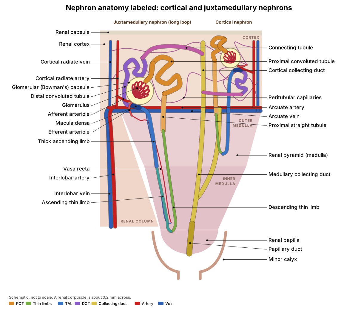
Notice where each part lives. All renal corpuscles and both convoluted tubules are in the cortex. Only the loops and the collecting ducts reach into the medulla. The long loop on the left turns near the papilla; the short loop on the right turns in the outer medulla and has no thin ascending limb.
Parts of the nephron and their functions
Each segment of the nephron has a different lining and a different job. Roughly, the proximal tubule reabsorbs most of everything, the loop of Henle builds the salt gradient, the distal tubule fine-tunes sodium and calcium, and the collecting duct sets the final water, sodium, potassium and acid content of the urine under hormone control. The percentages below are of the amount filtered, for a typical adult.
| Segment | Reabsorbs | Secretes | Key transporters | Hormones and drugs |
|---|---|---|---|---|
| Renal corpuscle | Nothing (filters plasma: ≈ 180 L/day) | – | None; driven by ≈ 10 mm Hg net filtration pressure | Angiotensin II, ANP and sympathetic nerves change GFR |
| Proximal convoluted tubule | ≈ 65–67% of water and Na+, ≈ 65% of K+, ≈ 50% of Cl−, 80–90% of HCO3−, almost all glucose and amino acids | H+, NH4+, creatinine, drugs, organic acids and bases | Na+/K+-ATPase, SGLT2/SGLT1, NHE3, carbonic anhydrase | PTH (cuts phosphate reabsorption, activates vitamin D); SGLT2 inhibitors |
| Descending thin limb | Water (up to ≈ 15%) | Urea enters | Aquaporin-1 | – |
| Ascending thin limb | NaCl (passively) | – | No aquaporins | – |
| Thick ascending limb | ≈ 25% of Na+, with K+ and Cl−; Ca2+ and Mg2+ between cells | K+ recycles to the lumen | NKCC2, ROMK, Na+/K+-ATPase | Loop diuretics (furosemide) block NKCC2 |
| Distal convoluted tubule | ≈ 5–10% of Na+ with Cl−; Ca2+; Mg2+ | – | NCC, TRPV5, TRPM6 | PTH (more Ca2+), aldosterone (late DCT); thiazides block NCC |
| Connecting tubule and collecting duct | ≈ 5% of Na+; water (variable, most of the last ≈ 10%); urea (inner medulla) | K+ (principal cells), H+ or HCO3− (intercalated cells) | ENaC, ROMK, aquaporin-2, H+-ATPase, pendrin, UT-A1/UT-A3 | Aldosterone, ADH, ANP; amiloride blocks ENaC |
Two points trip students up. First, the macula densa is the last stretch of the thick ascending limb, at the point where the tubule touches its own glomerulus; many books place it "at the start of the DCT". Second, the collecting duct is not part of the nephron in the strict sense, because one duct collects from many nephrons, but most courses teach it with the nephron.
Internal anatomy of the kidney, including the nephron
Cut a kidney lengthwise and you see three regions: the outer, granular renal cortex; the striped, cone-shaped renal pyramids of the medulla, separated by renal columns of cortex-like tissue; and the renal sinus, the central space that holds the calyces, renal pelvis, vessels and fat. The tip of each pyramid, the renal papilla, drains into a minor calyx. Minor calyces join into two or three major calyces, which join into the renal pelvis and then the ureter. Vessels, nerves and the pelvis pass through the hilum.
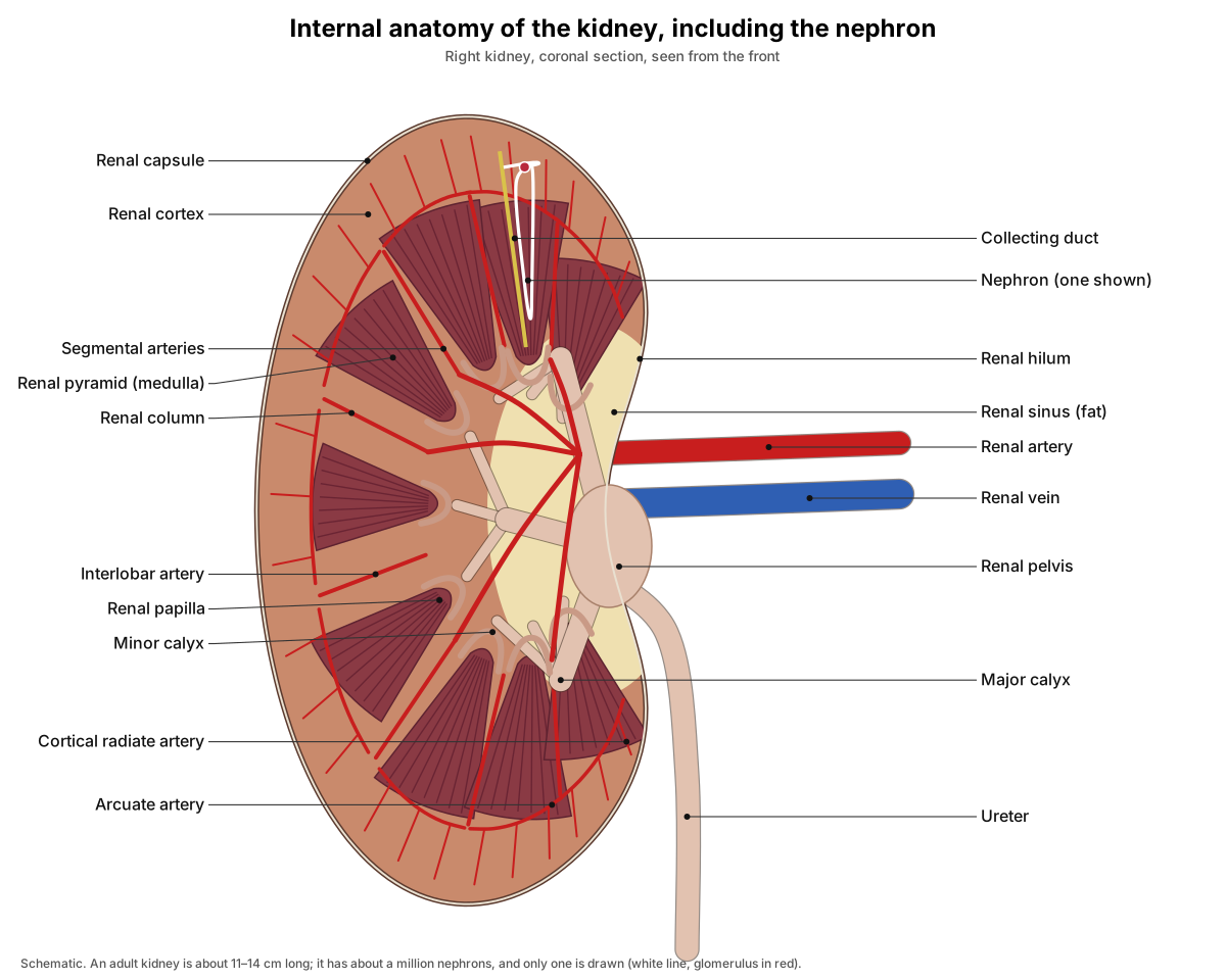
A nephron spans these regions. Its corpuscle and convoluted tubules sit in the cortex; its loop dips into a pyramid; and its collecting duct runs down through the pyramid to a papilla. The stripes of a pyramid are thousands of loops, collecting ducts and vasa recta running side by side, which is why the medulla looks striated and the cortex, packed with corpuscles, looks granular. Renal columns divide each kidney into 6–8 lobes; a lobe is one pyramid with its cap of cortex, and it is a lobe that the 3D block shows.
- Renal capsule: Holds the kidney's shape and protects it. The fat pad and renal fascia outside it cushion and anchor the kidney.
- Renal cortex: Site of filtration (glomeruli) and of most reabsorption (proximal and distal convoluted tubules). Peritubular capillaries here return reabsorbed fluid to the blood.
- Renal pyramid (medulla): Holds the loops of Henle, collecting ducts and vasa recta that build and use the osmotic gradient (about 300 to 1,200 mOsm/kg) needed to make concentrated urine.
- Renal column: Carries the interlobar arteries and veins between the renal sinus and the cortex.
- Renal papilla: Where the papillary ducts open and release urine into the minor calyx.
- Minor calyx: First part of the urinary collecting passages outside the nephron. Smooth muscle in its wall moves urine on by peristalsis.
- Major calyx: Carries urine from the minor calyces to the renal pelvis.
- Renal pelvis: Funnels urine from the calyces into the ureter (OpenStax A&P 2e).
- Renal hilum: Entry and exit point for the renal artery, renal vein, lymphatics, nerves and the renal pelvis/ureter.
For the kidneys in the context of the whole body, see the urinary system in our 3D human anatomy model.
Blood supply of the nephron: the renal vascular path
Blood reaches a nephron by this route: abdominal aorta → renal artery → segmental arteries → interlobar arteries (in the renal columns) → arcuate arteries (along the bases of the pyramids) → cortical radiate (interlobular) arteries → afferent arteriole → glomerulus → efferent arteriole → peritubular capillaries or vasa recta → cortical radiate veins → arcuate veins → interlobar veins → renal vein → inferior vena cava.
- Two capillary beds in series. The glomerulus is a high-pressure bed between two arterioles, built for filtration. The peritubular capillaries and vasa recta that follow are low-pressure beds, built for taking back what the tubules reabsorb.
- The afferent arteriole is wider than the efferent. This, and the tone of both arterioles, keeps pressure in the glomerulus high.
- Cortical nephrons feed peritubular capillaries; juxtamedullary nephrons feed the vasa recta. The descending vasa recta come from the efferent arterioles of juxtamedullary glomeruli and are the main blood supply of the medulla.
- Veins run beside the arteries in the same order, cortical radiate → arcuate → interlobar → renal vein, but unlike the arteries they connect freely with one another.
- Afferent arteriole: Controls how much blood enters the glomerulus. It constricts when stretched (myogenic response) or when the macula densa signals (tubuloglomerular feedback), keeping GFR steady.
- Efferent arteriole: Its resistance keeps glomerular pressure high. In cortical nephrons it feeds the peritubular capillaries; in juxtamedullary nephrons it gives rise to the vasa recta.
- Peritubular capillaries: Take up the water and solutes reabsorbed by the tubules and supply substances for secretion.
- Vasa recta: Supply the medulla and act as a countercurrent exchanger: they remove reabsorbed water without washing out the medullary salt and urea gradient.
The renal corpuscle: glomerulus and Bowman's capsule
The renal corpuscle is the filtering unit of the nephron. It is a tuft of capillaries, the glomerulus, wrapped by the double-walled glomerular (Bowman's) capsule. The outer, parietal layer is simple squamous epithelium. At the vascular pole, where the arterioles enter and leave, it folds back to form the inner, visceral layer, made of podocytes that cling to the capillaries. Between the two layers is the capsular space, which collects the filtrate and opens into the proximal tubule at the urinary pole.
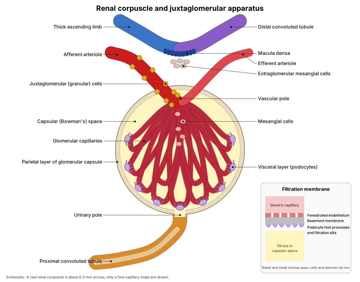
Filtrate crosses a three-layer filtration membrane: the fenestrated endothelium of the capillary (pores about 70 nm across), a shared basement membrane, and the filtration slits between the interlocking foot processes (pedicels) of podocytes. Molecules smaller than about 4 nm pass freely; blood cells, platelets and large proteins such as albumin are held back. Negative charges in the barrier also repel negatively charged proteins. Mesangial cells in the core of the tuft support the capillaries and can contract to change the filtration rate.
Juxtaglomerular apparatus
The juxtaglomerular apparatus (JGA) is where the end of the thick ascending limb touches the vascular pole of its own glomerulus. It has three parts: the macula densa, a plaque of crowded tubule cells that senses NaCl in the passing fluid; the juxtaglomerular (granular) cells, modified smooth muscle cells in the afferent arteriole wall that release renin; and the extraglomerular mesangial cells in the angle between the two arterioles.
The JGA links each nephron's output to its own input. When fluid reaching the macula densa is too salty (GFR too high), it releases ATP and adenosine, which constrict the afferent arteriole and bring GFR back down: this is tubuloglomerular feedback. When salt delivery or blood pressure falls, renin release rises and starts the renin–angiotensin–aldosterone system.
- Macula densa: Senses NaCl in the tubular fluid. High NaCl triggers tubuloglomerular feedback (ATP and adenosine constrict the afferent arteriole); low NaCl promotes renin release.
- Juxtaglomerular (granular) cells: Release renin when blood pressure or NaCl delivery falls, or with sympathetic stimulation. Renin starts the renin–angiotensin–aldosterone system.
- Extraglomerular mesangial cells: Thought to relay signals from the macula densa to the arterioles and JG cells.
Nephron loop (loop of Henle) anatomy
The nephron loop, or loop of Henle, is the U-shaped part of the tubule that dips from the cortex into the medulla and back. It has a descending limb (the thick proximal straight tubule, then the descending thin limb), a hairpin bend, and an ascending limb (the ascending thin limb in long loops, then the thick ascending limb). Thin segments are lined by flat, simple squamous cells; thick segments by cuboidal cells.
- Proximal straight tubule: Continues proximal reabsorption and is a main site of organic anion and cation secretion (the S3 segment).
- Descending thin limb: Freely permeable to water (aquaporin-1) but not to most solutes, so water leaves into the salty medulla and the filtrate becomes more concentrated as it descends.
- Ascending thin limb: Impermeable to water but permeable to NaCl, so salt leaves passively and the filtrate starts to become dilute.
- Thick ascending limb (TAL): Actively reabsorbs about 25% of filtered Na+ through the Na+–K+–2Cl− cotransporter (NKCC2) but no water, so it dilutes the filtrate and builds the medullary gradient (the 'single effect').
Collecting system: collecting duct and papillary duct
The collecting system starts where several connecting tubules join a cortical collecting duct. The duct runs straight down through the medulla, merging with others, and ends as a wide papillary duct (duct of Bellini) that opens on the sieve-like tip of the papilla, the area cribrosa. Its lining is a "salt and pepper" mix of principal cells, which handle Na+, K+ and water, and darker intercalated cells, which handle acid and base. Principal cells outnumber intercalated cells about 3:1 in the cortical duct and 2:1 in the outer medulla.
- Connecting tubule: Reabsorbs Na+ and secretes K+ under aldosterone, and can reabsorb water under ADH, like the collecting duct.
- Cortical collecting duct: Principal cells reabsorb Na+ (ENaC) and secrete K+ (ROMK) under aldosterone, and reabsorb water through aquaporin-2 when ADH is present. Intercalated cells secrete H+ or HCO3−.
- Medullary collecting duct: Final water reabsorption into the concentrated medulla when ADH is present. The inner medullary part also returns urea to the interstitium (UT-A1, UT-A3).
- Papillary duct: Carries the final urine to the minor calyx. After this point, no more reabsorption happens.
- Area cribrosa: Lets urine drip from the papillary ducts into the minor calyx.
Cortical vs juxtamedullary nephrons
There are two types of nephron. Cortical nephrons, about 85% of the total, have their glomeruli in the outer cortex and short loops. Juxtamedullary nephrons, about 15%, have their glomeruli deep in the cortex beside the medulla ("juxta" means next to) and long loops that reach the inner medulla. The 3D model shows one of each.
| Feature | Cortical nephron | Juxtamedullary nephron |
|---|---|---|
| Share of nephrons (human) | ≈ 85% | ≈ 15% |
| Glomerulus position | Outer and middle cortex | Inner cortex, next to the medulla |
| Loop of Henle | Short; turns in the outer medulla | Long; reaches deep into the inner medulla |
| Thin ascending limb | Absent | Present |
| Efferent arteriole feeds | Peritubular capillaries | Vasa recta (and some peritubular capillaries) |
| Main role | Most filtration and reabsorption | Builds the medullary gradient to concentrate urine |
Follow the filtrate: interactive nephron function
Step through the nephron in the order fluid flows. Each step shows the lining, what is reabsorbed or secreted, the main transporters, the hormones that act, and how concentrated the fluid is as it leaves. Switch ADH off to see how the same nephron makes dilute urine.
Renal corpuscle: filtration
- Lining
- Fenestrated capillary endothelium, basement membrane, podocytes
- Fluid leaving
- ≈ 300 mOsm/kg, the same as plasma
- Transporters
- None: filtration is passive, driven by a net filtration pressure of about 10 mm Hg
- Hormones
- Angiotensin II, ANP and sympathetic nerves change GFR by changing arteriole tone
- Plasma water, ions, glucose, amino acids, urea and creatinine are filtered
- Blood cells and large proteins such as albumin stay in the blood
GFR is about 125 mL/min in men and 105 mL/min in women: about 180 L and 150 L of filtrate a day.
Values are typical for an adult human and rounded; textbooks differ by a few percent. Sources: OpenStax Anatomy & Physiology 2e, Sands & Layton (2009), Curthoys & Moe (2014), Subramanya & Ellison (2014).
Filtration, reabsorption and secretion: how a nephron makes urine
A nephron makes urine in three steps. Glomerular filtration pushes plasma out of the glomerular capillaries into the capsular space. Tubular reabsorption moves the useful parts of that filtrate, such as water, sodium, glucose and amino acids, back into the blood. Tubular secretion moves extra waste and ions, such as H+, K+, creatinine and many drugs, from the blood into the tubule. What is left at the end is urine.
Filtration is driven by pressure. In OpenStax's worked example, the blood pressure in the glomerular capillaries is about 55 mm Hg. Two forces oppose it: fluid pressure in the capsule (about 15 mm Hg) and the pull of plasma proteins (colloid osmotic pressure, about 30 mm Hg). That leaves a net filtration pressure of about 10 mm Hg. It is enough to filter about 125 mL of plasma a minute in men and 105 mL in women, or about 180 L and 150 L a day. Because about 99% of that is reabsorbed, only 1–2 L of urine are made each day. (OpenStax Anatomy & Physiology 2e, 25.5 Physiology of Urine Formation.)
The numbers show how selective reabsorption is. Each day the kidneys filter about 180 L of water and keep 179 L, filter 180 g of glucose and keep all of it, filter 540 g of sodium and keep 537 g, but keep none of the 1.4 g of filtered creatinine. Urea is in between: about 53 g are filtered and about 25 g leave in the urine. (OpenStax Anatomy & Physiology 2e, 25.6 Tubular Reabsorption.)
Most reabsorption happens early. The proximal tubule takes back about two-thirds of the filtered water and NaCl, most of the bicarbonate and almost all of the glucose and amino acids. Its key glucose transporter is SGLT2 in the S1 and S2 segments, which accounts for about 97% of glucose reabsorption; SGLT1 in the S3 segment recovers the rest. This is why SGLT2-blocking drugs make people lose glucose in their urine. (Curthoys & Moe (2014): Proximal tubule function and response to acidosis, Clin J Am Soc Nephrol.)
Countercurrent multiplier: how the loop of Henle concentrates urine
The countercurrent multiplier is the mechanism by which the loop of Henle builds a salt gradient in the renal medulla, from about 300 mOsm/kg near the cortex to about 1,200 mOsm/kg at the papilla in humans. 'Countercurrent' means the fluid flows down one limb and back up the other. 'Multiplier' means a small difference between the two limbs at any level is multiplied, along the length of the loop, into a large difference from top to bottom. (OpenStax Anatomy & Physiology 2e, 25.6 Tubular Reabsorption.)
It works because the two limbs have opposite permeabilities. The thick ascending limb pumps NaCl out into the interstitium through the Na+–K+–2Cl− cotransporter (NKCC2) but does not let water follow. The descending limb lets water out but not much salt, so its fluid becomes as concentrated as the salty interstitium around it. In the outer medulla, NaCl reabsorbed by thick ascending limbs is the main solute in the gradient. In the inner medulla, urea recycled from the collecting ducts adds a large share; exactly how the inner medullary gradient is made is still debated. (Sands & Layton (2009): The physiology of urinary concentration: an update, Semin Nephrol.)
The vasa recta keep the gradient from being washed away. These hairpin capillaries come from the efferent arterioles of juxtamedullary glomeruli and run beside the long loops. As blood flows down, it picks up salt and urea and loses water; as it flows back up, it gives the salt and urea back and picks up water. This countercurrent exchange removes the water reabsorbed in the medulla while leaving most of the solute behind. (Edwards, Silldorff & Pallone (2000): The renal medullary microcirculation, Frontiers in Bioscience.)
The gradient is then used by the collecting duct. When ADH is present, aquaporin-2 channels in the collecting duct let water move out into the concentrated medulla, and urine can reach about 1,200 mOsm/kg, about four times the concentration of plasma. After drinking a lot of water, ADH falls and urine can be as dilute as about 50 mOsm/kg. Desert rodents with very long loops can do much better: Sands and Layton list about 3,000 mOsm/kg for rats and about 7,600 for chinchillas. (Sands & Layton (2009): The physiology of urinary concentration: an update, Semin Nephrol.)
Hormonal control of the nephron: RAAS, aldosterone, ADH and ANP
The renin–angiotensin–aldosterone system (RAAS) raises blood volume and pressure. When extracellular fluid volume falls, the juxtaglomerular cells of the afferent arteriole release renin. Renin cuts angiotensinogen, made by the liver, into angiotensin I. Angiotensin-converting enzyme (ACE), made mainly in the lungs and bound to vessel walls, turns angiotensin I into angiotensin II. Angiotensin II is a powerful vasoconstrictor and constricts both the afferent and efferent arterioles. It also triggers release of aldosterone from the adrenal cortex. (OpenStax Anatomy & Physiology 2e, 25.8 Endocrine Regulation of Kidney Function.)
Aldosterone makes the late distal tubule, connecting tubule and collecting duct keep more Na+ and lose more K+. Water follows the retained sodium. It does this by increasing Na+/K+-ATPase pumps on the basolateral side and Na+ and K+ channels on the luminal side of principal cells. Aldosterone is also released directly in response to high plasma K+. (OpenStax Anatomy & Physiology 2e, 25.6 Tubular Reabsorption.)
Antidiuretic hormone (ADH, vasopressin) is a 9-amino-acid peptide released by the posterior pituitary when plasma becomes too concentrated. In collecting duct principal cells it binds V2 receptors, raises cAMP, and moves aquaporin-2 water channels into the luminal membrane, so more water is reabsorbed. Osmoreceptors respond to a rise in plasma osmolality of only about 2 mOsm/kg. (Sands & Layton (2009): The physiology of urinary concentration: an update, Semin Nephrol.)
Atrial natriuretic peptide (ANP) works the other way. The heart's atria release it when they are over-stretched by too much blood volume. ANP raises GFR by dilating the afferent arteriole and constricting the efferent arteriole, and it reduces sodium recovery by inhibiting aldosterone release, sodium reabsorption in the DCT, and ADH release. The result is more salt and water in the urine. (OpenStax Anatomy & Physiology 2e, 25.8 Endocrine Regulation of Kidney Function.)
Parathyroid hormone (PTH) is the other hormone students meet here. Released when blood calcium falls, it blocks phosphate reabsorption in the proximal tubule, so phosphate is lost in the urine, and it stimulates the proximal tubule to make calcitriol, the active form of vitamin D. (OpenStax Anatomy & Physiology 2e, 25.8 Endocrine Regulation of Kidney Function.)
In the distal convoluted tubule, PTH increases calcium reabsorption: it boosts production of the TRPV5 calcium channels in the luminal membrane and keeps them there longer. Calcium entering the cell is carried across by calbindin and pushed out into the blood by a Na+/Ca2+ exchanger and calcium pumps. (Subramanya & Ellison (2014): Distal convoluted tubule, Clin J Am Soc Nephrol.)
The kidney also controls its own filtration rate. In the myogenic response, the afferent arteriole constricts when stretched by higher pressure. In tubuloglomerular feedback, the macula densa senses high NaCl in the fluid reaching it and releases ATP and adenosine, which constrict the afferent arteriole and lower GFR; nitric oxide has the opposite effect. (OpenStax Anatomy & Physiology 2e, 25.7 Regulation of Renal Blood Flow.)
Hormones that act on the nephron: summary table
| Hormone | Made by | Released when | Acts on | Effect on urine |
|---|---|---|---|---|
| ADH (vasopressin) | Hypothalamus; released from posterior pituitary | Plasma too concentrated; low blood volume | Collecting duct principal cells (aquaporin-2) | Less, more concentrated urine |
| Aldosterone | Adrenal cortex | Angiotensin II; high plasma K+ | Late DCT, connecting tubule, collecting duct (ENaC, Na+/K+-ATPase) | Less Na+, more K+ in urine; water follows Na+ |
| Angiotensin II | Made in blood from angiotensin I by ACE | Renin release (low volume or pressure) | Arterioles; adrenal cortex | Keeps GFR up in low pressure; less Na+ and water lost |
| Renin (an enzyme) | Juxtaglomerular cells | Low pressure, low NaCl at macula densa, sympathetic input | Angiotensinogen | Starts RAAS |
| ANP | Heart atria | Atria over-stretched (high volume) | Arterioles, DCT, collecting duct; inhibits aldosterone and ADH | More Na+ and water lost |
| PTH | Parathyroid glands | Low blood Ca2+ | DCT (Ca2+), PCT (phosphate, vitamin D) | Less Ca2+, more phosphate in urine |
Microscopic anatomy of the nephron (histology)
In a kidney section, you tell the tubules apart by their lining. The proximal convoluted tubule is the most common profile in the cortex: tall, pink-staining (eosinophilic) cuboidal cells packed with mitochondria, with a fuzzy brush border of microvilli that often fills the lumen. The distal convoluted tubule has smaller, paler cells with no brush border, so its lumen looks wide and clean and more nuclei show in each cross-section. Collecting ducts have pale cuboidal to columnar cells with clear cell borders. Thin limbs look like capillaries but carry no blood cells.
| Structure | Epithelium | How to recognise it |
|---|---|---|
| Parietal layer of capsule | Simple squamous | Thin outline around the capsular space |
| Podocytes | Specialised visceral epithelium | Nuclei on the outside of the capillary loops |
| Proximal convoluted tubule | Simple cuboidal, brush border | Tall pink cells, many mitochondria, fuzzy small lumen; most common cortical tubule |
| Thin limbs | Simple squamous | Flat cells, thin wall, no red cells in the lumen (unlike capillaries) |
| Thick ascending limb | Simple cuboidal | Low cuboidal cells, no brush border, in medulla and medullary rays |
| Macula densa | Tall, crowded cells | Nuclei packed close together where the tubule touches the vascular pole |
| Distal convoluted tubule | Simple cuboidal | Smaller, paler cells, no brush border, wide clear lumen |
| Collecting duct | Cuboidal to columnar | Pale cells with distinct borders; light principal and darker intercalated cells; wider toward the papilla |
| Papillary duct | Tall columnar | Wide lumen at the papilla tip; opens on the area cribrosa |
| Minor calyx | Transitional epithelium (urothelium) | Lines the cup around the papilla |
Sources for this table: OpenStax A&P 2e (25.4), Kidney Pathology's tubule histology tutorial and Digital Histology's renal papilla page, all listed below.
Kidney and nephron structures: full labeled list
This is the text version of the 3D model: every labeled structure, grouped by system, with what it does. Use it as a study list or to check a labeling activity.
Kidney regions
- Kidney (coronal section)
- The whole right kidney cut lengthwise, shown small on the left so you can see where the magnified block of tissue comes from. Filters the blood, makes urine, and regulates water, salt, potassium, acid–base balance and blood pressure. It also makes renin, erythropoietin and calcitriol.
- Renal hilum
- The notch on the medial, concave border of the kidney where vessels, nerves and the ureter enter or leave. Entry and exit point for the renal artery, renal vein, lymphatics, nerves and the renal pelvis/ureter.
- Renal capsule (Fibrous capsule)
- A thin, tough layer of dense irregular connective tissue that wraps the kidney. Holds the kidney's shape and protects it. The fat pad and renal fascia outside it cushion and anchor the kidney.
- Renal cortex
- The outer, granular region of the kidney. It holds every renal corpuscle and the convoluted tubules. Site of filtration (glomeruli) and of most reabsorption (proximal and distal convoluted tubules). Peritubular capillaries here return reabsorbed fluid to the blood.
- Renal column (Column of Bertin)
- A band of cortex-like tissue that runs down between two renal pyramids. Carries the interlobar arteries and veins between the renal sinus and the cortex.
- Renal pyramid (medulla) (Medullary pyramid)
- A cone-shaped mass of medulla with its broad base toward the cortex and its tip, the papilla, toward the renal sinus. Holds the loops of Henle, collecting ducts and vasa recta that build and use the osmotic gradient (about 300 to 1,200 mOsm/kg) needed to make concentrated urine.
- Renal papilla
- The rounded tip of a renal pyramid, which pokes into a minor calyx. Where the papillary ducts open and release urine into the minor calyx.
- Minor calyx
- A cup-shaped tube that fits over one renal papilla and collects the urine dripping from it. First part of the urinary collecting passages outside the nephron. Smooth muscle in its wall moves urine on by peristalsis.
- Major calyx
- One of the two or three large branches of the renal pelvis, each formed by several minor calyces. Carries urine from the minor calyces to the renal pelvis.
- Renal pelvis
- The funnel-shaped space formed where the major calyces join. It narrows into the ureter. Funnels urine from the calyces into the ureter (OpenStax A&P 2e).
- Ureter
- The muscular tube that carries urine from the renal pelvis to the urinary bladder. Moves urine by peristaltic waves of smooth muscle.
Renal corpuscle
- Glomerulus (Glomerular capillaries)
- A tuft of looping, fenestrated capillaries where blood plasma is filtered into the nephron. Filtration. Blood pressure pushes water and small solutes through the filtration membrane into the capsular space, making about 180 L of filtrate a day in men and 150 L in women.
- Glomerular capsule (parietal layer) (Bowman's capsule)
- The cup-shaped start of the nephron that surrounds the glomerulus. Its outer, parietal layer is simple squamous epithelium. Catches the filtrate and passes it to the proximal convoluted tubule at the urinary pole.
- Capsular space (Bowman's space, urinary space)
- The narrow space between the parietal and visceral layers of the capsule. It fills with filtrate. Receives the glomerular filtrate before it enters the proximal convoluted tubule.
- Visceral layer (podocytes) (Podocytes, pedicels, filtration slits)
- Octopus-like cells that wrap the glomerular capillaries. Their interlocking foot processes (pedicels) leave thin filtration slits. Form the last layer of the filtration membrane. The slits, bridged by a slit diaphragm, hold back albumin and other large proteins.
- Mesangial cells (intraglomerular)
- Contractile support cells in the core (stalk) of the glomerular tuft, between the capillary loops. Support the capillary loops, clear trapped material, and can contract to change the filtration surface and so GFR.
Renal tubule
- Proximal convoluted tubule (PCT)
- The long, coiled first part of the renal tubule, lined by cuboidal cells with a brush border of microvilli. Reabsorbs about 65–67% of filtered water and Na+, nearly all glucose and amino acids, and 80–90% of bicarbonate. Secretes H+, NH4+, creatinine and many drugs.
- Proximal straight tubule (Pars recta, thick descending limb)
- The straight end of the proximal tubule, which runs down out of the cortex into the outer medulla. Continues proximal reabsorption and is a main site of organic anion and cation secretion (the S3 segment).
- Descending thin limb (Descending limb of the nephron loop)
- A thin tube of flat (simple squamous) cells that runs down into the medulla. Freely permeable to water (aquaporin-1) but not to most solutes, so water leaves into the salty medulla and the filtrate becomes more concentrated as it descends.
- Ascending thin limb
- A thin, flat-celled segment that climbs from the hairpin bend back up through the inner medulla. Impermeable to water but permeable to NaCl, so salt leaves passively and the filtrate starts to become dilute.
- Thick ascending limb (TAL)
- The cuboidal-celled segment that pumps salt out of the filtrate on its way back to the cortex. Actively reabsorbs about 25% of filtered Na+ through the Na+–K+–2Cl− cotransporter (NKCC2) but no water, so it dilutes the filtrate and builds the medullary gradient (the 'single effect').
- Distal convoluted tubule (DCT)
- A short, coiled cortical segment after the macula densa, lined by cuboidal cells without a brush border. Reabsorbs about 5–10% of filtered Na+ through the thiazide-sensitive Na+–Cl− cotransporter (NCC), and Ca2+ under PTH control (TRPV5 channels). Remains impermeable to water.
- Connecting tubule (Connecting segment)
- The short link between the distal convoluted tubule and a collecting duct. Reabsorbs Na+ and secretes K+ under aldosterone, and can reabsorb water under ADH, like the collecting duct.
- Cortical nephron (short loop)
- The common nephron type (about 85%): glomerulus in the outer cortex and a short loop that turns in the outer medulla. Does most of the kidney's filtration and reabsorption. Its efferent arteriole feeds the peritubular capillaries around the cortical tubules.
Juxtaglomerular apparatus
- Macula densa
- A plaque of crowded, tall cells in the wall of the thick ascending limb where it touches its own glomerulus. Senses NaCl in the tubular fluid. High NaCl triggers tubuloglomerular feedback (ATP and adenosine constrict the afferent arteriole); low NaCl promotes renin release.
- Juxtaglomerular (granular) cells (JG cells)
- Modified smooth muscle cells in the wall of the afferent arteriole that store and release renin. Release renin when blood pressure or NaCl delivery falls, or with sympathetic stimulation. Renin starts the renin–angiotensin–aldosterone system.
- Extraglomerular mesangial cells (Lacis cells, polkissen)
- A small cushion of cells in the angle between the afferent arteriole, efferent arteriole and macula densa. Thought to relay signals from the macula densa to the arterioles and JG cells.
Collecting system
- Cortical collecting duct
- The first part of the collecting duct, in the cortex. Several nephrons drain into it. Principal cells reabsorb Na+ (ENaC) and secrete K+ (ROMK) under aldosterone, and reabsorb water through aquaporin-2 when ADH is present. Intercalated cells secrete H+ or HCO3−.
- Medullary collecting duct
- The collecting duct as it runs through the renal pyramid, joining others as it goes. Final water reabsorption into the concentrated medulla when ADH is present. The inner medullary part also returns urea to the interstitium (UT-A1, UT-A3).
- Papillary duct (Duct of Bellini)
- The wide end of the collecting system, formed by several merged collecting ducts, that opens at the tip of the papilla. Carries the final urine to the minor calyx. After this point, no more reabsorption happens.
- Area cribrosa
- The sieve-like tip of the renal papilla, dotted with the openings of the papillary ducts. Lets urine drip from the papillary ducts into the minor calyx.
Arteries & arterioles
- Renal artery
- The large artery that brings blood from the abdominal aorta to the kidney. Supplies about 20–25% of the resting cardiac output to the two kidneys (OpenStax A&P 2e).
- Segmental arteries
- The first branches of the renal artery, each supplying one segment of the kidney. Distribute blood to the kidney's segments. They are end arteries, so a blocked one kills its segment.
- Interlobar artery
- An artery that runs up a renal column between two pyramids. Carries blood from a segmental artery to the arcuate arteries.
- Arcuate artery
- An artery that arches along the base of a renal pyramid, at the border of cortex and medulla. Gives off the cortical radiate arteries that climb into the cortex.
- Cortical radiate artery (Interlobular artery)
- A small artery that runs straight out through the cortex toward the capsule. Gives off the afferent arterioles, one to each glomerulus.
- Afferent arteriole
- The arteriole that carries blood into the glomerulus. It is wider than the efferent arteriole. Controls how much blood enters the glomerulus. It constricts when stretched (myogenic response) or when the macula densa signals (tubuloglomerular feedback), keeping GFR steady.
- Efferent arteriole
- The narrower arteriole that carries blood out of the glomerulus. The blood in it is still oxygen-rich. Its resistance keeps glomerular pressure high. In cortical nephrons it feeds the peritubular capillaries; in juxtamedullary nephrons it gives rise to the vasa recta.
Capillaries
- Peritubular capillaries
- A low-pressure capillary network around the cortical tubules, fed by efferent arterioles. Take up the water and solutes reabsorbed by the tubules and supply substances for secretion.
- Vasa recta
- Long, hairpin-shaped capillaries that run beside the loops of Henle deep into the medulla. Descending vessels shown red, ascending blue. Supply the medulla and act as a countercurrent exchanger: they remove reabsorbed water without washing out the medullary salt and urea gradient.
Veins
- Cortical radiate vein (Interlobular vein)
- A vein that drains the peritubular capillaries of the cortex. Carries blood from the cortex to the arcuate vein.
- Arcuate vein
- A vein along the base of the pyramid. It receives the cortical radiate veins and the ascending vasa recta. Drains the cortex and medulla into the interlobar vein.
- Interlobar vein
- A vein that runs down a renal column toward the renal sinus. Carries blood from the arcuate veins toward the renal vein.
- Renal vein
- The large vein that returns blood from the kidney to the inferior vena cava. Drains the filtered, adjusted blood back into the general circulation.
MCAT nephron summary: high-yield points
For the MCAT and similar exams, these are the nephron facts you are most likely to be tested on. They match the content covered in MCAT biology and biochemistry review books, written here in our own words.
- Order of flow: glomerulus → Bowman's capsule → PCT → descending limb → ascending limb → DCT → collecting duct → renal pelvis → ureter → bladder → urethra.
- Filtration is by size and charge: cells and large proteins stay in the blood. Protein or blood in urine points to damage at the glomerulus.
- Starling forces: glomerular hydrostatic pressure pushes fluid out; capsular hydrostatic pressure and plasma oncotic pressure push back.
- PCT: reabsorbs most of everything, isosmotically; all glucose and amino acids under normal conditions. Glucose in urine means the transport maximum was exceeded (for example, uncontrolled diabetes).
- Loop of Henle: descending limb permeable to water only; ascending limb permeable to salt only (active pumping in the thick part). Medulla gets saltier with depth. Fluid leaving the loop is hypotonic.
- Countercurrent multiplier builds the gradient; vasa recta preserve it by countercurrent exchange.
- DCT: aldosterone and PTH act here; it reabsorbs Na+ and Ca2+.
- Collecting duct: ADH adds aquaporins (water reabsorption, concentrated urine); aldosterone increases Na+ reabsorption and K+ secretion. ADH changes water only; aldosterone changes salt and water together, so it changes blood volume without changing osmolarity much.
- RAAS: low blood pressure → renin (JG cells) → angiotensin I → ACE (lungs) → angiotensin II → vasoconstriction and aldosterone. ANP does the opposite.
- Acid–base: the kidney secretes H+ and reabsorbs HCO3− (PCT and type A intercalated cells), the slow, metabolic side of pH control.
How many nephrons are in a kidney?
The usual textbook answer is about a million nephrons per kidney; OpenStax gives about 1.3 million. Careful counts in autopsy kidneys show a much wider range between healthy people, from about 210,000 to 2,700,000 per kidney, with an average of about 900,000 in a large study of 398 kidneys. (Charlton et al. (2021): Nephron number and its determinants: a 2020 update, Pediatr Nephrol.)
Humans make all their nephrons before birth: nephron endowment is fixed at full-term birth, and no new nephrons form afterward. A low nephron number at birth has been associated with high blood pressure, and chronic kidney disease is, by definition, a state of having too few working nephrons. People born preterm may be at higher risk of chronic kidney disease because their nephrons formed in altered conditions, although this has not been studied enough. (Charlton et al. (2021): Nephron number and its determinants: a 2020 update, Pediatr Nephrol.)
Label the nephron: unlabeled diagram and answer key
Test yourself before you check. Print or save the unlabeled diagram, write the name of each numbered part, then open the answer key. You can also use the 3D model: turn on "All labels" to check, or click a part to read what it does.
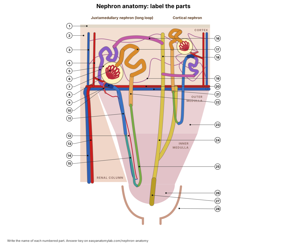
Show the answer key (1–28)
- Renal capsule
- Renal cortex
- Cortical radiate (interlobular) vein
- Cortical radiate (interlobular) artery
- Glomerular (Bowman's) capsule
- Distal convoluted tubule
- Glomerulus
- Afferent arteriole
- Macula densa
- Efferent arteriole
- Thick ascending limb
- Vasa recta
- Interlobar artery
- Interlobar vein
- Ascending thin limb
- Connecting tubule
- Proximal convoluted tubule
- Cortical collecting duct
- Peritubular capillaries
- Arcuate artery
- Arcuate vein
- Proximal straight tubule
- Renal pyramid (medulla)
- Medullary collecting duct
- Descending thin limb
- Renal papilla
- Papillary duct
- Minor calyx
Nephron anatomy quiz
Eight short questions on nephron structure and function. Click each one for the answer.
1. Which part of the nephron is lined by cuboidal cells with a brush border?
The proximal convoluted tubule. The microvilli of the brush border give it a huge surface for reabsorption.
2. Which segment of the loop of Henle is permeable to water but not to salt?
The descending (thin) limb.
3. Which segment pumps NaCl out but is impermeable to water?
The thick ascending limb, using the Na+–K+–2Cl− cotransporter (NKCC2).
4. Name the three parts of the juxtaglomerular apparatus.
The macula densa, the juxtaglomerular (granular) cells and the extraglomerular mesangial cells.
5. Which vessel carries blood out of the glomerulus?
The efferent arteriole. It leads to the peritubular capillaries (cortical nephrons) or the vasa recta (juxtamedullary nephrons).
6. Put these in order: arcuate artery, segmental artery, afferent arteriole, interlobar artery, cortical radiate artery.
Segmental → interlobar → arcuate → cortical radiate → afferent arteriole.
7. Which hormone inserts aquaporin-2 into collecting duct cells?
ADH (antidiuretic hormone, vasopressin), from the posterior pituitary.
8. Which nephrons have long loops and make the vasa recta?
Juxtamedullary nephrons, about 15% of the total.
Frequently asked questions
What are the parts of a nephron?
A nephron has two main parts: the renal corpuscle (the glomerulus inside the glomerular, or Bowman's, capsule) and the renal tubule. In order, the tubule is the proximal convoluted tubule, the nephron loop (loop of Henle: descending limb, thin ascending limb in long loops, and thick ascending limb), and the distal convoluted tubule, which drains through a connecting tubule into a collecting duct. The collecting duct is usually counted as part of the collecting system, not the nephron.
What is the function of the nephron?
The nephron makes urine and so controls the volume and composition of the blood. It does this in three steps: glomerular filtration of plasma into the capsule, tubular reabsorption of the useful parts (water, salts, glucose, amino acids) back into the blood, and tubular secretion of extra waste, H+ and K+ into the tubule.
How many nephrons are in a kidney?
On average about 900,000 per kidney, but the number varies widely between healthy people: autopsy studies have counted from about 210,000 to 2,700,000 per kidney (Charlton et al. 2020). The often-quoted 'one million' is a rounded average. No new nephrons form after birth.
Where are nephrons located in the kidney?
Every renal corpuscle and both convoluted tubules are in the renal cortex. The loops of Henle dip into the renal medulla (pyramids): short loops of cortical nephrons only reach the outer medulla, while long loops of juxtamedullary nephrons reach deep into the inner medulla. Collecting ducts run down through the pyramids to the papilla.
What is the difference between cortical and juxtamedullary nephrons?
Cortical nephrons (about 85%) have glomeruli in the outer cortex, short loops of Henle that stay in the outer medulla, and efferent arterioles that feed peritubular capillaries. Juxtamedullary nephrons (about 15%) have glomeruli next to the medulla, long loops reaching the inner medulla, and efferent arterioles that form the vasa recta. Juxtamedullary nephrons build the gradient used to concentrate urine.
What does the loop of Henle do?
The loop of Henle creates a salt gradient in the medulla so the kidney can make urine more concentrated than blood. The descending limb lets water out but not salt; the ascending limb pumps salt out (NKCC2 in the thick part) but keeps water in. This countercurrent multiplier raises the medullary interstitium from about 300 to about 1,200 mOsm/kg.
Which part of the nephron reabsorbs the most?
The proximal convoluted tubule. It reabsorbs about two-thirds of the filtered water and sodium, almost all of the glucose and amino acids, and most of the bicarbonate.
What is the juxtaglomerular apparatus?
It is the contact point between the end of the thick ascending limb and its own glomerulus's arterioles. It has three parts: the macula densa (salt-sensing tubule cells), the juxtaglomerular or granular cells (renin-secreting cells in the afferent arteriole wall), and the extraglomerular mesangial cells. It controls GFR and starts the renin–angiotensin–aldosterone system.
How do ADH and aldosterone act on the nephron?
ADH (vasopressin) from the posterior pituitary inserts aquaporin-2 water channels into principal cells of the collecting duct, so more water is reabsorbed and urine gets concentrated. Aldosterone from the adrenal cortex acts on the late distal tubule, connecting tubule and collecting duct to increase Na+ reabsorption (ENaC, Na+/K+-ATPase) and K+ secretion.
References
- OpenStax. Anatomy and Physiology 2e, section 25.3: Gross Anatomy of the Kidney. Rice University.
- OpenStax. Anatomy and Physiology 2e, section 25.4: Microscopic Anatomy of the Kidney. Rice University.
- OpenStax. Anatomy and Physiology 2e, section 25.5: Physiology of Urine Formation. Rice University.
- OpenStax. Anatomy and Physiology 2e, section 25.6: Tubular Reabsorption. Rice University.
- OpenStax. Anatomy and Physiology 2e, section 25.7: Regulation of Renal Blood Flow. Rice University.
- OpenStax. Anatomy and Physiology 2e, section 25.8: Endocrine Regulation of Kidney Function. Rice University.
- Curthoys, N. P. & Moe, O. W. (2014). Proximal tubule function and response to acidosis. Clinical Journal of the American Society of Nephrology 9(9), 1627–1638.
- Subramanya, A. R. & Ellison, D. H. (2014). Distal convoluted tubule. Clinical Journal of the American Society of Nephrology 9(12), 2147–2163.
- Roy, A., Al-bataineh, M. M. & Pastor-Soler, N. M. (2015). Collecting duct intercalated cell function and regulation. Clinical Journal of the American Society of Nephrology.
- Marcoux, A.-A. et al. (2021). Molecular characteristics and physiological roles of Na+–K+–Cl− cotransporter 2. Journal of Cellular Physiology 236(3), 1712–1729.
- Sands, J. M. & Layton, H. E. (2009). The physiology of urinary concentration: an update. Seminars in Nephrology 29(3), 178–195.
- Charlton, J. R., Baldelomar, E. J., Hyatt, D. M. & Bennett, K. M. (2021). Nephron number and its determinants: a 2020 update. Pediatric Nephrology 36(4), 797–807.
- Edwards, A., Silldorff, E. P. & Pallone, T. L. (2000). The renal medullary microcirculation. Frontiers in Bioscience 5, D36–52.
- Kidney Pathology. Tubules: histology (normal renal tubules and collecting ducts).
- Digital Histology, Virginia Commonwealth University. Renal papilla.
More interactive anatomy models
- Action potential diagramDepolarization, repolarization and ion channels of a neuron action potential.
- Skin layersEpidermis, dermis and hypodermis with hair follicles, glands and receptors, in 3D.
- Sarcomere diagramZ discs, A and I bands, H zone, actin and myosin of a sarcomere, in 3D.
- Heart blood flowStep-by-step path of blood through the heart's chambers and valves, in 3D.
- Human anatomy1,000+ bones, muscles, organs, vessels and nerves from real anatomical data.
- Octopus anatomyThree hearts, a ring-shaped brain, eight arms and a beak, in 3D.