

Euglena diagram: the essentials
A Euglena diagram shows a single spindle-shaped cell with a flagellum at the front, a red eyespot beside a pocket called the reservoir, a contractile vacuole, many green chloroplasts, white paramylon granules, a central nucleus and a flexible outer pellicle. These are the parts every school and university diagram asks you to label.
Euglena is a genus of protists: single-celled eukaryotes that are neither plants, animals nor fungi. It lives in fresh water, especially in ponds and ditches rich in decaying organic matter, where dense populations can turn the water green. This page describes Euglena gracilis, the best-studied species and the one used in most laboratories. Where Euglena viridis, the type species of the genus and the one in many older textbooks, differs, the text says so. (Zoltner & Field (2022): Microbe Profile: Euglena gracilis, Microbiology; Connecticut College Silica Secchi Disk key: Euglenoids.)
The name means "beautiful eyeball", after the orange-red eyespot. Antonie van Leeuwenhoek probably saw Euglena among his "animalcules" in lake water in the late 17th century, and Christian Gottfried Ehrenberg named the genus around 1830. (Zoltner & Field (2022): Microbe Profile: Euglena gracilis, Microbiology.)
Labeled diagram of a Euglena
A labeled diagram of a Euglena names, from front to back: the long flagellum with its fine hairs (mastigonemes); the canal and reservoir at the anterior end; the paraflagellar body, a swelling on the flagellum inside the reservoir; the eyespot beside the reservoir; the short second flagellum and the two basal bodies; the contractile vacuole; then, through the rest of the cell, the chloroplasts with their pyrenoids, the paramylon granules, the nucleus with its large nucleolus, the mitochondria and Golgi bodies, all inside the pellicle.
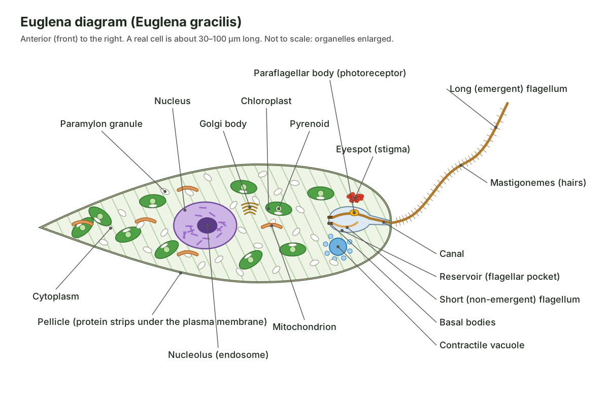
For a labeled diagram of a Euglena with 12 structures, the usual set is: flagellum, canal, reservoir, eyespot, paraflagellar body (photoreceptor), contractile vacuole, chloroplast, pyrenoid, paramylon granule, nucleus, nucleolus and pellicle. The quiz near the end of the page uses exactly these twelve. In the 3D model above, switch on All labels to see every structure named at once, or click any part to zoom to it and read what it does.
Two points are often drawn wrongly. First, the eyespot is not inside a chloroplast: in euglenids it lies free in the cytoplasm next to the reservoir. Second, the canal is not a mouth. Older books call it a cytopharynx or gullet, but photosynthetic Euglena does not swallow food through it. (Leander (2012): Euglenida, Tree of Life Web Project.)
Structure of Euglena and functions
Each part of Euglena has one main job: the flagellum moves the cell, the eyespot and paraflagellar body steer it towards light, the chloroplasts make food, paramylon stores it, the contractile vacuole removes water, the nucleus controls the cell and the pellicle gives it a flexible shape.
| Structure | Where it is | Function |
|---|---|---|
| Pellicle | Under the plasma membrane, all over the cell | Protein strips that hold the shape but let the cell bend; strips slide for metaboly |
| Long (emergent) flagellum | Leaves the canal at the front | Swimming; carries the photoreceptor |
| Short (non-emergent) flagellum | Inside the reservoir | Reduced; no clear role in swimming |
| Basal bodies | At the base of each flagellum | Anchor the flagella |
| Canal | Anterior tip | Opening to the outside for the flagellum and for water leaving the reservoir |
| Reservoir | Behind the canal | Pocket that holds the flagellar bases; receives water from the contractile vacuole |
| Eyespot (stigma) | Beside the reservoir, in the cytoplasm | Shades the photoreceptor so the cell can tell light direction |
| Paraflagellar body | Swelling on the long flagellum in the reservoir | The photoreceptor: detects light |
| Contractile vacuole | Next to the reservoir | Pumps out excess water (osmoregulation) |
| Chloroplasts | Throughout the cell, mostly near the surface | Photosynthesis |
| Pyrenoids | In each chloroplast (E. gracilis) | CO₂ fixation centre; paramylon forms against them |
| Paramylon granules | Cytoplasm | Food store (β-1,3-glucan) |
| Nucleus and nucleolus | Centre of the cell | Controls the cell; nucleolus makes ribosomes |
| Mitochondrion | Network through the cytoplasm | Aerobic respiration (ATP) |
| Golgi bodies and ER | Around the nucleus | Make, process and package proteins and membranes |
Parts of Euglena: every structure in the 3D model
This is the text version of the 3D model: every structure of the Euglena gracilis cell, grouped by system, with where it is and what it does. Use it as a study list or to check a Euglena labeling worksheet.
Pellicle
- Pellicle (Pellicula; often mislabeled as a cell wall)
- The flexible outer covering of Euglena: protein strips lying just beneath the plasma membrane, wound in a spiral around the cell. Holds the cell's spindle shape while still letting it bend. Neighbouring strips can slide against each other, which lets the cell squirm and change shape (metaboly).
- Plasma membrane (Cell membrane)
- The outermost layer of the cell, a lipid bilayer that covers the pellicle strips. Controls what enters and leaves the cell, and carries the channels and pumps that control its salt and water balance.
- Pellicular microtubules (Subpellicular microtubules)
- Rows of microtubules that run along the underside of the pellicle strips. Part of the cytoskeleton that supports the strips and is thought to take part in euglenoid movement.
Flagella & reservoir
- Canal (Cytopharynx or gullet in older textbooks)
- A narrow tube at the front of the cell that leads from the outside into the reservoir. The passage through which the long flagellum leaves the cell and through which the reservoir empties. It is not a mouth: photosynthetic Euglena does not swallow food through it.
- Reservoir (Flagellar pocket; ampulla)
- A flask-shaped pocket, an inward fold of the plasma membrane, at the front of the cell. Houses the bases of both flagella and the paraflagellar body, and receives the water pumped out by the contractile vacuole before it leaves through the canal.
- Long (emergent) flagellum (Locomotory flagellum; dorsal flagellum)
- The one long, whip-like flagellum that sticks out of the canal and pulls the cell along. Beats to swim. It also carries the paraflagellar body, so it is part of the light-steering system.
- Short (non-emergent) flagellum (Second flagellum; ventral flagellum)
- A second, very short flagellum that never leaves the reservoir. It has no clear role in swimming. It is a reduced flagellum, a leftover of the two-flagella body plan of euglenids.
- Basal bodies (Kinetosomes; blepharoplasts in older books)
- Two short cylinders of microtubules that anchor the two flagella. Anchor each flagellum and organise the growth of its axoneme.
- Paraflagellar rod (PFR; paraxial rod)
- A lattice-like rod that runs alongside the microtubules inside the long flagellum. Stiffens the flagellum and supports its signalling; it also connects the axoneme to the paraflagellar body.
- Mastigonemes (Flagellar hairs)
- Very fine hairs that cover the long flagellum. Thought to change how the beating flagellum pushes on the water, helping to pull the cell in the direction of the flagellar tip.
Eyespot & photoreceptor
- Eyespot (stigma) (Stigma)
- A cluster of orange-red carotenoid granules beside the reservoir, near the base of the long flagellum. Acts as a shade. As the cell rotates while it swims, the eyespot shades the paraflagellar body on and off when light comes from the side, which tells the cell which way the light is.
- Paraflagellar body (photoreceptor) (PFB; paraflagellar swelling; photoreceptor)
- A small swelling on the long flagellum inside the reservoir. It is the cell's light sensor. Detects light. Its photoreceptor protein makes the signal molecule cyclic AMP when lit, which changes how the flagellum beats and steers the cell towards or away from light.
Contractile vacuole
- Contractile vacuole
- A small bubble beside the reservoir that fills with water and then squeezes it out into the reservoir. Osmoregulation. Fresh water keeps leaking into the cell by osmosis; the contractile vacuole collects it and pumps it out so the cell does not swell and burst.
- Accessory vacuoles (Contractile vacuole complex)
- Tiny vesicles around the contractile vacuole that collect water and merge into it. Gather water from the cytoplasm and fuse to refill the contractile vacuole after each emptying.
Chloroplasts & paramylon
- Chloroplasts (Plastids; chromatophores in older books)
- Many green, disc-shaped chloroplasts that carry out photosynthesis. Photosynthesis: they capture light with chlorophylls a and b and use it to make sugar from carbon dioxide and water.
- Pyrenoids
- Dense protein regions inside the chloroplasts where carbon dioxide is fixed. Concentrate the CO₂-fixing enzyme RuBisCO; paramylon is laid down against them, outside the chloroplast.
- Paramylon granules (Paramylum; paramylon bodies; reserve granules)
- White grains of paramylon, the food store of Euglena, scattered through the cytoplasm. Store glucose made by photosynthesis, to be used in the dark or when food is short.
Nucleus
- Nucleus (Nuclear envelope)
- The large round-to-oval nucleus in the middle of the cell, wrapped in a double nuclear envelope with pores. Holds the cell's chromosomes and controls the cell. It divides by mitosis before the cell splits.
- Nucleolus (endosome) (Endosome (in euglenid and protist literature))
- A large, dense body in the centre of the nucleus. Makes ribosomal RNA and assembles ribosome subunits.
- Chromosomes (Condensed chromatin)
- Thread-like chromosomes that stay condensed even when the cell is not dividing. Carry the genes.
Cytoplasm & organelles
- Cytoplasm
- The jelly-like inside of the cell in which the organelles sit. Where most of the cell's chemistry happens. The whole cytoplasm also acts as the cell's gravity sensor by pressing on the lower membrane.
- Mitochondrion (Mitochondrial reticulum)
- A branching network of tubes that makes ATP by aerobic respiration. Aerobic respiration: releases energy from sugar as ATP, in the light and in the dark.
- Golgi bodies (Dictyosomes)
- Stacks of flattened membrane sacs that package proteins and membrane. Modify, sort and package proteins and lipids, for example membrane for the reservoir and mucus.
- Endoplasmic reticulum (ER)
- A network of membrane sheets and tubes, joined to the nuclear envelope. Makes proteins (rough ER) and lipids, and carries them to the Golgi.
Euglena cell structure: pellicle, reservoir and flagella
The cell surface of Euglena is a pellicle: a set of protein strips lying just beneath the plasma membrane and linked to microtubules underneath. The strips are made of euglenid-specific proteins called articulins, and their number is characteristic of each species. Euglena has no cell wall. (Zoltner & Field (2022): Microbe Profile: Euglena gracilis, Microbiology.) In photosynthetic euglenids the strip count ranges from about 16 to about 120, and species with more than about 20 strips can usually change shape. (Leander (2012): Euglenida, Tree of Life Web Project.)
- Pellicle: Holds the cell's spindle shape while still letting it bend. Neighbouring strips can slide against each other, which lets the cell squirm and change shape (metaboly).
- Plasma membrane: Controls what enters and leaves the cell, and carries the channels and pumps that control its salt and water balance.
- Pellicular microtubules: Part of the cytoskeleton that supports the strips and is thought to take part in euglenoid movement.
At the front, the plasma membrane folds inward to form the flask-shaped reservoir (flagellar pocket), which opens to the outside through the narrow canal. Two flagella rise from basal bodies at the floor of the reservoir. Only one, the long emergent flagellum, leaves the cell; the other is short and stays inside. (Häder & Hemmersbach (2022): Euglena, a gravitactic flagellate of multiple usages, Life; Hammond et al. (2021): The distinctive flagellar proteome of Euglena gracilis, New Phytologist.)
- Canal: The passage through which the long flagellum leaves the cell and through which the reservoir empties. It is not a mouth: photosynthetic Euglena does not swallow food through it.
- Reservoir: Houses the bases of both flagella and the paraflagellar body, and receives the water pumped out by the contractile vacuole before it leaves through the canal.
- Long (emergent) flagellum: Beats to swim. It also carries the paraflagellar body, so it is part of the light-steering system.
- Short (non-emergent) flagellum: It has no clear role in swimming. It is a reduced flagellum, a leftover of the two-flagella body plan of euglenids.
- Basal bodies: Anchor each flagellum and organise the growth of its axoneme.
- Paraflagellar rod: Stiffens the flagellum and supports its signalling; it also connects the axoneme to the paraflagellar body.
- Mastigonemes: Thought to change how the beating flagellum pushes on the water, helping to pull the cell in the direction of the flagellar tip.
Like all eukaryotic flagella, each Euglena flagellum has a 9 + 2 axoneme of microtubules. Beside it, the long flagellum also contains a paraflagellar rod, a lattice that runs beside the axoneme, and is covered with about 10,000 mastigonemes, fine hairs of two lengths. (Hammond et al. (2021): The distinctive flagellar proteome of Euglena gracilis, New Phytologist; Häder & Hemmersbach (2022): Euglena, a gravitactic flagellate of multiple usages, Life.) Note that some textbooks, including OpenStax Biology 2e, say euglenoids swim with "two long flagella". That is true of some euglenids, but in Euglena only one flagellum emerges. (OpenStax Biology 2e, 23.3 Groups of protists.)
How does Euglena move? Flagellar swimming and metaboly
Euglena moves in two ways. It swims by beating its long flagellum, and it crawls or squeezes by metaboly (euglenoid movement), in which the pellicle strips slide past each other so the cell changes shape like a worm.
Flagellar swimming. The long flagellum points forward from the front end and beats so that it pulls the cell along. The mastigonemes are thought to help propel the cell in the direction of the flagellar tip. As it swims, the cell rotates around its long axis, so its path through the water is a corkscrew (helix). (Hammond et al. (2021): The distinctive flagellar proteome of Euglena gracilis, New Phytologist; Häder & Hemmersbach (2022): Euglena, a gravitactic flagellate of multiple usages, Life.)
Metaboly (euglenoid movement). Neighbouring pellicle strips can slide against one another, supported by the microtubules beneath them, so the cell rounds up at one end, a bulge travels backwards, and the cell stretches out again. Euglena uses metaboly mainly in confined spaces, such as mud or crowded water. (Zoltner & Field (2022): Microbe Profile: Euglena gracilis, Microbiology.) In species such as Euglena mutabilis, where neither flagellum leaves the reservoir, metaboly and gliding are the only ways to move. (Häder & Hemmersbach (2022): Euglena, a gravitactic flagellate of multiple usages, Life.)
| Flagellar swimming | Metaboly (euglenoid movement) | |
|---|---|---|
| Driven by | Beating of the long flagellum | Sliding of pellicle strips |
| Cell shape | Stays spindle-shaped | Changes constantly: bulges, rounds and stretches |
| Used | Open water | Tight spaces, sediment, crowded cultures |
| Path | Helical (corkscrew), rotating | Slow, crawling |
Compare this with the very different movement of other single cells: the pseudopodia of the amoeba, and the rotary motor of a bacterial flagellum in the bacterial cell diagram.
Euglena eyespot and phototaxis
Euglena steers towards light with two structures that work as a pair: the eyespot (stigma), a cluster of orange-red carotenoid granules beside the reservoir, and the paraflagellar body, a swelling on the long flagellum next to it. The paraflagellar body is the light detector. The eyespot is a shade that is not light-sensitive itself. (Häder & Hemmersbach (2022): Euglena, a gravitactic flagellate of multiple usages, Life; Zoltner & Field (2022): Microbe Profile: Euglena gracilis, Microbiology.)
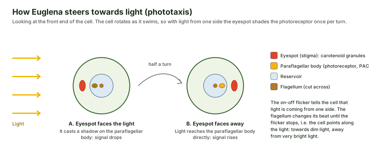
Because the cell rotates as it swims, light coming from one side is blocked by the eyespot once in every turn, so the signal from the paraflagellar body flickers. The cell changes its flagellar beat until the flicker stops, which happens when it is pointing along the light. At low light intensity cells swim towards the light (positive phototaxis); in very strong light they swim away (negative phototaxis), which protects their chloroplasts. Colourless mutants that lack the paraflagellar body cannot do this. (Häder & Hemmersbach (2022): Euglena, a gravitactic flagellate of multiple usages, Life.)
- Eyespot (stigma): Acts as a shade. As the cell rotates while it swims, the eyespot shades the paraflagellar body on and off when light comes from the side, which tells the cell which way the light is.
- Paraflagellar body (photoreceptor): Detects light. Its photoreceptor protein makes the signal molecule cyclic AMP when lit, which changes how the flagellum beats and steers the cell towards or away from light.
Euglena vs Chlamydomonas eyespot. In the green alga Chlamydomonas, the eyespot is made of layers of carotenoid granules inside the chloroplast, under photoreceptors in the plasma membrane. (Mittelmeier et al. (2013): Independent localization of plasma membrane and chloroplast components during eyespot assembly, Eukaryotic Cell.) In euglenids the photoreceptive apparatus is extra-plastidic, that is, outside the chloroplast; some colourless euglenids have an eyespot but never had chloroplasts. (Leander (2012): Euglenida, Tree of Life Web Project.) So a Euglena diagram that draws the eyespot inside a chloroplast is wrong.
Chloroplasts, pyrenoids and paramylon
Euglena gracilis has several disc-shaped green chloroplasts, each with a pyrenoid, spread through the cytoplasm. They contain chlorophylls a and b, like plant chloroplasts, but are surrounded by three membranes instead of two, because they came from a green alga taken in by an ancestral euglenid (secondary endosymbiosis). Their thylakoids are stacked in threes. (Häder & Hemmersbach (2022): Euglena, a gravitactic flagellate of multiple usages, Life; Leander (2012): Euglenida, Tree of Life Web Project.) The captured alga was probably related to the Pyramimonadales, a group of prasinophyte green algae. (Jackson, Knoll, Chan & Verbruggen (2018): Plastid phylogenomics and the origins of secondary green plastids, Scientific Reports.)
- Chloroplasts: Photosynthesis: they capture light with chlorophylls a and b and use it to make sugar from carbon dioxide and water.
- Pyrenoids: Concentrate the CO₂-fixing enzyme RuBisCO; paramylon is laid down against them, outside the chloroplast.
- Paramylon granules: Store glucose made by photosynthesis, to be used in the dark or when food is short.
Pyrenoid. In the Euglena gracilis cell, the pyrenoids are rich in the CO₂-fixing machinery: antibodies to RuBisCO activase stain them strongly. (Konupková, Peña-Diaz & Hampl (2025): Visualisation of Euglena gracilis organelles and cytoskeleton using expansion microscopy, Life Science Alliance.) In euglenids with disc-shaped plastids, each pyrenoid bulges towards the cell centre and is capped on its cytoplasmic side by a grain of paramylon. (Brown, Zakryś & Farmer (2003): Plastid morphology, ultrastructure and development in Colacium and the loricate euglenophytes, Journal of Phycology.)
Paramylon bodies. Paramylon is the storage carbohydrate of euglenids, a β-1,3-glucan found in no other group. It is laid down as granules in the cytoplasm and can reach 85% of the cell's dry weight. (Zoltner & Field (2022): Microbe Profile: Euglena gracilis, Microbiology; Kostygov et al. (2021): Euglenozoa: taxonomy, diversity and ecology, symbioses and viruses, Open Biology.) Plants store starch, which is α-1,4-linked and made inside the chloroplast, so finding paramylon outside the chloroplasts is one way to tell a euglenid from a green alga.
Euglena viridis chloroplast. E. viridis is different: each cell has a single, irregular, star-shaped (stellate) chloroplast in the middle, with pyrenoids, and a mass of paramylon grains at its centre, with more grains scattered through the cell. This "axial stellate chloroplast with a paramylon centre" is the main feature used to recognise it. (Wikipedia: Euglena viridis (species description, citing John et al., Freshwater Algal Flora of the British Isles).) If your textbook shows one star-shaped chloroplast, it is drawing E. viridis; if it shows many discs, it is drawing E. gracilis or a similar species. Compare the two-membraned plant organelle in our chloroplast structure model.
Euglena nutrition: is Euglena a plant or an animal?
Euglena is mixotrophic: in the light it is a photoautotroph that makes its own food by photosynthesis, and in the dark it becomes a heterotroph that lives on organic substances dissolved in the water. In the dark its chloroplasts shrink and stop working for a while. (OpenStax Biology 2e, 23.3 Groups of protists; Zoltner & Field (2022): Microbe Profile: Euglena gracilis, Microbiology.)
Photosynthetic euglenids have only a highly reduced feeding apparatus, unlike their relatives that hunt bacteria or other cells, so heterotrophic Euglena mainly absorbs dissolved nutrients rather than swallowing particles. (Leander (2012): Euglenida, Tree of Life Web Project.) Ponds rich in decaying organic matter, which release sugars, fatty acids and vitamins B1 and B12, suit it well. (Connecticut College Silica Secchi Disk key: Euglenoids.)
The plant-or-animal debate. Euglena photosynthesises like a plant but has no cell wall, swims and can feed heterotrophically like an animal. Early naturalists working with Linnaeus's two kingdoms could not place it: zoologists classed it with the protozoa and botanists with the algae, and euglenids are still named under both the zoological and the botanical codes. It was one of the organisms that led Ernst Haeckel to propose a third kingdom, Protista, in the 1860s.
Modern classification. Euglena belongs to the euglenids (Euglenida, class Euglenoidea or Euglenophyceae) in the phylum Euglenozoa. Euglenozoa also contains the kinetoplastids, such as the trypanosomes that cause sleeping sickness, and the diplonemids. (Zoltner & Field (2022): Microbe Profile: Euglena gracilis, Microbiology; Kostygov et al. (2021): Euglenozoa: taxonomy, diversity and ecology, symbioses and viruses, Open Biology.) Euglenozoa, Heterolobosea and the jakobids form a major clade named Discoba, traditionally placed in the supergroup Excavata. (Hampl et al. (2009): Phylogenomic analyses support the monophyly of Excavata, PNAS.) Whether Excavata is one natural group is still debated, so many newer schemes simply use Discoba.
| Rank | Name |
|---|---|
| Domain | Eukaryota |
| Supergroup | Excavata (Discoba) |
| Phylum | Euglenozoa |
| Class | Euglenoidea (Euglenophyceae) |
| Order | Euglenales |
| Family | Euglenaceae |
| Genus | Euglena |
| Species | Euglena gracilis Klebs; Euglena viridis Ehrenberg (type species) |
Osmoregulation in Euglena: the contractile vacuole
Euglena controls its water content with a contractile vacuole beside the reservoir. Fresh water has fewer dissolved particles than the cytoplasm, so water constantly enters the cell by osmosis. With no cell wall to resist the pressure, the cell would swell and burst unless the water were pumped out.
Small accessory vacuoles collect water from the cytoplasm and fuse to form the contractile vacuole, which then empties into the reservoir; the water leaves the cell through the canal. Freshwater euglenoids have a contractile vacuole, and so do other freshwater protists such as Paramecium. (Connecticut College Silica Secchi Disk key: Euglenoids; OpenStax Microbiology, 5.1 Unicellular eukaryotic parasites.)
- Contractile vacuole: Osmoregulation. Fresh water keeps leaking into the cell by osmosis; the contractile vacuole collects it and pumps it out so the cell does not swell and burst.
- Accessory vacuoles: Gather water from the cytoplasm and fuse to refill the contractile vacuole after each emptying.
Euglena reproduction: longitudinal binary fission
Euglena reproduces asexually by longitudinal binary fission: the nucleus divides first, then the cell splits lengthwise, starting at the front end and finishing at the tail, to give two daughter cells. Sexual reproduction has never been observed. (Häder & Hemmersbach (2022): Euglena, a gravitactic flagellate of multiple usages, Life.)
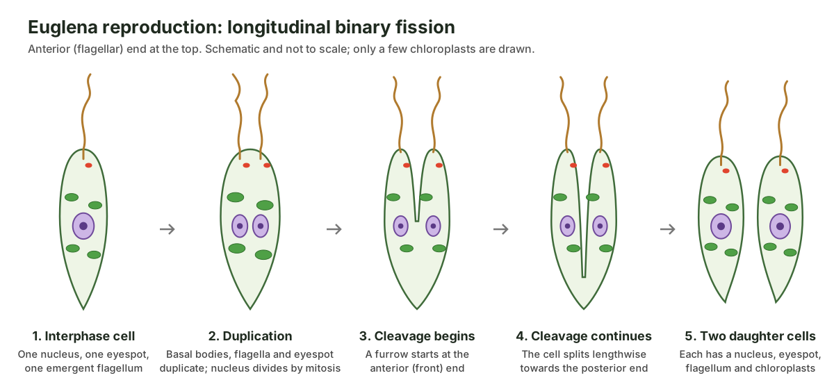
- Duplication. The basal bodies, flagellar apparatus and pellicle strips are duplicated. The eyespot disappears during division and two new ones form, one for each daughter. (Häder & Hemmersbach (2022): Euglena, a gravitactic flagellate of multiple usages, Life; Connecticut College Silica Secchi Disk key: Euglena reproduction.)
- Mitosis. The nucleus divides by mitosis. Because euglenid chromosomes are permanently condensed, they can often be seen with a light microscope. (Leander (2012): Euglenida, Tree of Life Web Project; Connecticut College Silica Secchi Disk key: Euglena reproduction.)
- Cleavage. A furrow forms at the anterior end and runs back along the cell, so a dividing cell looks "two-headed" until it finishes. (Connecticut College Silica Secchi Disk key: Euglena reproduction.)
- Separation. The two daughter cells separate. Each has a nucleus, an eyespot, a flagellar apparatus and a share of the chloroplasts, which go on dividing as the cell grows. (Brown, Zakryś & Farmer (2003): Plastid morphology, ultrastructure and development in Colacium and the loricate euglenophytes, Journal of Phycology.)
This is different from bacterial binary fission, which has no nucleus and no mitosis, and from the transverse (crosswise) fission of Paramecium.
Euglena life cycle: palmella stage and cysts
The life cycle of Euglena is simple: an active swimming cell grows and divides by binary fission, over and over. When conditions turn bad it can switch to one of two resting forms, the palmella stage or a cyst, and switch back when conditions improve. There is no sexual stage.
- Palmella stage. Cells lose or stop using their flagella, become embedded in mucilage (slime) and go on dividing inside it, forming a jelly-like mass of cells. (Connecticut College Silica Secchi Disk key: Euglena reproduction.)
- Resting cyst. When nutrients are short, cells round up, lose their flagella, store more paramylon and surround themselves with a protective mucilaginous wall. The cyst can survive until conditions improve, then the cell emerges and swims again. (Connecticut College Silica Secchi Disk key: Euglena reproduction.)
Cysts help explain why Euglena appears quickly in puddles and new ponds. Encystment, the process of forming a cyst, is common among protists generally. (OpenStax Microbiology, 5.1 Unicellular eukaryotic parasites.)
Euglena vs amoeba vs paramecium vs Chlamydomonas
Euglena, amoeba, paramecium and Chlamydomonas are the four single-celled organisms most often compared in school biology. Euglena moves with one flagellum and photosynthesises; amoeba crawls with pseudopodia; paramecium swims with cilia; Chlamydomonas swims with two flagella and is a true green alga with a cell wall.
| Feature | Euglena | Amoeba | Paramecium | Chlamydomonas |
|---|---|---|---|---|
| Group | Euglenozoa (euglenid) | Amoebozoa | Ciliate (alveolate) | Green alga (Chlorophyta) |
| Shape | Spindle; flexible | No fixed shape | Slipper-shaped, fixed | Oval to pear-shaped, fixed |
| Outer covering | Pellicle of protein strips; no wall | Plasma membrane only | Pellicle | Cell wall |
| Movement | One long flagellum; metaboly | Pseudopodia | Thousands of cilia | Two equal flagella at the front |
| Nutrition | Mixotrophic | Heterotrophic (phagocytosis) | Heterotrophic, via an oral groove | Photoautotrophic |
| Chloroplasts | Many, three membranes | None | None | One cup-shaped, two membranes |
| Eyespot | Yes, outside the chloroplast | No | No | Yes, inside the chloroplast |
| Food store | Paramylon | Glycogen and food vacuoles | Glycogen and food vacuoles | Starch, in the chloroplast |
| Contractile vacuole | Yes, by the reservoir | Yes | Yes, usually two | Yes, usually two |
| Nucleus | One, with a large nucleolus | One | Macronucleus and micronucleus | One |
The amoeba, paramecium and Euglena features are from OpenStax. (OpenStax Microbiology, 5.1 Unicellular eukaryotic parasites; OpenStax Biology 2e, 23.3 Groups of protists.) The Chlamydomonas eyespot sits in the chloroplast at the cell's equator, near its two anterior flagella. (Mittelmeier et al. (2013): Independent localization of plasma membrane and chloroplast components during eyespot assembly, Eukaryotic Cell.) See the structures of the amoeba in 3D in our amoeba diagram.
Euglena vs Spirogyra. Spirogyra, another pond organism often drawn alongside Euglena, is a filamentous green alga: a chain of walled cells, each with ribbon-shaped chloroplasts wound in a spiral, and no flagella or eyespot. Euglena is a single, wall-less, swimming cell.
Euglena gracilis vs Euglena viridis and other species
The genus Euglena contains many species. The two in textbooks are E. gracilis, the laboratory model, and E. viridis, the type species and a common indicator of polluted, organic-rich water.
| Feature | Euglena gracilis | Euglena viridis | Euglena sanguinea |
|---|---|---|---|
| Chloroplasts | Several, disc-shaped, each with a pyrenoid | One central star-shaped (stellate) chloroplast | Several; cells contain red astaxanthin |
| Paramylon | Many granules through the cytoplasm | Mass at the centre of the chloroplast, plus scattered grains | Granules in the cytoplasm |
| Typical length | About 20–100 µm, depending on shape | About 40–65 µm | Varies |
| Notes | Model organism; easy to culture | Type species; common in organic-rich water | Red blooms; some strains make euglenophycin |
Sources: E. gracilis size and features from Zoltner & Field; E. viridis description and size from the Wikipedia species account; E. sanguinea pigment from the US Geological Survey. (Zoltner & Field (2022): Microbe Profile: Euglena gracilis, Microbiology; Wikipedia: Euglena viridis (species description, citing John et al., Freshwater Algal Flora of the British Isles); US Geological Survey: Euglena sanguinea bloom.) Euglenoids as a group number around 800–900 species in about 40 or more genera, most of them in fresh water; about a third of the genera are photosynthetic. (Zoltner & Field (2022): Microbe Profile: Euglena gracilis, Microbiology; Connecticut College Silica Secchi Disk key: Euglenoids.)
Euglena under the microscope
Under a light microscope, Euglena looks like a small, green, spindle-shaped cell that swims in a spiral and sometimes stops to squirm and change shape. At 100× you can see it swimming; at 400× you can usually make out the red eyespot at the front, the green chloroplasts, colourless paramylon grains, and sometimes the clear reservoir and the pulsing contractile vacuole. The flagellum is hard to see without phase contrast or a stain because it is so thin.
- Find it: look in a drop of green pond water or a purchased culture. Cells gather at the lit side of the container because of phototaxis.
- Slow it down: a drop of methyl cellulose or a thin wisp of cotton slows fast swimmers.
- Tell it from Chlamydomonas: Chlamydomonas is smaller and rounder, has two flagella and does not change shape. Euglena has one visible flagellum and shows metaboly.
- Size: a Euglena gracilis cell is a few tens of micrometres long, several times smaller than a large Amoeba proteus.
What you can see in the light microscope matches the school diagram. The mastigonemes, paraflagellar rod, pellicle strips and organelle membranes in the 3D model are electron-microscope details.
How to draw a Euglena: a simple labeled diagram
To draw a simple Euglena diagram, draw a long spindle shape, rounded at the front and tapering to a point at the back, then add a long wavy flagellum from the front, a red eyespot and a small bubble-like reservoir at the front end, green oval chloroplasts through the cell, a round nucleus in the middle and a few white paramylon granules.
- Draw the outline: a spindle about three to five times longer than it is wide. Label it pellicle.
- At the front end, draw a small flask-shaped reservoir joined to the tip by a narrow canal.
- Draw the flagellum from inside the reservoir, out through the canal, about as long as the cell.
- Add the red eyespot beside the reservoir, not inside a chloroplast, and the contractile vacuole next to it.
- Draw a round nucleus near the middle with a dark nucleolus.
- Fill the cell with green chloroplasts and small white paramylon granules.
- Rule label lines that do not cross, write labels outside the cell, and add a title and magnification.
Euglena for Class 9 and KS3: simple summary
In simple terms, Euglena is a one-celled organism that lives in ponds. It has a long whip-like flagellum to swim, a red eyespot to find light, chloroplasts to make food by photosynthesis, a contractile vacuole to remove extra water and a nucleus to control the cell. It can make its own food in light and absorb food in the dark, so it shows features of both plants and animals.
- Kingdom: Protista (in the five-kingdom scheme used in many school books).
- Movement: flagellum; also changes shape (euglenoid movement).
- Nutrition: autotrophic in light, heterotrophic in the dark (mixotrophic).
- Food store: paramylon, not starch.
- Reproduction: asexual, by longitudinal binary fission.
- Common mistakes: giving it a cell wall, drawing two long flagella, or putting the eyespot inside a chloroplast.
Euglena blooms and Euglena sanguinea
Euglena can multiply into blooms that colour still water green or, in the case of Euglena sanguinea, red. Euglenoids thrive in water with high nutrient levels, so they are used as bioindicators of organic pollution. (Connecticut College Silica Secchi Disk key: Euglenoids.)
The red colour of E. sanguinea comes from the carotenoid pigment astaxanthin in its cells. (US Geological Survey: Euglena sanguinea bloom.) Some strains also make euglenophycin, an alkaloid toxin with a structure similar to solenopsin from fire-ant venom. In the summer of 2002 a North Carolina aquaculture facility lost more than 21,000 striped bass, and researchers later linked further fish kills of striped bass, tilapia and channel catfish to euglenoid blooms. (USDA Agricultural Research Service (2009): Algal toxin kills fish, may also fight cancer.) A later survey found euglenophycin in six of seven E. sanguinea strains and in several other euglenoid species, with E. sanguinea and E. socialis making by far the most. (Zimba et al. (2017): Euglenophycin is produced in at least six species of euglenoid algae, Harmful Algae.)
Euglena blooms are a sign of nutrient-rich, often polluted water. Most Euglena species, including E. gracilis, are harmless to people; members of the genus are typically not pathogenic. (OpenStax Microbiology, 5.1 Unicellular eukaryotic parasites.)
Inside the Euglena photoreceptor: a light-switched enzyme
The light sensor of Euglena is an enzyme, not a pigment spot. The paraflagellar body holds photoactivated adenylyl cyclase (PAC), a 400 kDa complex that absorbs blue light with bound flavin molecules and, when lit, makes cyclic AMP (cAMP). cAMP is a signal molecule that switches on protein kinase A, which in turn changes how the flagellar motor proteins work. (Zoltner & Field (2022): Microbe Profile: Euglena gracilis, Microbiology.)
Silencing the PAC genes, and studying mutant strains, showed that PAC makes cells swim faster when they meet strong light (photoavoidance), while PAC in the paraflagellar body, working with pterins made by the enzyme GTP cyclohydrolase I, coordinates both positive and negative phototaxis. Each PAC complex contains two α and two β subunits. (Hammond et al. (2021): The distinctive flagellar proteome of Euglena gracilis, New Phytologist.)
Why the Euglena chloroplast has three membranes
A plant chloroplast has two envelope membranes because it descends from a cyanobacterium swallowed by an early eukaryote (primary endosymbiosis). The Euglena chloroplast has three because it came one step later: an ancestral euglenid, which ate other cells, swallowed a green alga and kept its chloroplast (secondary endosymbiosis). (Leander (2012): Euglenida, Tree of Life Web Project.)
Comparisons of whole chloroplast genomes point to a group of prasinophyte green algae, the order Pyramimonadales, as the closest living relatives of the alga Euglena's ancestor captured. The same studies show that euglenids, chlorarachniophytes and the dinoflagellate Lepidodinium acquired their green plastids in three separate events. (Jackson, Knoll, Chan & Verbruggen (2018): Plastid phylogenomics and the origins of secondary green plastids, Scientific Reports.)
The green plastid is found only in euglenids within the Euglenozoa. Their relatives the kinetoplastids, which include the trypanosomes that cause sleeping sickness, have none. Nuclear genes of both green and red algal origin serve the Euglena plastid, which suggests a long history of borrowing genes from several algal partners. (Zoltner & Field (2022): Microbe Profile: Euglena gracilis, Microbiology.)
Gravitaxis: how a single cell knows which way is up
Euglena also orients to gravity. Its whole cytoplasm, slightly denser than water (about 1.05 g/mL), acts as the sensor: it presses on mechanosensitive TRP channels in the lower cell membrane with a force of roughly 0.6 to 1.1 piconewtons. The resulting calcium influx works through calmodulin, cAMP and protein kinase A to change the flagellar beat. (Häder & Hemmersbach (2022): Euglena, a gravitactic flagellate of multiple usages, Life.)
The direction of the response is not fixed: Häder and Hemmersbach report that it reverses between young and old cultures, with a threshold of about 0.08 to 0.12 g. Combined with phototaxis, gravitaxis helps the cell hold a position in the water column where light is right for photosynthesis. (Häder & Hemmersbach (2022): Euglena, a gravitactic flagellate of multiple usages, Life.)
Euglena gracilis as a model organism
Euglena gracilis is the best-studied euglenid because it is easy to grow. Its nuclear genome is large, estimated at 500 million base pairs or more, with over 36,000 protein-coding genes, much repeated sequence and unusual RNA splicing. Its mitochondrial genome is extremely reduced, coding for only seven proteins. (Zoltner & Field (2022): Microbe Profile: Euglena gracilis, Microbiology.)
The cell can store paramylon up to 85% of its dry weight, and without oxygen it converts its stores into wax esters. These traits, together with vitamins and polyunsaturated fatty acids it accumulates, have made E. gracilis a candidate for food, feed and biofuel production. (Zoltner & Field (2022): Microbe Profile: Euglena gracilis, Microbiology.)
Experiments with E. gracilis were also historically important for plant biology: cells can be made to lose their chloroplasts permanently and survive as colourless heterotrophs, and these 'bleached' strains helped prove that chloroplasts carry their own genetic continuity rather than forming afresh in each cell. (Brown, Zakryś & Farmer (2003): Plastid morphology, ultrastructure and development in Colacium and the loricate euglenophytes, Journal of Phycology.)
Label the Euglena: unlabeled diagram and answer key
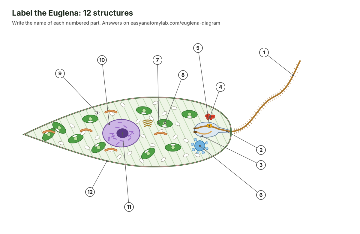
Test yourself with this blank Euglena diagram. Write the name of each of the 12 numbered parts, then open the answer key. For a quick quiz, turn off the labels in the 3D model, click parts at random and name them before you read the panel.
Show the answer key
- Long (emergent) flagellum
- Canal
- Reservoir (flagellar pocket)
- Eyespot (stigma)
- Paraflagellar body (photoreceptor)
- Contractile vacuole
- Chloroplast
- Pyrenoid
- Paramylon granule
- Nucleus
- Nucleolus (endosome)
- Pellicle
Short-answer questions:
- Which structure detects light, and which one shades it? (The paraflagellar body detects light; the eyespot shades it.)
- Why does Euglena need a contractile vacuole? (Water enters by osmosis from fresh water and must be pumped out.)
- What is the storage carbohydrate of Euglena, and how does it differ from starch? (Paramylon, a β-1,3-glucan stored outside the chloroplasts.)
- How many membranes surround a Euglena chloroplast, and why? (Three, because it came from an engulfed green alga by secondary endosymbiosis.)
- How does Euglena reproduce? (Asexually, by longitudinal binary fission starting at the front end.)
- Name two ways Euglena can move. (Flagellar swimming and metaboly.)
Frequently asked questions
Is Euglena a plant or an animal?
Neither. Euglena is a single-celled protist. It has chloroplasts and photosynthesises like a plant, but it has no cell wall, swims with a flagellum and can live on dissolved food in the dark like an animal cell. For that reason early naturalists put it in both kingdoms, and in 1866 Ernst Haeckel proposed a third kingdom, Protista, for organisms like it. Today it is placed in the Euglenozoa, part of the Discoba group of eukaryotes.
Where is the eyespot of Euglena and what does it do?
The eyespot (stigma) is a cluster of orange-red carotenoid granules at the front of the cell, beside the reservoir and near the base of the long flagellum. It lies in the cytoplasm, not in a chloroplast. It is a shade rather than an eye: the real light sensor is the paraflagellar body on the flagellum, and as the cell rotates the eyespot shades it, so the cell can tell which way the light is coming from.
How does Euglena move?
Euglena swims by beating its single long flagellum, rotating as it goes so that it follows a spiral path. It can also move by metaboly (euglenoid movement): the strips of its flexible pellicle slide past each other so the cell squeezes and stretches like a worm, which is useful in tight spaces.
How does Euglena get its food?
Euglena is mixotrophic. In the light it is a photoautotroph and makes its own food by photosynthesis in its chloroplasts. In the dark it switches to heterotrophy, absorbing dissolved organic substances from the water. It stores extra glucose as paramylon granules.
How does Euglena reproduce?
By longitudinal binary fission: the nucleus divides by mitosis, the flagellar apparatus and eyespot are duplicated, and the cell splits lengthwise from the front end to the back, making two daughter cells. Sexual reproduction has never been confirmed in Euglena. In bad conditions cells can round up into a palmella stage or a resting cyst.
What is the function of the contractile vacuole in Euglena?
It removes excess water. Euglena lives in fresh water, which has fewer dissolved particles than its cytoplasm, so water keeps entering by osmosis. The contractile vacuole collects this water and empties it into the reservoir, from where it leaves through the canal.
What is paramylon?
Paramylon is the storage carbohydrate of Euglena, a β-1,3-linked glucose polymer found only in euglenids. It is kept as white granules in the cytoplasm, outside the chloroplasts. Plants store starch instead, which is α-1,4-linked and is made inside the chloroplast.
Does Euglena have a cell wall?
No. Its shape is held by the pellicle, a set of flexible protein strips just under the plasma membrane. Because the pellicle is inside the membrane and the strips can slide, Euglena can bend and change shape, which a walled cell cannot do.
How big is Euglena?
Euglena gracilis is a few tens of micrometres long. Its length varies with conditions, from about 20 µm when it rounds up in the dark to about 100 µm when stretched out in bright light. That is too small to see clearly with the naked eye but easy to see at 100× to 400× under a light microscope.
Why is Euglena green, and why are some ponds red?
Euglena is green because its chloroplasts contain chlorophylls a and b. Dense blooms of Euglena can turn a pond green. Euglena sanguinea also contains the red carotenoid astaxanthin, so its blooms can turn water red. Some strains of E. sanguinea make a fish-killing toxin, euglenophycin.
References
- Zoltner, M. & Field, M. C. (2022). Microbe Profile: Euglena gracilis: photogenic, flexible and hardy. Microbiology 168(9), 001241.
- Hammond, M., Zoltner, M., Garrigan, J., Butterfield, E., Varga, V., Lukeš, J. & Field, M. C. (2021). The distinctive flagellar proteome of Euglena gracilis illuminates the complexities of protistan flagella adaptation. New Phytologist 232, 1323–1336.
- Häder, D.-P. & Hemmersbach, R. (2022). Euglena, a gravitactic flagellate of multiple usages. Life 12(10), 1522.
- Leander, B. S. (2012). Euglenida: euglenids or euglenoids. Version 10 November 2012. The Tree of Life Web Project.
- Brown, P. J. P., Zakryś, B. & Farmer, M. A. (2003). Plastid morphology, ultrastructure, and development in Colacium and the loricate euglenophytes (Euglenophyceae). Journal of Phycology 39, 115–121.
- Konupková, A., Peña-Diaz, P. & Hampl, V. (2025). Visualisation of Euglena gracilis organelles and cytoskeleton using expansion microscopy. Life Science Alliance 8(4), e202403110.
- Jackson, C., Knoll, A. H., Chan, C. X. & Verbruggen, H. (2018). Plastid phylogenomics with broad taxon sampling further elucidates the distinct evolutionary origins and timing of secondary green plastids. Scientific Reports 8, 1523.
- Kostygov, A. Y. et al. (2021). Euglenozoa: taxonomy, diversity and ecology, symbioses and viruses. Open Biology 11, 200407.
- Hampl, V. et al. (2009). Phylogenomic analyses support the monophyly of Excavata and resolve relationships among eukaryotic 'supergroups'. PNAS 106, 3859–3864.
- Mittelmeier, T. M., Thompson, M. D., Öztürk, E. & Dieckmann, C. L. (2013). Independent localization of plasma membrane and chloroplast components during eyespot assembly. Eukaryotic Cell 12, 1258–1270.
- Zimba, P. V., Huang, I.-S., Gutierrez, D., Shin, W., Bennett, M. S. & Triemer, R. E. (2017). Euglenophycin is produced in at least six species of euglenoid algae and six of seven strains of Euglena sanguinea. Harmful Algae 63, 79–84.
- USDA Agricultural Research Service (2009). News release on euglenophycin and catfish and striped bass kills.
- US Geological Survey. Euglena sanguinea bloom (image and description).
- Clark, M. A., Douglas, M. & Choi, J. (2018). Biology 2e, section 23.3: Groups of protists. OpenStax.
- Parker, N. et al. (2016). Microbiology, section 5.1: Unicellular eukaryotic parasites. OpenStax.
- Connecticut College, Silica Secchi Disk freshwater key: Euglenoids; Euglena reproduction.
More interactive anatomy models
- Bacterial cell diagramLabeled prokaryotic cell: cell wall, membrane, nucleoid, ribosomes, pili and flagellum.
- Amoeba diagramLabeled Amoeba proteus: pseudopodia, nucleus, contractile and food vacuoles.
- Bacteriophage diagramT4 phage structure in 3D: head, collar, tail sheath, baseplate and tail fibres.
- Virus structureCapsid, envelope, spikes and genome of helical, icosahedral and enveloped viruses.
- Human anatomy1,000+ bones, muscles, organs, vessels and nerves from real anatomical data.
- Octopus anatomyThree hearts, a ring-shaped brain, eight arms and a beak, in 3D.