

What are stomata? The stoma at a glance
Stomata (singular stoma) are tiny pores in the epidermis of leaves and young stems. Each pore is surrounded by a pair of guard cells that open and close it. When the pore is open, carbon dioxide diffuses into the leaf for photosynthesis, and oxygen and water vapour diffuse out. When it is closed, the leaf saves water but cannot take in CO₂.
Stomata are microscopic. In broad bean each guard cell is about 40–50 µm long, and the open pore is roughly 8–16 µm wide, a tenth of the width of a human hair. All the pores together cover typically less than 3% of the leaf surface, yet some 98% of the CO₂ a leaf takes up and the water it loses passes through them. (Lawson & Blatt (2014))
Most eudicot leaves have more stomata on the lower surface than on the upper surface. In a typical dicot the guard cells are kidney-shaped (bean-shaped); in grasses they are dumbbell-shaped and flanked by subsidiary cells. (NCERT Biology Class XI, Chapter 6) For the whole leaf around them, see the leaf cross-section.
Labeled diagram of stomata and guard cells
A labeled stomata diagram shows two kidney-shaped guard cells around a central pore, surrounded by epidermal cells. The diagram has two views, because textbooks and exams use both: the surface view you see when you peel the epidermis off a leaf, and a section (T.S.) cut across the middle of the stoma, which shows the walls, the cuticle and the air space underneath.
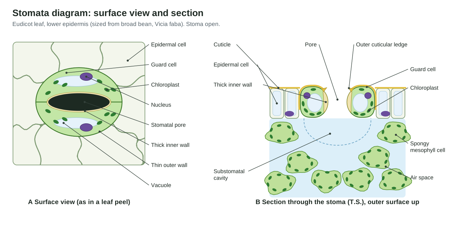
Describe the structure of stomata with a labelled diagram (model answer). A stoma is a pore in the epidermis bounded by two kidney-shaped guard cells (dumbbell-shaped in grasses). The guard cells are joined at their ends. Their inner walls, which line the pore, are thick and inelastic; their outer walls, next to the epidermal cells, are thin and elastic. Each guard cell is a living cell with a nucleus, cytoplasm, a vacuole and chloroplasts, unlike the surrounding epidermal cells, which have few or none. In some plants the guard cells are surrounded by subsidiary cells. The guard cells, the pore and the subsidiary cells together make up the stomatal apparatus. Below the pore is the substomatal cavity, an air space that connects with the air spaces of the spongy mesophyll. The whole surface, including the guard cells, is covered by a waxy cuticle. (NCERT Biology Class XI, Chapter 6)
Parts of a stoma and their functions
Each part of a stoma has a job. The table lists what to label on a stomata diagram and what each part does.
| Part | What it is | Function |
|---|---|---|
| Guard cells | Two curved cells around the pore | Swell to open the pore and shrink to close it |
| Stomatal pore (aperture) | The gap between the guard cells | Lets CO₂ in and O₂ and water vapour out |
| Thick inner (ventral) wall | Wall lining the pore | Strengthens the guard cell where it bends; resists stretching |
| Thin outer (dorsal) wall | Wall next to the epidermal cells | Stretches as the guard cell swells, so the cell curves |
| Cellulose microfibrils | Hoops of cellulose in the wall, radiating from the pore | Stop the cell getting fatter, so it bends instead |
| Chloroplasts | Green plastids in the guard cells | Make ATP, sugars and starch used in stomatal movement |
| Nucleus | Control centre of each guard cell | Controls the cell; responds to signals such as ABA |
| Vacuole | Large sap-filled space | Stores K⁺, Cl⁻ and malate; swells to open the stoma |
| Subsidiary cells | Special epidermal cells beside the guard cells (not in all plants) | Supply and store ions and water; help the guard cells move |
| Epidermal (pavement) cells | The ordinary cells of the epidermis | Protect the leaf; push back on the guard cells |
| Cuticle | Waxy film over the surface | Cuts water loss through the epidermis |
| Substomatal cavity | Air space under the pore | Connects the pore to the leaf's air spaces |
Click any part in the 3D model for its full description. The stomatal apparatus (or stomatal complex) means the pore, the guard cells and any subsidiary cells together.
How stomata open and close
Stomata open when the guard cells take up solutes and water and swell, and close when they lose them and shrink. NCERT’s Class 10 summary is exactly this: the guard cells swell when water flows into them, causing the pore to open; the pore closes if the guard cells shrink. (NCERT Science Class X, Chapter 5) The rest of the story is how the guard cells move the solutes, and why swelling makes them bend apart.
How guard cells open and close the pore
Step 1 of 6
Blue light is detected
At dawn, blue-light receptors in the guard cells (phototropins) absorb light. Red light, used in guard-cell and mesophyll photosynthesis, adds to the signal. The stoma is still closed.
Use the arrow keys or the buttons. The 3D model above follows the same steps.
1. Ions are pumped in: K⁺, Cl⁻ and malate
Opening starts at the plasma membrane. Blue light, red light and the hormone auxin activate a proton pump, the plasma-membrane H⁺-ATPase, which uses ATP to push protons (H⁺) out of the guard cell. This makes the inside of the cell strongly negative (hyperpolarised), which drives potassium ions in through inward K⁺ channels. Chloride ions enter with protons, and the guard cell makes malate, a negatively charged organic acid, partly from starch broken down in its chloroplasts. Later in the day sugars add to the solute load. (Schroeder et al. (2001))
The amounts are large. In broad bean guard cells, vacuolar K⁺ rises from about 38–92 mM when closed to 181–454 mM when open, and the total change in solute often exceeds 300–400 mosmol per litre. (Jezek & Blatt (2017)) Malate can supply 50–90% of the counter-ions for K⁺, depending on how much chloride is available. (Santelia & Lawson (2016))
2. Water follows: water potential and turgor
Dissolved solutes lower the water potential (Ψ) of the guard cells, making it more negative than in the cells around them. Water always moves from higher to lower water potential, so it flows into the guard cells by osmosis. The cell wall resists the swelling protoplast, and the pressure that builds up is turgor pressure. (OpenStax Biology 2e, 30.5 Transport of Water and Solutes in Plants) Guard-cell volume rises from about 2.7–3.7 pL to 4.0–7.5 pL as the stoma opens, and most of that is the vacuole, which makes up 80–90% of an open guard cell. (Jezek & Blatt (2017))
3. The wall makes the cells bend: microfibrils and stiff ends
A swelling cell would normally just get rounder. Guard cells bend instead because of their walls. Cellulose microfibrils wrap round each guard cell like hoops (in surface view they seem to radiate from the pore, which is called radial micellation), so the cell cannot get much fatter. The two cells are also joined and stiffened at their ends, so they cannot simply get longer. Pinned at both ends and unable to bulge, each cell bows outwards as it swells, and the gap between them opens.
The classic textbook explanation adds that the thick inner wall stretches less than the thin outer wall, so the outer side lengthens more and the cell curves away from the pore. Research suggests this is not the whole story. Atomic force microscopy found that young Arabidopsis guard cells open normally before their inner walls are stiffer, while the ends of the stoma are stiff in Arabidopsis, tomato and maize; the authors propose a ‘fix and flex’ model in which pectin-rich, stiff poles matter more than the thick inner wall. (Carter et al. (2017))
4. Closing: ions out, water out
Closing reverses the process. Abscisic acid (ABA), darkness, high CO₂ and pathogen signals raise the free calcium level in the guard-cell cytoplasm. Ca²⁺ blocks the proton pump and the inward K⁺ channels and opens anion channels, so Cl⁻ and malate leave. The membrane depolarises, which opens outward K⁺ channels, and K⁺ leaves too. Water follows the ions out, turgor falls, and the guard cells straighten and close the pore. (Schroeder et al. (2001))
What makes stomata open or close
| Signal | Effect | Why it makes sense |
|---|---|---|
| Light, especially blue light | Opens | Blue light is sensed by phototropins; light means photosynthesis can use CO₂ |
| Low CO₂ inside the leaf | Opens | The leaf needs more CO₂ |
| High CO₂ inside the leaf | Closes | Enough CO₂ is available; saves water |
| Darkness | Closes | No photosynthesis, so no point losing water |
| ABA (drought hormone) | Closes | Made when the plant is short of water |
| Very dry air or heat stress | Usually closes | Cuts transpiration when water loss would be fastest |
The blue-light response saturates at a low light level, around 10 µmol m⁻² s⁻¹, so stomata start opening early in the morning. (Santelia & Lawson (2016)) The old starch–sugar hypothesis (that light makes guard cells turn starch into sugar, lowering their water potential) is no longer accepted as the main mechanism; potassium uptake is. Starch still matters, as a source of malate and sugar.
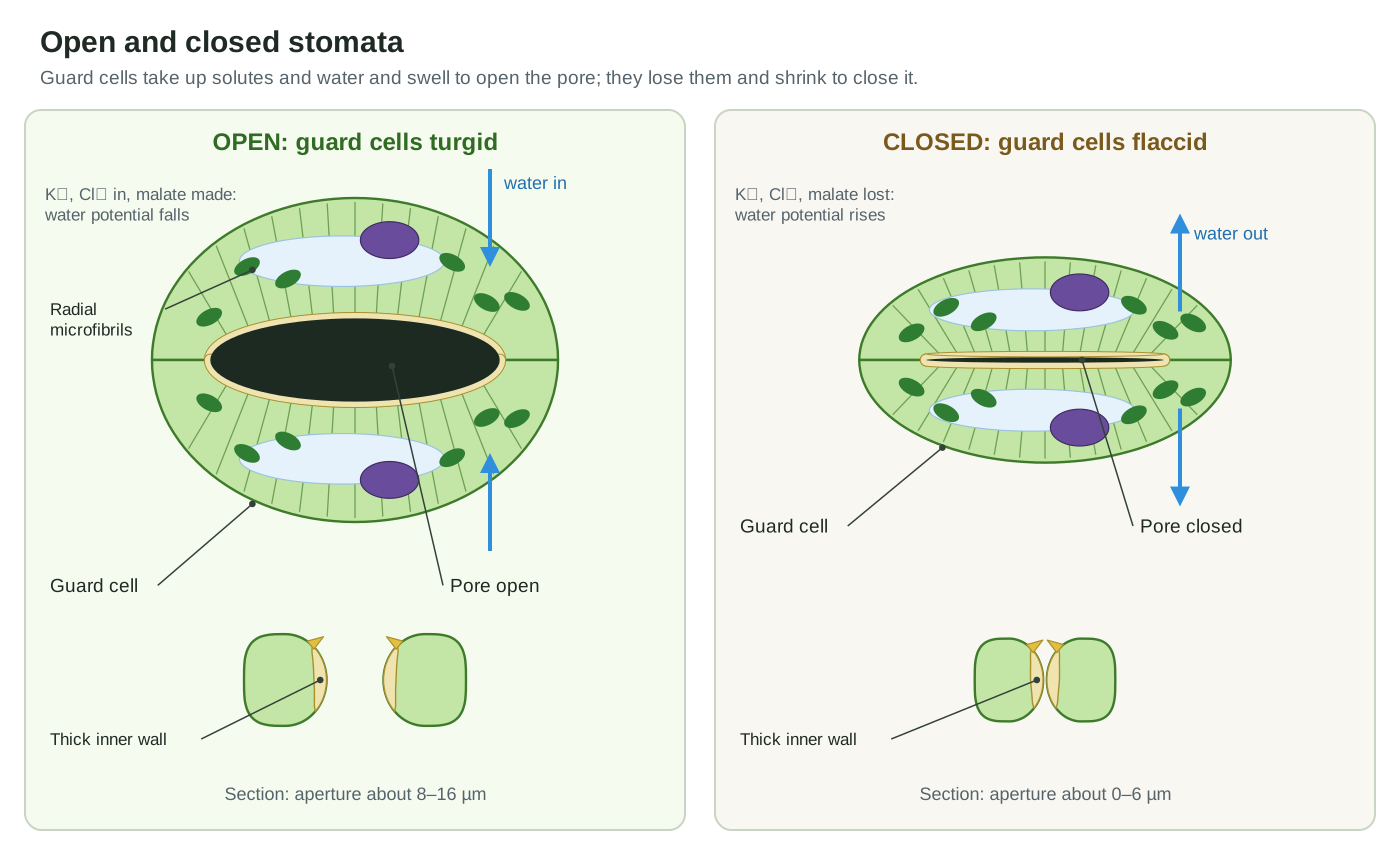
Open and closed stomata diagram
An open stoma has turgid, curved guard cells and a wide, oval pore; a closed stoma has flaccid, straighter guard cells pressed together, leaving a thin slit or no gap. Use the Open and Close buttons above the 3D model to see the change in 3D, or compare the two labeled diagrams.
| Open stoma | Closed stoma | |
|---|---|---|
| Guard cells | Turgid (swollen), curved apart | Flaccid (limp), straighter, touching |
| Solutes in guard cells | High K⁺, Cl⁻, malate | Low |
| Water potential of guard cells | Lower (more negative) than neighbours | Higher than neighbours |
| Pore width in broad bean | About 8–16.5 µm | About 0–6 µm |
| When | Daytime, enough water | Night, drought (ABA), high CO₂ |
| Gas exchange | CO₂ in; O₂ and water vapour out | Very little |
Stomata and transpiration
Transpiration is the loss of water vapour from a plant, mostly through its stomata. Water evaporates from the wet walls of the mesophyll cells into the air spaces, collects in the substomatal cavity, and diffuses out of the open pore into the drier air outside. The cuticle is nearly waterproof, so when the stomata are open they are the main exit. Up to 90% of the water a plant's roots take up can be lost this way. (OpenStax Biology 2e, 30.5 Transport of Water and Solutes in Plants.)
Transpiration is not just a loss. Evaporation from the leaf creates a pull that draws water, and the minerals dissolved in it, up the xylem from the roots. It also cools the leaf. NCERT's Class 10 text puts it this way: during the day, when the stomata are open, the transpiration pull becomes the major driving force for water movement in the xylem; at night root pressure matters more. (NCERT Science Class X, Chapter 5: Life Processes (PDF).)
The plant controls transpiration mainly by changing the stomatal aperture. Wider pores let water out faster. Closing the stomata in drought, in the dark, or in very dry air saves water. Trichomes (leaf hairs), a thick cuticle and sunken stomata all slow transpiration further by keeping a layer of still, humid air over the pores. (OpenStax Biology 2e, 30.4 Leaves.)
Stomata, photosynthesis and gas exchange
Stomata are where a leaf exchanges gases with the air. In the light, CO₂ diffuses in through the pores to the mesophyll chloroplasts, where it is fixed into sugar; O₂ made in photosynthesis diffuses out by the same route. At night the leaf only respires, so it takes in a little O₂ and gives out CO₂. NCERT's Class 10 book describes stomata as the tiny pores through which massive amounts of gas exchange take place for photosynthesis. (NCERT Science Class X, Chapter 5: Life Processes (PDF).)
The pores cover typically less than 3% of the leaf surface, yet some 98% of all the CO₂ taken up and water lost passes through them. This is the central trade-off of plant life: every opening that lets CO₂ in also lets water out. Plants balance the two by opening stomata when light makes photosynthesis possible and closing them when water is short or CO₂ inside the leaf is high. (Lawson & Blatt (2014): Stomatal size, speed, and responsiveness impact on photosynthesis and water use efficiency, Plant Physiology.)
Stomatal speed matters too. Stomata respond in minutes, but light under a moving canopy changes in seconds, so slow stomata waste water when they lag behind shade and lose photosynthesis when they lag behind sun. Smaller stomata generally respond faster than larger ones, and stomatal density is negatively correlated with stomatal size: leaves tend to have either many small stomata or fewer large ones. (Lawson & Blatt (2014): Stomatal size, speed, and responsiveness impact on photosynthesis and water use efficiency, Plant Physiology.)
For where the CO₂ that enters through the stomata is actually fixed, see chloroplast structure.
Types of stomata: anomocytic, anisocytic, paracytic, diacytic and grass
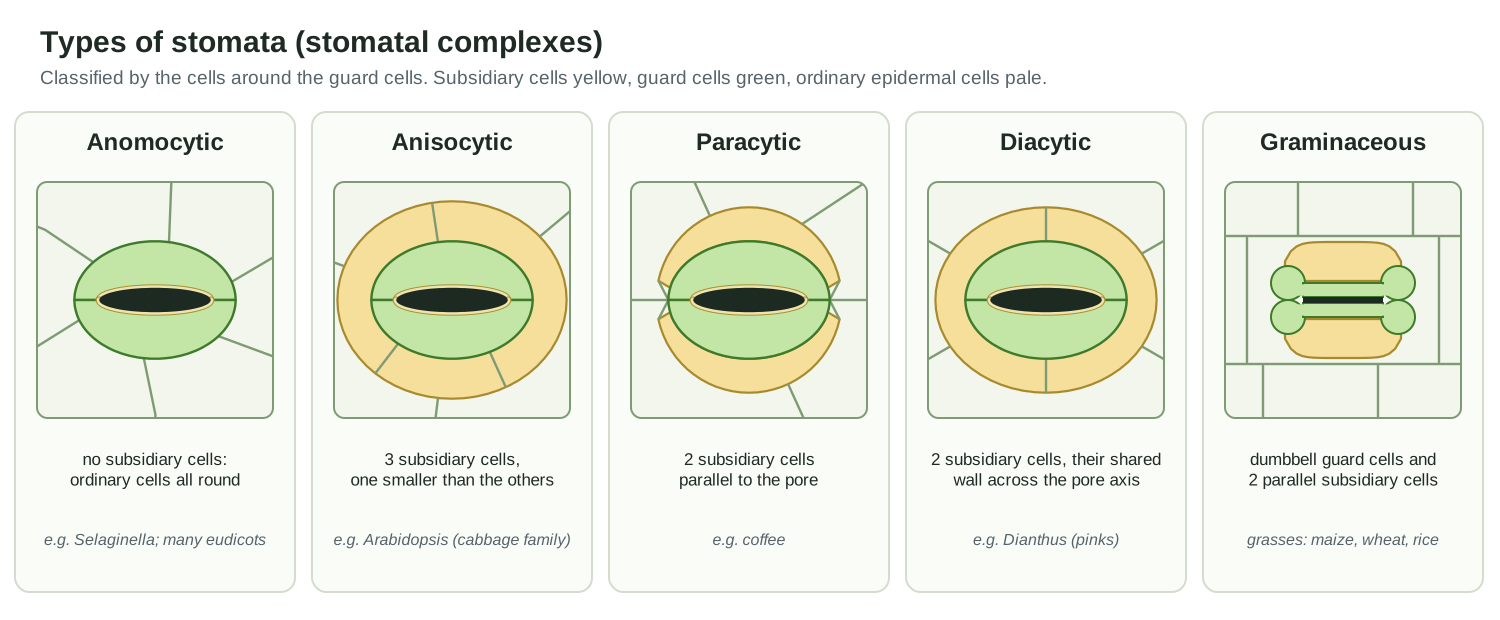
Stomata are classified by the cells around the guard cells. Subsidiary cells are epidermal cells next to the guard cells that differ in shape or size from ordinary epidermal cells. The number and arrangement of subsidiary cells vary widely between plant groups, which makes stomatal type useful for identifying plants. (Gray, Liu & Facette (2020))
| Type | Cells around the guard cells | Example |
|---|---|---|
| Anomocytic (irregular-celled) | No distinct subsidiary cells; ordinary epidermal cells all round | Selaginella; many eudicots |
| Anisocytic (unequal-celled) | Three subsidiary cells, one smaller than the other two | Arabidopsis and the cabbage family |
| Paracytic (parallel-celled) | Two subsidiary cells parallel to the pore and guard cells | Coffee |
| Diacytic (cross-celled) | Two subsidiary cells whose shared wall is at right angles to the guard cells | Dianthus (pinks, carnations) |
| Graminaceous (grass type) | Dumbbell guard cells, each with one parallel subsidiary cell | Grasses: maize, wheat, rice |
| Tetracytic, cyclocytic and others | Four, or a ring of many, subsidiary cells | Agave (tetracytic); Ginkgo (cyclocytic) |
The grass type is a special paracytic stoma. Its guard cells are narrow, thick-walled rods with bulbous ends. When the ends swell they push the rods apart, so the pore opens as a parallel-sided slit. In maize, potassium shuttles back and forth: it is high in the guard cells when stomata are open and high in the subsidiary cells when they are closed. (Gray, Liu & Facette (2020)) Brachypodium mutants that lack subsidiary cells have slower stomata with a smaller range of apertures, which supports the idea that subsidiary cells make grass stomata better. (Raissig et al. (2017))
Monocot vs dicot stomata
The textbook contrast is between a grass (the usual monocot example) and a typical eudicot. Grass stomata have dumbbell-shaped guard cells with subsidiary cells and lie in parallel rows; dicot stomata have kidney-shaped guard cells scattered over the leaf. NCERT also notes that an isobilateral (monocot) leaf usually has stomata on both surfaces, while a dorsiventral (dicot) leaf has more on the lower surface. (NCERT Biology Class XI, Chapter 6)
| Dicot (eudicot), e.g. bean | Grass (monocot), e.g. maize, wheat | |
|---|---|---|
| Guard cell shape | Kidney- or bean-shaped | Dumbbell-shaped: thick rods with bulbous ends |
| Subsidiary cells | Often none (anomocytic) or other types | Two, one beside each guard cell |
| Arrangement | Scattered in a net-veined leaf | In rows parallel to the veins, all pointing the same way |
| Distribution | Mostly on the lower surface | Often on both surfaces |
| Pore shape when open | Oval | Slit with parallel sides |
| Speed | Slower | About ten times faster in one comparison (wheat) |
Two cautions. First, ‘monocot’ does not always mean dumbbell: lilies, onions and many other monocots have kidney-shaped guard cells. Second, the speed difference comes from a careful comparison of four species, in which wheat stomata moved about an order of magnitude faster than kidney-type stomata. (Franks & Farquhar (2007))
Stomatal distribution: hypostomatic, amphistomatic and epistomatic leaves
Leaves are classed by where their stomata are. A hypostomatic leaf has stomata only on the lower (abaxial) surface; an amphistomatic leaf has them on both surfaces; an epistomatic leaf has them only on the upper (adaxial) surface. NCERT notes that in a dorsiventral (typical dicot) leaf the lower epidermis generally bears more stomata than the upper, which may lack them, while in an isobilateral (monocot) leaf stomata are present on both surfaces. (NCERT Biology Class XI, Chapter 6: Anatomy of Flowering Plants (PDF).)
Floating leaves are the classic epistomatic case. In the water lily (Nymphaea) the lower surface lies on the water, so stomata are restricted to the upper surface, where they can reach the air. The leaf also has large air chambers that help it float. Submerged leaves often have few or no stomata. (UBC BIOL 210: Hydrophyte leaves.)
The two surfaces of one leaf are not independent copies. In Arabidopsis, stomatal patterning is regulated differently in the upper and lower epidermis, and the ratio of upper to lower stomata depends on genotype and growing conditions such as humidity. (Jalakas et al. (2024): Stomatal patterning is differently regulated in adaxial and abaxial epidermis in Arabidopsis, Journal of Experimental Botany.)
Sunken stomata: xerophytic leaf adaptations
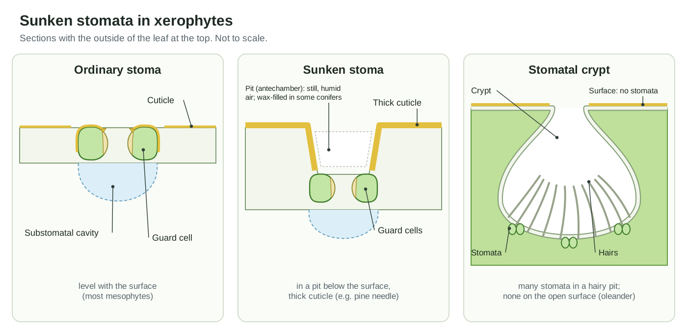
In xerophytes (plants of dry places) stomata are often sunken in pits, protected by hairs or wax, and covered by a thick cuticle. These features trap a layer of still, humid air over each pore.
Sunken stomata in xerophytes: pine and oleander
Sunken stomata are stomata set in pits below the general leaf surface. They are a classic feature of xerophytes, plants adapted to dry habitats. Conifer needles such as pine have sunken stomata and a small surface area, two features OpenStax lists as reducing water loss. Xerophytes usually also have a much thicker cuticle and often a dense covering of trichomes. (OpenStax Biology 2e, 30.4 Leaves.)
Oleander (Nerium oleander) goes further: its stomata lie in stomatal crypts, large chambers in the leaf lined with epidermis that bears stomata and hairs. The leaf surface between the crypts has no stomata. The narrow crypt opening and the hairs keep the air inside still, so a water molecule leaving a pore has a good chance of diffusing back in rather than escaping. (Plant anatomy image notes: Stomatal crypts in a transverse section of an oleander leaf (Constantine the Philosopher University, Nitra).)
In some conifers the chamber above each sunken stoma is filled with a porous plug of wax. How much water these features save is still debated. A model of crypts found they cut transpiration by less than 15% when stomata are open, and a 2022 review points out that sunken stomata also occur in wet habitats and may protect the pore from water films, fog and fungi as much as from drought. (Šantrůček (2022): The why and how of sunken stomata, Annals of Botany.)
Stomatal index and stomatal density
Stomatal density is the number of stomata per square millimetre of leaf surface. Stomatal index is the percentage of epidermal cells that are stomata:
Stomatal index (SI) = S ÷ (S + E) × 100
Here S is the number of stomata and E the number of ordinary epidermal (pavement) cells in the same area. Research papers often write it as SD ÷ (SD + PCD), where SD is stomatal density and PCD is pavement-cell density, and may leave out the × 100. (Jalakas et al. (2024)) Count each stoma once (one pair of guard cells = one stoma).
Worked example. On the upper surface of Arabidopsis rosette leaves, one study counted about 101 stomata and 243 pavement cells per mm². Stomatal density = 101 per mm². Stomatal index = 101 ÷ (101 + 243) × 100 ≈ 29%. (Inan et al. (2004))
Why have two measures? Stomatal density changes when leaf cells grow bigger or smaller (for example in shade or drought), even if the same proportion of cells became stomata. Stomatal index measures that proportion, so it is more stable and tells you whether the plant actually made more or fewer stomata. Both are used to study how plants respond to CO₂ and climate.
Method. Make an epidermal impression (see the next section), view it at a magnification where you can see individual cells, and count stomata and epidermal cells inside a known area: either the squares of an eyepiece graticule or the whole field of view. Calibrate that area with a stage micrometer. Count several fields or squares and take the mean. (Science & Plants for Schools (SAPS))
Stomata under the microscope: the nail varnish epidermal peel
The quickest way to see stomata is to make a nail varnish impression of the lower surface of a leaf. Clear nail varnish copies the surface in fine detail, including the outlines of the guard cells and epidermal cells.
- Choose a mature leaf with a smooth lower surface and paint a patch, about 1 cm², between the larger veins with clear (or pale) nail varnish.
- Let it dry completely.
- Press a strip of clear sticky tape over the patch and peel it off: the dried varnish comes away with it.
- Stick the tape onto a clean glass slide.
- Look at it under a light microscope, first at low power and then at about ×400. Count with a graticule, or count whole fields of view. (Rothamsted Research Bioimaging)
With some plants you can instead tear off a strip of real epidermis (an epidermal peel) and mount it in a drop of water. Broad bean, onion leaves and Tradescantia work well. A real peel shows living guard cells with green chloroplasts, which an impression cannot. Some leaves are damaged by the solvent in nail varnish, absorbing it and turning brown; test a leaf first, or use water-based varnish, which can even be applied to a leaf still on the plant. (Science & Plants for Schools (SAPS))
What you see: pairs of curved guard cells, each pair around a dark slit or oval, scattered among jigsaw-shaped or polygonal epidermal cells. In grass leaves the stomata lie in neat rows. Compare the lower and upper surfaces of the same leaf, or leaves from sun and shade, and calculate the stomatal density and index for each. Never eat or taste plant material in a lab, and wash your hands after handling nail varnish.
Stomata in bryophytes and other plant groups
Almost all land plants have stomata, but where they sit differs. In mosses and hornworts, stomata occur only on the sporophyte capsule (the spore-producing part), not on the leafy plant. Liverworts are the only living land plants that lack stomata entirely; their thalloid forms have simple air pores instead. (Renzaglia, Browning & Merced (2020): With over 60 independent losses, stomata are expendable in mosses, Frontiers in Plant Science.)
Bryophyte stomata seem to work differently from those of flowering plants. Renzaglia and colleagues argue that moss and hornwort stomata do not open and close in response to light, water status or ABA, and that they mainly help the capsule dry out so spores can be released. Some laboratory studies have reported ABA responses in moss stomata, so this is an active debate. (Renzaglia, Browning & Merced (2020): With over 60 independent losses, stomata are expendable in mosses, Frontiers in Plant Science.)
Ferns and lycophytes have kidney-shaped guard cells without subsidiary cells. Many gymnosperms have sunken stomata; in Ginkgo the guard cells are recessed under a ring of subsidiary cells (a cyclocytic stoma). (Gray, Liu & Facette (2020): Flanking support: how subsidiary cells contribute to stomatal form and function, Frontiers in Plant Science.)
Stomata for CBSE Class 10, Class 9 and Class 7
These are the key points school syllabuses expect, from simplest to most detailed.
Class 7 (nutrition in plants). Leaves have tiny pores called stomata, mostly on the lower surface. Each pore is surrounded by guard cells. Carbon dioxide for photosynthesis enters through the stomata, and oxygen and water vapour leave through them.
Class 9 (plant tissues). Stomata are pores in the epidermis, the outer protective tissue of the leaf. Each stoma is enclosed by two kidney-shaped guard cells. Stomata are needed for gas exchange with the atmosphere and for transpiration, the loss of water as vapour. Learn to draw guard cells and epidermal cells in surface view.
Class 10 (life processes). Gas exchange for photosynthesis happens through the stomata. Because a lot of water can be lost through them, the plant closes the pores when it does not need CO₂. Opening and closing is a function of the guard cells: they swell when water flows into them and the pore opens; they shrink and the pore closes. The diagram to know is ‘open and closed stomatal pore’. Transpiration pulls water up the xylem and is the main driving force during the day, when the stomata are open. (NCERT Science Class X, Chapter 5)
Class 11 (anatomy of flowering plants). Bean-shaped guard cells, dumbbell-shaped in grasses; thin outer walls and highly thickened inner walls; guard cells have chloroplasts; subsidiary cells; and the stomatal apparatus (pore + guard cells + subsidiary cells). (NCERT Biology Class XI, Chapter 6)
Every structure in the 3D stomata model
The text version of the 3D diagram: every structure you can click, grouped as in the viewer.
Epidermis and cuticle
- Cuticle (Waxy cuticle)
- A thin, waxy, waterproof film that covers the outer surface of the epidermis, including the guard cells. Cuts water loss from the leaf surface, so almost all the water and CO₂ that a leaf exchanges has to pass through the stomata instead.
- Epidermal cells (pavement cells) (Ordinary epidermal cells)
- The flat, tightly fitted cells that make up most of the epidermis. In many eudicot leaves their walls are wavy, so they fit together like jigsaw pieces. Protect the leaf and hold the guard cells in place. Their turgor pushes back against the guard cells, so they affect how far a stoma can open.
- Epidermal cell nucleus
- The nucleus of an epidermal cell, seen here where the front cut face slices through a cell. Controls the cell. Like any living plant cell, an epidermal cell has a nucleus, cytoplasm and a large vacuole.
- Epidermal cell vacuole (Central vacuole)
- The large, water-filled central vacuole that takes up most of an epidermal cell. Keeps the cell turgid (firm). Epidermal cells and guard cells exchange water and ions, so the epidermis takes part in stomatal movement.
Guard cells and pore
- Guard cells (Kidney-shaped (bean-shaped) guard cells)
- The pair of curved, kidney-shaped cells that surround each stomatal pore and open or close it. Take up ions and water to swell and open the pore, and lose them to shrink and close it. They sense light, CO₂, humidity and the drought hormone ABA.
- Stomatal pore (aperture) (Stoma, stomatal opening)
- The gap between the two guard cells, through which CO₂ enters the leaf and water vapour and O₂ leave. The adjustable opening for gas exchange. Its width (the stomatal aperture) sets how fast gases can diffuse in and out.
- Thick inner (ventral) wall (Ventral wall, pore wall)
- The thick, stiff part of each guard cell wall that lines the pore. Drawn in cream. Strengthens the guard cell where it bends most. The classic textbook explanation is that the thick inner wall stretches less than the thin outer wall, so the cell bows outwards when it swells.
- Thin outer (dorsal) wall (Dorsal wall)
- The thinner, more stretchy wall on the side of each guard cell away from the pore, next to the epidermal cells. Stretches and bulges into the neighbouring cells as the guard cell swells, letting the guard cells curve apart.
- Guard cell ends (polar regions) (Poles of the stoma)
- The two ends of the stoma, where the guard cells are joined together. Stiff, pectin-rich ends pin the guard cells down so they cannot simply get longer when they swell; they bow apart instead and the pore opens.
- Cellulose microfibrils (radial micellation) (Radially arranged microfibrils)
- Bundles of cellulose in the guard-cell wall that wrap round each guard cell like hoops. In surface view they seem to radiate out from the pore. The hoops stop the guard cell getting fatter in girth, so when it takes up water it can only lengthen along its outer edge, which makes it curve and open the pore.
- Outer cuticular ledge (Stomatal ledge, front cavity rim)
- A narrow ridge of wall and cuticle along the top inner edge of each guard cell, over the pore. Forms the lip of the small front cavity (vestibule) above the pore. The ledges can meet to seal a closed stoma.
Inside a guard cell
- Guard cell cytoplasm
- The living contents of the guard cell around the vacuole, packed with organelles and ion transporters. Its plasma membrane carries the proton pump and channels that move ions in and out to open and close the stoma.
- Guard cell vacuole
- The large vacuole inside each guard cell, where most of the K⁺, Cl⁻ and malate taken up during opening is stored. Swells as solutes and water enter and shrinks as they leave; its volume change drives most of the guard cell's change in shape.
- Guard cell nucleus
- The nucleus of each guard cell. Holds the cell's DNA. Guard cells switch genes on and off in response to signals such as ABA.
- Guard cell chloroplasts
- The chloroplasts inside the guard cells. Guard cells have obvious chloroplasts; other epidermal cells have few, small ones or none. Make ATP and sugars and store starch. Guard-cell photosynthesis helps power stomatal movement, and in ABA-induced closure the chloroplasts produce reactive oxygen signals (Iwai et al. 2019); the exact role is still debated.
- Starch grains
- Granules of starch stored in the guard-cell chloroplasts. In the morning, blue light triggers guard cells to break down starch; the products help make malate and sugars that draw water in.
Cavity and mesophyll
- Substomatal cavity (Substomatal chamber, air chamber)
- The air space just inside each stoma, between the guard cells and the spongy mesophyll. Connects the pore to the network of air spaces in the leaf. Water evaporating from mesophyll cell walls collects here as vapour and diffuses out of the pore; CO₂ diffuses in.
- Spongy mesophyll (Spongy parenchyma)
- Loosely packed, irregular, lobed photosynthetic cells with large air spaces between them, lying just inside the lower epidermis. Photosynthesises and gives a large, wet surface for gas exchange with the air spaces.
- Mesophyll chloroplasts
- The chloroplasts lining the walls of the spongy mesophyll cells. Carry out most of the leaf's photosynthesis, using the CO₂ that enters through the stomata.
- Intercellular air spaces
- The connected network of air between the mesophyll cells. Lets CO₂ spread from the stomata to every mesophyll cell and lets water vapour and O₂ diffuse out.
Gas exchange
- CO₂ diffusing in
- Carbon dioxide from the air diffuses in through the open pore to the mesophyll, where it is used in photosynthesis. Supplies the carbon for photosynthesis. Closing the stomata saves water but cuts off this supply.
- Water vapour diffusing out (transpiration)
- Water vapour diffuses out of the leaf through the open pore: transpiration. Transpiration pulls water and minerals up the xylem from the roots and cools the leaf, but too much dries the plant out.
Grass stoma (maize type)
- Dumbbell guard cell: thick-walled middle (Rod-shaped centre)
- The narrow, thick-walled middle part of each dumbbell-shaped grass guard cell, which lines the slit-like pore. Stays straight and rigid. When the bulbous ends swell, they push the two rods apart in parallel, so the pore opens as a slit.
- Dumbbell guard cell: bulbous ends
- The swollen, thin-walled ends of each grass guard cell. Swell when the guard cells take up water and push the rods apart; shrink to let the pore close.
- Subsidiary cells (Accessory cells)
- The two special epidermal cells that lie beside the guard cells in a grass stoma, one on each side. Act as a reservoir of K⁺ and water. Ions shuttle from subsidiary cells into the guard cells during opening and back during closing, which speeds up both.
- Grass stomatal pore
- The slit-shaped pore between the rods of a grass stoma. Opens as a parallel-sided slit rather than an oval.
- Grass long epidermal cells
- Long, narrow epidermal cells arranged in rows parallel to the leaf veins. Form the grass leaf surface. Grass stomata lie in rows between them, all pointing the same way.
Stomata diagram worksheet: label it yourself (with answers)
Test yourself with this unlabeled stomata diagram. Choose the correct name for each of the 12 numbers. It also works as a black and white printout: colour the guard cells green, the vacuoles blue, the nuclei purple and the cuticle yellow.
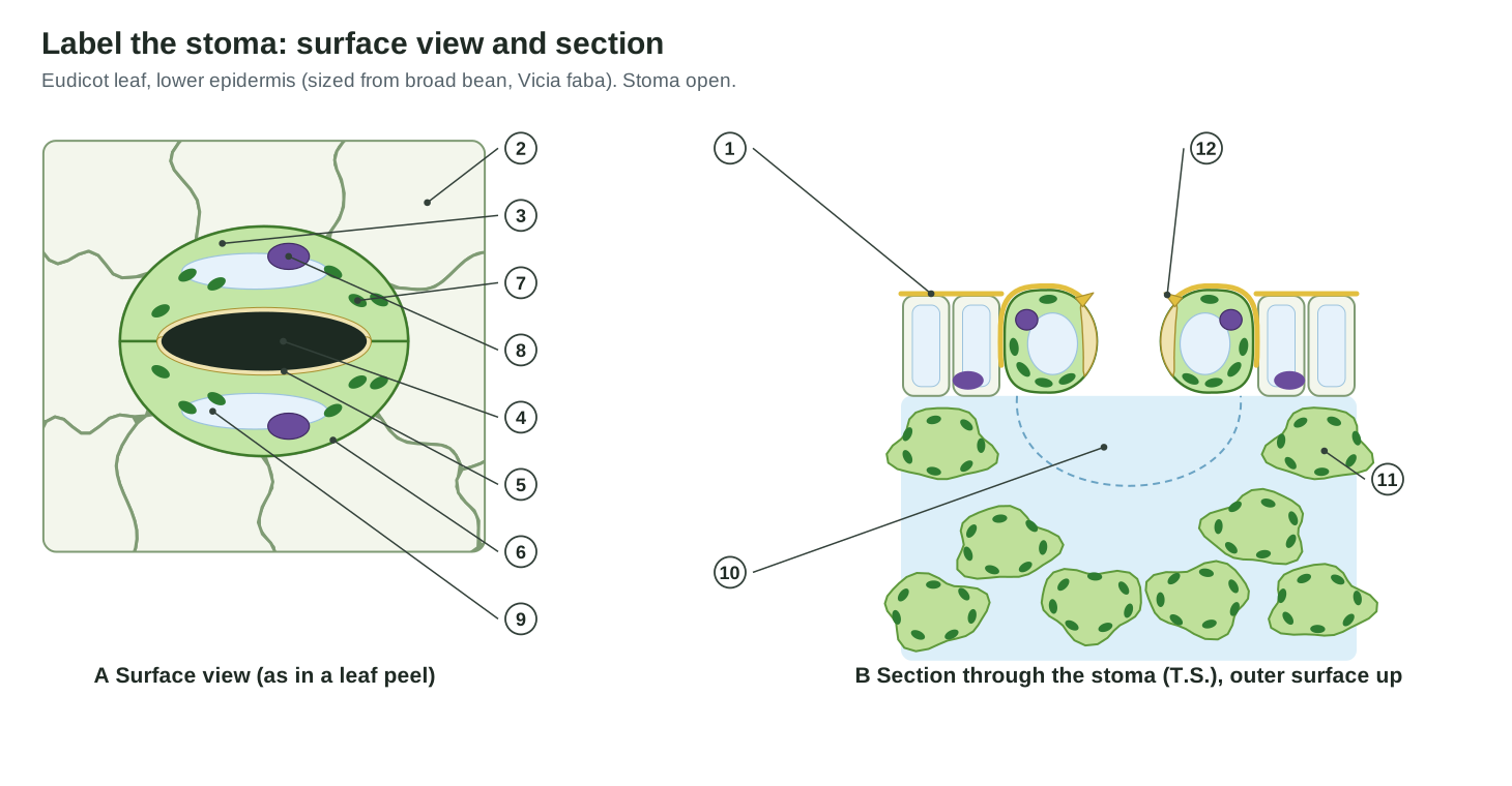
Answer key: stomata labeling worksheet
- Cuticle
- Epidermal (pavement) cell
- Guard cell
- Stomatal pore
- Thick inner (ventral) wall
- Thin outer (dorsal) wall
- Chloroplast
- Nucleus
- Vacuole
- Substomatal cavity
- Spongy mesophyll cell
- Outer cuticular ledge
Stomata practice questions
Questions of the kind set in tests and exams, with answers.
- Why are the inner walls of guard cells thick? To strengthen the guard cell where it bends and to resist stretching on the pore side, so the cell curves outwards when turgid (the classic explanation; stiff ends also matter).
- Which ion moves into guard cells to open stomata? Potassium (K⁺), balanced by chloride and malate.
- What does the proton pump do in stomatal opening? It pumps H⁺ out of the guard cell, making the inside negative so that K⁺ is drawn in.
- Name the hormone that closes stomata in drought. Abscisic acid (ABA).
- A leaf peel shows 24 stomata and 96 epidermal cells in one field. What is the stomatal index? 24 ÷ (24 + 96) × 100 = 20%.
- Why does a water lily have stomata only on its upper surface? Its lower surface lies on the water, so only the upper surface is in contact with air.
- How are grass stomata different from bean stomata? Dumbbell-shaped guard cells with subsidiary cells, in parallel rows, opening as a slit; bean stomata have kidney-shaped guard cells, usually without subsidiary cells, scattered over the leaf.
- Why do guard cells have chloroplasts? To make ATP and sugars and store starch, which supply energy and solutes for stomatal movement.
Frequently asked questions
What is a stoma?
A stoma (plural stomata) is a tiny pore in the epidermis of a leaf or young stem, surrounded by two guard cells that open and close it. Stomata let carbon dioxide into the leaf for photosynthesis and let oxygen and water vapour out.
What are the parts of a stoma?
A stoma has two guard cells, the pore (aperture) between them, and in many plants subsidiary cells around them. Each guard cell has a thick inner wall next to the pore, a thin outer wall, chloroplasts, a nucleus and a large vacuole. Below the pore is the substomatal cavity, an air space that leads into the spongy mesophyll.
Why do stomata open in the light and close in the dark?
Blue light switches on proton pumps in the guard-cell membrane. Potassium ions, chloride and malate then build up in the guard cells, water follows by osmosis, the cells swell and bow apart, and the pore opens. In the dark the pumps stop, ions leak out, water leaves and the pore closes, so the leaf does not lose water when it cannot photosynthesise.
How does ABA close stomata during drought?
When a plant is short of water it makes the hormone abscisic acid (ABA). ABA raises calcium in the guard cells, which opens anion channels and blocks the proton pump. The membrane depolarises, potassium flows out through outward channels, water follows, turgor falls and the stoma closes.
Why are stomata mostly on the lower surface of a leaf?
The lower surface is shaded and cooler, so less water evaporates from stomata there. Many eudicot leaves are hypostomatic, with stomata only or mainly on the lower side. Grass leaves often have stomata on both sides, and floating leaves such as water lilies have them only on the upper side.
What is the difference between monocot and dicot stomata?
Dicots (eudicots) usually have kidney-shaped guard cells scattered in a net-veined leaf, often with no subsidiary cells or with irregular ones. Grasses, the best-known monocots, have dumbbell-shaped guard cells with two subsidiary cells, arranged in parallel rows. Not all monocots are grass-like; lilies and onions have kidney-shaped guard cells.
What is the stomatal index?
The stomatal index is the percentage of epidermal cells that are stomata: SI = S ÷ (S + E) × 100, where S is the number of stomata and E the number of other epidermal cells in the same area. Unlike stomatal density (stomata per mm²), it does not change much when leaf cells expand.
What are sunken stomata?
Sunken stomata sit in pits below the leaf surface. They are common in xerophytes such as pine needles and oleander. The pit traps still, humid air over the pore, which is usually said to slow water loss, though measurements suggest the saving can be small.
Do all plants have stomata?
Nearly all land plants have them. Liverworts are the only living land plants with no stomata at all; mosses and hornworts have them only on the spore capsule. Submerged water plants often have few or none.
References
- OpenStax. Biology 2e, section 30.4: Leaves. Rice University.
- OpenStax. Biology 2e, section 30.5: Transport of Water and Solutes in Plants. Rice University.
- NCERT. Biology, Class XI, Chapter 6: Anatomy of Flowering Plants (2026–27 reprint).
- NCERT. Science, Class X, Chapter 5: Life Processes (2026–27 reprint).
- Jezek, M. & Blatt, M. R. (2017). The membrane transport system of the guard cell and its integration for stomatal dynamics. Plant Physiology 174, 487–519.
- Santelia, D. & Lawson, T. (2016). Rethinking guard cell metabolism. Plant Physiology 172, 1371–1392.
- Schroeder, J. I., Allen, G. J., Hugouvieux, V., Kwak, J. M. & Waner, D. (2001). Guard cell signal transduction. Annual Review of Plant Physiology and Plant Molecular Biology 52, 627–658.
- Carter, R. et al. (2017). Stomatal opening involves polar, not radial, stiffening of guard cells. Current Biology 27, 2974–2983.
- Franks, P. J. & Farquhar, G. D. (2007). The mechanical diversity of stomata and its significance in gas-exchange control. Plant Physiology 143, 78–87.
- Gray, A., Liu, L. & Facette, M. (2020). Flanking support: how subsidiary cells contribute to stomatal form and function. Frontiers in Plant Science 11, 881.
- Raissig, M. T. et al. (2017). Mobile MUTE specifies subsidiary cells to build physiologically improved grass stomata. Science 355, 1215–1218.
- Lawson, T. & Blatt, M. R. (2014). Stomatal size, speed, and responsiveness impact on photosynthesis and water use efficiency. Plant Physiology 164, 1556–1570.
- Šantrůček, J. (2022). The why and how of sunken stomata: does the behaviour of encrypted stomata and the leaf cuticle matter? Annals of Botany 130, 285–300.
- Roth-Nebelsick, A., Hassiotou, F. & Veneklaas, E. J. (2009). Stomatal crypts have small effects on transpiration: a numerical model analysis. Plant Physiology 151, 2018–2027.
- Renzaglia, K. S., Browning, W. B. & Merced, A. (2020). With over 60 independent losses, stomata are expendable in mosses. Frontiers in Plant Science 11, 567.
- Barton, K. A. et al. (2016). Epidermal pavement cells of Arabidopsis have chloroplasts. Plant Physiology 171, 723–726.
- Iwai, S. et al. (2019). Guard cell photosynthesis is crucial in abscisic acid-induced stomatal closure. Plant Direct 3, e00137.
- Jalakas, P., Tulva, I., Bērziņa, N. M. & Hõrak, H. (2024). Stomatal patterning is differently regulated in adaxial and abaxial epidermis in Arabidopsis. Journal of Experimental Botany 75, 6476–6488.
- Inan, G. et al. (2004). Salt cress. A halophyte and cryophyte Arabidopsis relative model system and its applicability to molecular genetic analyses of growth and development of extremophiles. Plant Physiology 135, 1718–1737.
- UBC BIOL 210. Hydrophyte leaves. University of British Columbia.
- Science & Plants for Schools (SAPS). Measuring stomatal density.
- Rothamsted Research Bioimaging. Measuring stomatal density using nail varnish.
More interactive anatomy models
- Flower anatomyParts of a flower in 3D: petals, sepals, stamens, pistil, ovary and ovules of a lily.
- Leaf cross sectionLabeled leaf cross section: cuticle, epidermis, palisade and spongy mesophyll, veins.
- Seed anatomyBean and corn seed anatomy: seed coat, cotyledons, endosperm and embryo.
- Chloroplast structureEnvelope, stroma, thylakoids, grana and lumen of a chloroplast, in 3D.
- Human anatomy1,000+ bones, muscles, organs, vessels and nerves from real anatomical data.
- Octopus anatomyThree hearts, a ring-shaped brain, eight arms and a beak, in 3D.