

Kidney at a glance
- What it is
- The kidney is a bean-shaped organ that filters blood to form urine and regulate fluid, electrolytes and blood pressure.
- Location
- It lies retroperitoneally on each side of the vertebral column, from about T12 to L3, with the right kidney slightly lower because of the liver.
- Function
- It filters blood, removes waste and excess water, regulates acid-base balance and blood pressure, and produces erythropoietin and activates vitamin D.
- Body system
- Urinary · paired (left and right)
- FMA ID
- FMA7205 (Foundational Model of Anatomy)
Key facts
- Functional unit: nephron (roughly 1 million per kidney, with wide variation).
- Divided into cortex and medulla, with renal pyramids, calyces and a renal pelvis.
- Supplied by the renal artery from the abdominal aorta at about L1 to L2.
- Each kidney is about 10–14 cm long, 5–7 cm wide and 3–4 cm thick, and weighs roughly 115–175 g (slightly less in women).
- At the hilum the renal vein lies in front, the renal artery in the middle and the renal pelvis behind.
Clinical relevance
Kidney stones can obstruct the ureter, causing severe loin-to-groin pain (renal colic).
Where are your kidneys?
The kidneys lie on either side of the spine in the retroperitoneal space, the area between the parietal peritoneum and the back wall of the abdomen. OpenStax places the left kidney at about the T12 to L3 vertebrae and notes that the right kidney is lower because the liver pushes it down. The upper parts of both kidneys are partly protected by the 11th and 12th ribs. (OpenStax Anatomy and Physiology 2e, 25.3: Gross Anatomy of the Kidney.)
Radiopaedia describes the same T12 to L3 range and adds which ribs lie behind each kidney: the 12th rib crosses the back of both kidneys, while the 11th rib is a posterior relation of the left kidney only, because the left kidney sits higher. The upper pole of each kidney is also covered behind by the diaphragm and the pleura. (Radiopaedia: Kidney.)
Because the diaphragm covers the back of the upper half of each kidney, the kidneys move up and down a little with breathing. They are deep organs, so an ultrasound scan of the kidneys is usually done from the patient's back. (Kenhub: Kidneys.)
A useful surface landmark is the transpyloric plane, an imaginary horizontal plane midway between the jugular notch and the top of the pubic symphysis, at about the level of the L1 vertebral body. The hila of both kidneys are traditionally placed on this plane, although individual variation is normal. (Radiopaedia: Transpyloric plane.)
Kidney location on the back
From behind, each kidney lies under the costovertebral angle, the angle between the paraspinal muscles and the 12th rib. The kidney sits in front of (deep to) this angle in the retroperitoneal space, and pressure at the angle moves the kidney. This is why doctors check for tenderness here when they suspect an inflamed kidney, for example acute pyelonephritis. (Loyola University Chicago, Physical Examination: Costovertebral angle.)
So, to point to your kidneys on your back, place your hands on the lowest ribs on each side of the spine with your thumbs towards the midline: the kidneys lie deep to this area, with the upper poles tucked under the ribs and the lower poles below them. The right kidney is a little lower than the left. (OpenStax Anatomy and Physiology 2e, 25.3: Gross Anatomy of the Kidney.)
Kidney location in a woman's body
The kidneys sit in the same place in women and men: on the back wall of the abdomen, either side of the spine, from about T12 to L3. They are not pelvic organs, so they lie well above the uterus and ovaries. Female kidneys are on average slightly smaller: OpenStax gives a mass of 115 to 155 g in females and 125 to 175 g in males. (OpenStax Anatomy and Physiology 2e, 25.3: Gross Anatomy of the Kidney.)
Radiopaedia gives typical lengths of 10 to 14 cm in males and 9 to 13 cm in females, with the left kidney usually slightly larger than the right. The 3D model on this page comes from BodyParts3D, which is based on an adult male body. (Radiopaedia: Kidney.)
Layers around the kidney: capsule, fat and renal fascia
Each kidney is wrapped in four layers. From deep to superficial they are: the renal (fibrous) capsule, a tough layer stuck to the kidney surface; the perirenal (perinephric) fat; the renal fascia (Gerota's fascia), which encloses the kidney and the suprarenal (adrenal) gland; and the pararenal fat, which lies mainly behind and to the side. (TeachMeAnatomy: The Kidneys.)
The adrenal gland sits on the upper pole of each kidney inside the renal fascia but in its own compartment. OpenStax describes the fat as a shock-absorbing renal fat pad, and the fascia and peritoneum as anchoring the kidney firmly to the back wall of the abdomen. (OpenStax Anatomy and Physiology 2e, 25.3: Gross Anatomy of the Kidney.)
What touches the kidneys? Anterior and posterior relations
The two kidneys have different organs in front of them. In front of the right kidney lie the right suprarenal gland, the liver, the second (descending) part of the duodenum and the right colic (hepatic) flexure. In front of the left kidney lie the left suprarenal gland, the spleen, the stomach, the pancreas, the left colic (splenic) flexure and loops of jejunum. (TeachMeAnatomy: The Kidneys.)
Radiopaedia adds the hepatorenal recess (the space between liver and right kidney) as an anterior relation on the right, and the lesser sac and the tail of the pancreas on the left. (Radiopaedia: Kidney.)
Behind, both kidneys rest on the diaphragm, psoas major, quadratus lumborum and transversus abdominis, with the subcostal, iliohypogastric and ilioinguinal nerves running between kidney and muscle. The 12th rib lies behind both kidneys and the 11th rib behind the left one as well. (TeachMeAnatomy: The Kidneys.)
Kenhub describes the lower half of the back of the kidney as touching three muscles from medial to lateral: psoas major, quadratus lumborum and transversus abdominis. (Kenhub: Kidneys.)
Relations of the right and left kidney
| Side | Right kidney | Left kidney |
|---|---|---|
| Superior | Right suprarenal gland | Left suprarenal gland |
| Anterior | Liver (with the hepatorenal recess), 2nd part of duodenum, right colic flexure | Stomach, spleen, pancreas (tail), left colic flexure, jejunum |
| Posterior | Diaphragm, 12th rib, psoas major, quadratus lumborum, transversus abdominis | Diaphragm, 11th and 12th ribs, psoas major, quadratus lumborum, transversus abdominis |
| Nerves behind | Subcostal, iliohypogastric and ilioinguinal nerves | Subcostal, iliohypogastric and ilioinguinal nerves |
Compiled from TeachMeAnatomy and Radiopaedia (see references).
Inside the kidney: cortex, medulla, pyramids and papillae
A kidney cut in half shows two regions of tissue (parenchyma): an outer, lighter renal cortex and an inner renal medulla. Strips of cortex, called renal columns, run inwards between the parts of the medulla. All the renal corpuscles (the filtering units) and the proximal convoluted tubules of the nephrons lie in the cortex. (OpenStax Anatomy and Physiology 2e, 25.3: Gross Anatomy of the Kidney.)
The medulla is made of cone-shaped renal pyramids. Kenhub gives 8 to 18 pyramids per kidney. The wide base of each pyramid faces the cortex and its tip, the renal papilla, points medially towards the renal sinus. (Kenhub: Kidneys.)
Renal papillae are bundles of collecting ducts that carry the urine made by the nephrons into the calyces. About 15 percent of nephrons, the juxtamedullary nephrons, have long loops of Henle that dip deep into the medulla. OpenStax estimates about 1.3 million nephrons in each kidney. (OpenStax Anatomy and Physiology 2e, 25.3: Gross Anatomy of the Kidney.)
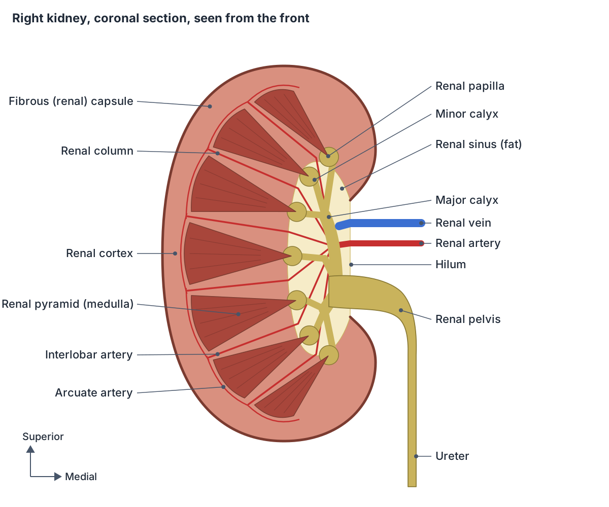
Minor calyx, major calyx and the renal pelvis
Urine leaves each papilla into a cup-shaped minor calyx (plural calyces). Minor calyces join to form major calyces, the major calyces drain into the renal pelvis, and the renal pelvis drains into the ureter. TeachMeAnatomy describes the pelvis as flattened and funnel-shaped. (TeachMeAnatomy: The Kidneys.)
Radiopaedia gives the usual numbers: four or five minor calyces unite to form a major calyx, and each kidney normally has two or three major calyces. Kenhub names the major calyces superior, middle and inferior when there are three. (Radiopaedia: Kidney.)
The renal pelvis (the 'pelvis in the kidney') connects the calyces to the ureter. It is formed either by the union of two or three major calyces or of seven to eleven minor calyces. It is triangular, lies at the back of the renal hilum surrounded by fat and vessels, and its shape ranges from a single wide sac to several funnels, all tapering into the ureter. (Radiopaedia: Renal pelvis.)
The renal pelvis narrows to become the ureter, which carries urine to the bladder by waves of peristalsis rather than by gravity. (OpenStax Anatomy and Physiology 2e, 25.2: Gross Anatomy of Urine Transport.)
Renal hilum (hilus) and the sinus of the kidney
The renal hilum (Latin hilum renale, older spelling hilus) is the deep slit on the concave medial border of each kidney. It is the doorway through which the renal artery enters and the renal vein and ureter leave, along with nerves and lymphatics. (TeachMeAnatomy: The Kidneys.)
From front to back, the main structures at the hilum are the renal vein, the renal artery and the renal pelvis (often remembered as vein, artery, pelvis, or VAP). Kenhub gives the same order from superior to inferior: renal vein, renal artery, ureter. Branches vary, so this is a typical pattern rather than a rule. (Radiopaedia: Kidney.)
The renal sinus is the fat-filled space inside the kidney that the hilum opens into. It lies in the medial part of the kidney, contains the hilum and is bordered laterally by renal parenchyma. Fat is its main component and increases with age and obesity. It also holds the major and minor calyces, the main branches of the renal artery and vein, lymphatics, autonomic nerves and fibrous tissue. (Radiopaedia: Renal sinus.)
On ultrasound the sinus looks brighter (more echogenic) than the surrounding kidney tissue. Checking it is part of looking for hydronephrosis (a swollen collecting system), and on older contrast studies (intravenous urography) for filling defects caused by tumours, clots or stones. (Radiopaedia: Renal sinus.)
Blood supply, lymph and nerves
Each kidney is supplied by a renal artery, a branch of the abdominal aorta that arises just below the origin of the superior mesenteric artery. The right renal artery is longer and passes behind the inferior vena cava. At the hilum it divides into an anterior division (about 75% of the supply) and a posterior division (about 25%), which give five segmental arteries. (TeachMeAnatomy: The Kidneys.)
Inside the kidney, segmental arteries give interlobar arteries that run through the renal columns, then arcuate arteries, cortical radiate arteries and finally afferent arterioles to the glomeruli. The kidneys receive about 25 percent of the cardiac output at rest. (OpenStax Anatomy and Physiology 2e, 25.3: Gross Anatomy of the Kidney.)
The segmental arteries are end arteries: they do not connect with each other, so a blocked branch causes loss of blood supply (ischaemia) and death of that segment. The renal veins leave the hilum in front of the arteries and drain straight into the inferior vena cava. The left renal vein is longer and crosses in front of the aorta, just below the superior mesenteric artery. (TeachMeAnatomy: The Kidneys.)
Because the left renal vein passes between the superior mesenteric artery and the aorta, it can be squeezed there, which is called the nutcracker phenomenon. Accessory renal arteries are also common. (Kenhub: Kidneys.)
Lymph from the kidney drains to the lateral aortic (para-aortic) lymph nodes, which lie around the origins of the renal arteries. (TeachMeAnatomy: The Kidneys.)
Sympathetic nerves reach the kidney through the renal and aorticorenal plexuses, carrying fibres from the lesser and least splanchnic nerves. Parasympathetic fibres come from the vagus nerve through the coeliac (celiac), renal and aorticorenal plexuses. (Radiopaedia: Kidney.)
Development and variants
The collecting system (ureter, pelvis and calyces) grows from the ureteric bud, which buds off the mesonephric duct in the fourth week. The rest of the kidney tissue comes from the metanephros, which appears in the fifth week. The kidney forms in the pelvis and moves up to its adult position, and the fetal kidneys begin filtering at about nine weeks. (Radiopaedia: Kidney.)
If a kidney does not ascend, it stays in the pelvis as a pelvic kidney, usually at the level of the common iliac artery. In a horseshoe kidney the lower poles are joined by an isthmus, which lies below the inferior mesenteric artery; it is usually symptomless but more prone to obstruction. Accessory renal arteries are found in about 25% of people. (TeachMeAnatomy: The Kidneys.)
Kenhub notes that a missing kidney on one side (unilateral renal agenesis) often goes unnoticed, while absence of both kidneys is fatal without treatment, and that a third (supernumerary) kidney, when present, is usually small and atrophic. (Kenhub: Kidneys.)
Clinical relevance
Kidney stones can pass from the kidney and lodge in the narrower ureter, causing severe pain. Acute kidney failure is often caused by poor blood flow (ischaemia) or by drugs that damage the kidney. (Kenhub: Kidneys.)
Because each segmental artery supplies its own part of the kidney, a small renal cell carcinoma can often be removed by partial nephrectomy, taking the tumour with a margin of healthy tissue and keeping the rest of the kidney. Surgeons also use Brödel's line, a relatively avascular plane along the lateral, slightly posterior border, to enter the kidney. (TeachMeAnatomy: The Kidneys.)
Structures next to the kidney
The nearest named structures in the 3D model, measured from the centre of each part:
- Anterior division of renal artery · cardiovascular
- Subcostal artery · cardiovascular
- Posterior division of renal artery · cardiovascular
- Subcostal vein · cardiovascular
- Inferior suprarenal artery · cardiovascular
- Renal vein · cardiovascular
- Eleventh rib · skeletal
- Twelfth rib · skeletal
- Hepatovenous segment VI · digestive
- Hepatovenous segment VII · digestive
Frequently asked questions
Where are your kidneys located on your back?
The kidneys lie deep in the upper part of the back, one on each side of the spine, between about the T12 and L3 vertebrae. Their upper poles are tucked under the 11th and 12th ribs, and each kidney sits in front of the costovertebral angle, the corner between the 12th rib and the back muscles. The right kidney is usually a little lower than the left.
Why is the right kidney lower than the left?
The liver takes up a lot of space in the upper right abdomen and pushes the right kidney slightly downwards. For this reason the 12th rib lies behind both kidneys, but the 11th rib lies behind only the higher left kidney. The difference is small, and both kidneys also move a little with breathing because the diaphragm lies behind their upper halves.
Are the kidneys in the same place in women?
Yes. In both women and men the kidneys lie on the back wall of the abdomen beside the spine, at about T12 to L3. They are well above the pelvis, so they are not near the uterus or ovaries. Female kidneys are on average slightly smaller and lighter, about 115 to 155 g compared with 125 to 175 g in males.
What is the difference between a minor calyx and a major calyx?
A minor calyx is a small cup that fits around one renal papilla and collects the urine dripping from it. Several minor calyces, usually four or five, join to make a larger major calyx. Most kidneys have two or three major calyces, which then join to form the renal pelvis, the funnel that continues as the ureter.
What is the renal sinus?
The renal sinus is the space inside the medial part of the kidney that opens out through the hilum. It is filled mainly with fat and also contains the calyces, the renal pelvis, the main branches of the renal artery and vein, lymphatics and nerves. The amount of sinus fat increases with age and obesity.
What passes through the renal hilum?
The renal hilum is the slit on the medial border of the kidney. The renal vein, the renal artery and the renal pelvis pass through it, usually in that order from front to back, together with lymph vessels and autonomic nerves. The renal pelvis narrows to become the ureter as it leaves the hilum.
What is the renal cortex?
The renal cortex is the outer, lighter layer of kidney tissue under the capsule. It holds the renal corpuscles, where blood is filtered, and the proximal convoluted tubules. Extensions of cortex called renal columns run inwards between the renal pyramids of the medulla and carry the interlobar blood vessels.
References
- OpenStax. Anatomy and Physiology 2e, §25.3 Gross Anatomy of the Kidney. Rice University, 2022.
- OpenStax. Anatomy and Physiology 2e, §25.2 Gross Anatomy of Urine Transport. Rice University, 2022.
- Jones O. The Kidneys. TeachMeAnatomy.
- Kidney. Radiopaedia reference article.
- Renal sinus. Radiopaedia reference article.
- Renal pelvis. Radiopaedia reference article.
- Transpyloric plane. Radiopaedia reference article.
- Kidneys. Kenhub anatomy library.
- Loyola University Chicago Stritch School of Medicine. Physical Examination: Costovertebral angle.
- Foundational Model of Anatomy: Kidney (FMA7205). EMBL-EBI Ontology Lookup Service.
- Mitsuhashi N. et al. (2009). BodyParts3D: 3D structure database for anatomical concepts. Nucleic Acids Research 37, D782–D785. Data © The Database Center for Life Science, CC BY 4.0.
Related anatomy
- UreterWhat do the ureters do? Ureter function, course, relations, the three narrowings where kidney stones lodge, blood…
- Adrenal glandAdrenal gland: The adrenal (suprarenal) gland is a small endocrine gland on top of each kidney. See where it is in an…
- Renal arteryRenal artery: A large paired branch of the abdominal aorta supplying the kidney. See where it is in an interactive 3D…
- Renal veinRenal vein: A large vein draining the kidney into the inferior vena cava. See where it is in an interactive 3D human…
- Abdominal aortaAbdominal aorta: The abdominal aorta is the part of the aorta that runs through the abdomen and supplies the abdominal…
- Urinary bladderUrinary bladder: The urinary bladder is a hollow muscular organ that stores urine. See where it is in an interactive…
Explore the whole body in the interactive 3D human anatomy model, or browse the A–Z list of human structures.