

What is the knee joint made of?
The knee has three articulations: the femoropatellar joint between the patella and the femur, and the medial and lateral tibiofemoral joints between the condyles of the femur and tibia. All three share a single joint capsule. (OpenStax Anatomy and Physiology 2e, 9.6: Anatomy of Selected Synovial Joints.)
The tibiofemoral joint is a synovial hinge joint; the patellofemoral joint is a plane joint. The joint surfaces of the femur and tibia do not fit closely together, and the menisci improve the fit. Kenhub describes the knee as the largest and most stressed joint in the body. (Kenhub: Knee joint.)
The fibula is not part of the knee joint itself; it lies below the lateral condyle of the tibia. It matters to the knee because the lateral collateral ligament attaches to the head of the fibula. (TeachMeAnatomy: The Knee Joint.)
Ligaments of the knee: the four main ligaments
Four main ligaments stabilise the knee. Two lie outside the joint capsule at the sides (extracapsular): the fibular (lateral) collateral ligament and the tibial (medial) collateral ligament. Two lie inside the capsule (intracapsular) and cross each other in an X: the anterior and posterior cruciate ligaments. (OpenStax Anatomy and Physiology 2e, 9.6: Anatomy of Selected Synovial Joints.)
The cruciate ligaments are inside the joint capsule but outside the synovial cavity. Several smaller ligaments add support: the patellar ligament in front, the oblique and arcuate popliteal ligaments behind, and the ligaments that anchor the menisci. (Kenhub: Knee joint.)
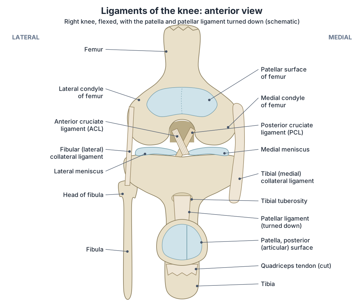
Ligaments of the knee
| Ligament | Attachments | Main job |
|---|---|---|
| Anterior cruciate (ACL) | Anterior intercondylar area of tibia to medial side of lateral femoral condyle | Stops the tibia sliding forward on the femur; resists hyperextension |
| Posterior cruciate (PCL) | Posterior intercondylar area of tibia to lateral side of medial femoral condyle | Stops the tibia sliding backward on the femur; supports the bent knee |
| Tibial (medial) collateral (MCL) | Medial femoral epicondyle to medial tibial condyle and shaft | Resists force from the outer side of the knee (valgus stress); attached to the medial meniscus |
| Fibular (lateral) collateral (LCL) | Lateral femoral epicondyle to head of fibula | Resists force from the inner side of the knee (varus stress) |
| Patellar ligament | Apex of patella to tibial tuberosity | Transmits the pull of quadriceps to the tibia |
| Oblique popliteal | Semimembranosus tendon to the back of the lateral femoral condyle | Reinforces the back of the capsule |
| Arcuate popliteal | Head of fibula to the back of the capsule | Reinforces the back of the capsule |
| Transverse (intermeniscal) | Anterior horn of medial meniscus to anterior horn of lateral meniscus | Links the two menisci |
| Meniscofemoral (Humphry and Wrisberg) | Posterior horn of lateral meniscus to medial femoral condyle | Steady the lateral meniscus |
From TeachMeAnatomy, Kenhub, OpenStax and Fox et al. (2012).
ACL and PCL: the cruciate ligaments
The anterior cruciate ligament (ACL) arises from the anterior intercondylar area of the tibia, close to the attachment of the medial meniscus. It runs upward, backward and laterally to the inner (posteromedial) side of the lateral femoral condyle, crossing lateral to the PCL. It is about 38 mm long. (Kenhub: Anterior cruciate ligament.)
The ACL stops the tibia sliding forward on the femur. The posterior cruciate ligament (PCL) runs from the posterior intercondylar area of the tibia to the medial femoral condyle and stops the tibia sliding backward on the femur. (TeachMeAnatomy: The Knee Joint.)
The ACL tightens as the knee straightens and resists hyperextension. The PCL is the stronger of the two cruciate ligaments. It supports the bent, weight-bearing knee and prevents the femur sliding forward off the tibia. (OpenStax Anatomy and Physiology 2e, 9.6: Anatomy of Selected Synovial Joints.)
Inside the intercondylar fossa of the femur, the ACL attaches to the medial side of the lateral condyle and the PCL to the lateral side of the medial condyle, which is why the two ligaments cross. (TeachMeAnatomy: The Femur.)
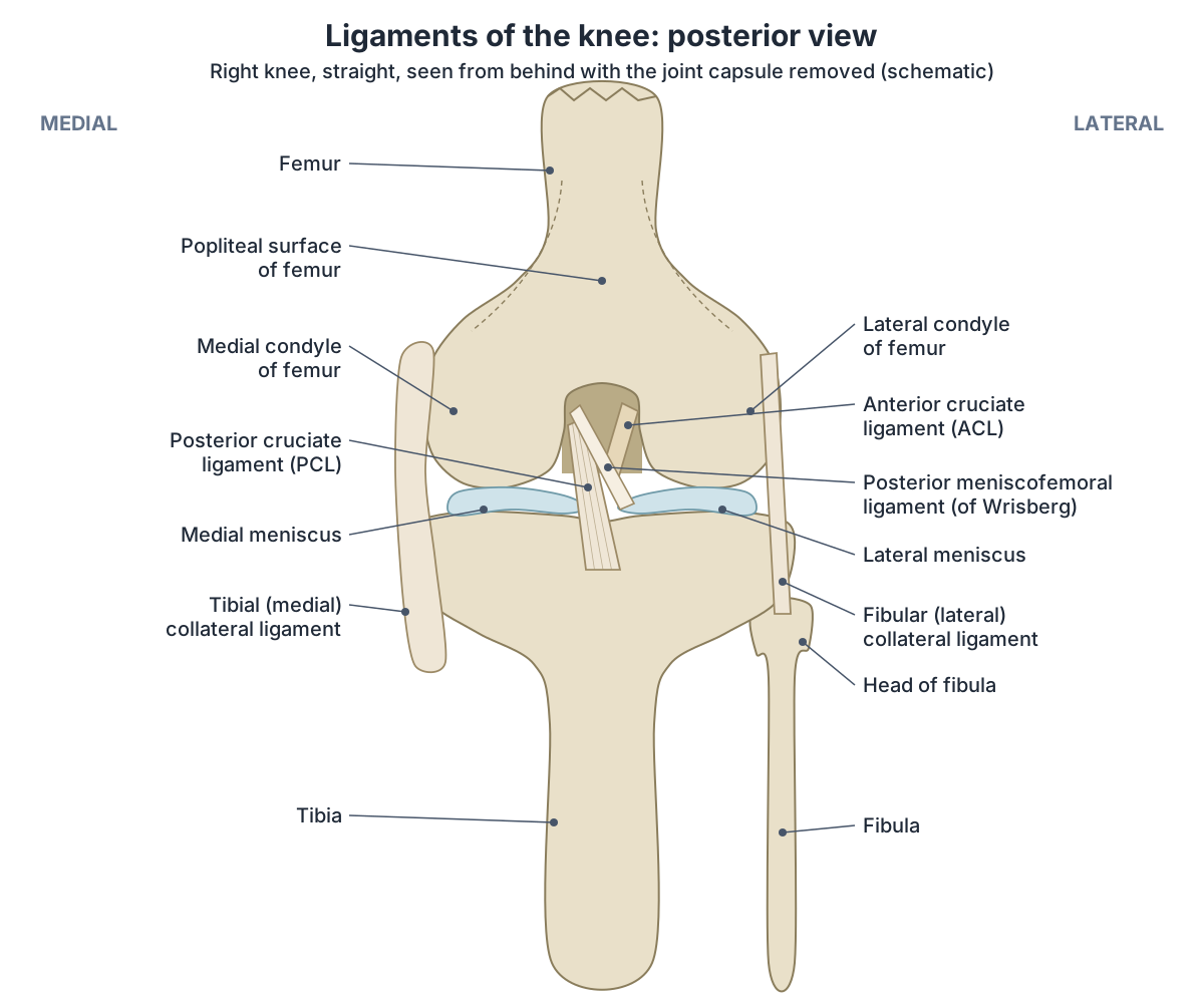
MCL and LCL: the collateral ligaments
The medial collateral ligament (MCL), also called the tibial collateral ligament, is wide and flat. It runs from the medial epicondyle of the femur to the medial condyle of the tibia. The lateral collateral ligament (LCL), or fibular collateral ligament, is thinner and rounder and runs from the lateral epicondyle of the femur to the lateral surface of the head of the fibula. (TeachMeAnatomy: The Knee Joint.)
The deep part of the MCL is a thickening of the joint capsule that is attached to the medial meniscus. The LCL splits the tendon of biceps femoris and lies superficial to the tendon of popliteus. (Kenhub: Knee joint.)
Both collateral ligaments are tight when the knee is straight. They stop the knee bending sideways and limit rotation. (OpenStax Anatomy and Physiology 2e, 9.6: Anatomy of Selected Synovial Joints.)
Because the medial meniscus is attached to the MCL and the lateral meniscus is not attached to the LCL, the lateral meniscus is free to move more. (Fox AJS, Bedi A, Rodeo SA. The Basic Science of Human Knee Menisci. Sports Health. 2012.)
Patellar ligament and patellar retinacula
The patellar ligament is the continuation of the quadriceps tendon below the patella and attaches to the tibial tuberosity. The patella increases the leverage of the quadriceps and reduces friction on the femoral condyles. (TeachMeAnatomy: The Knee Joint.)
On each side of the patella, the medial and lateral patellar retinacula, expansions of vastus medialis and vastus lateralis, blend with the patellar ligament. Behind the knee, the oblique popliteal ligament, an expansion of the semimembranosus tendon, and the arcuate popliteal ligament from the head of the fibula reinforce the capsule and help prevent hyperextension. (Kenhub: Knee joint.)
OpenStax also describes quadriceps femoris, the muscle that extends the knee, as a 'dynamic ligament' that supports the knee. (OpenStax Anatomy and Physiology 2e, 9.6: Anatomy of Selected Synovial Joints.)
Medial and lateral meniscus
The menisci are two crescent-shaped wedges of fibrocartilage on top of the tibia. The medial meniscus is roughly semicircular (C-shaped) and the lateral meniscus is nearly circular. Each is thick at its outer edge, where it attaches to the capsule, and thins to a free inner edge. The upper surface is concave to fit the femoral condyle and the lower surface is flat. (Fox AJS, Bedi A, Rodeo SA. The Basic Science of Human Knee Menisci. Sports Health. 2012.)
The anterior horn of the medial meniscus attaches in front of the ACL, and its posterior horn between the lateral meniscus and the PCL. A transverse ligament joins the two anterior horns. The posterior and anterior meniscofemoral ligaments (of Wrisberg and Humphry) run from the back of the lateral meniscus to the medial femoral condyle, beside the PCL; their number varies from none to four. (Fox AJS, Bedi A, Rodeo SA. The Basic Science of Human Knee Menisci. Sports Health. 2012.)
The lateral meniscus covers about 80% of the lateral tibial joint surface and the medial meniscus about 60% of the medial surface. The menisci carry about 50% of the load in the medial compartment and about 70% in the lateral compartment, and they also absorb shock and help lubricate and nourish the joint. (Fox AJS, Bedi A, Rodeo SA. The Basic Science of Human Knee Menisci. Sports Health. 2012.)
Only the outer part of each meniscus has a good blood supply, from the genicular arteries: about 10 to 30% of the width of the medial meniscus and 10 to 25% of the lateral. The rest of the meniscus is nourished by synovial fluid instead. (Fox AJS, Bedi A, Rodeo SA. The Basic Science of Human Knee Menisci. Sports Health. 2012.)
The medial meniscus is anchored to the capsule and the tibial collateral ligament, so it moves less. It is about 20 times more likely to be injured than the lateral meniscus, and the parts of each meniscus that lack an arterial supply heal poorly. (OpenStax Anatomy and Physiology 2e, 9.6: Anatomy of Selected Synovial Joints.)
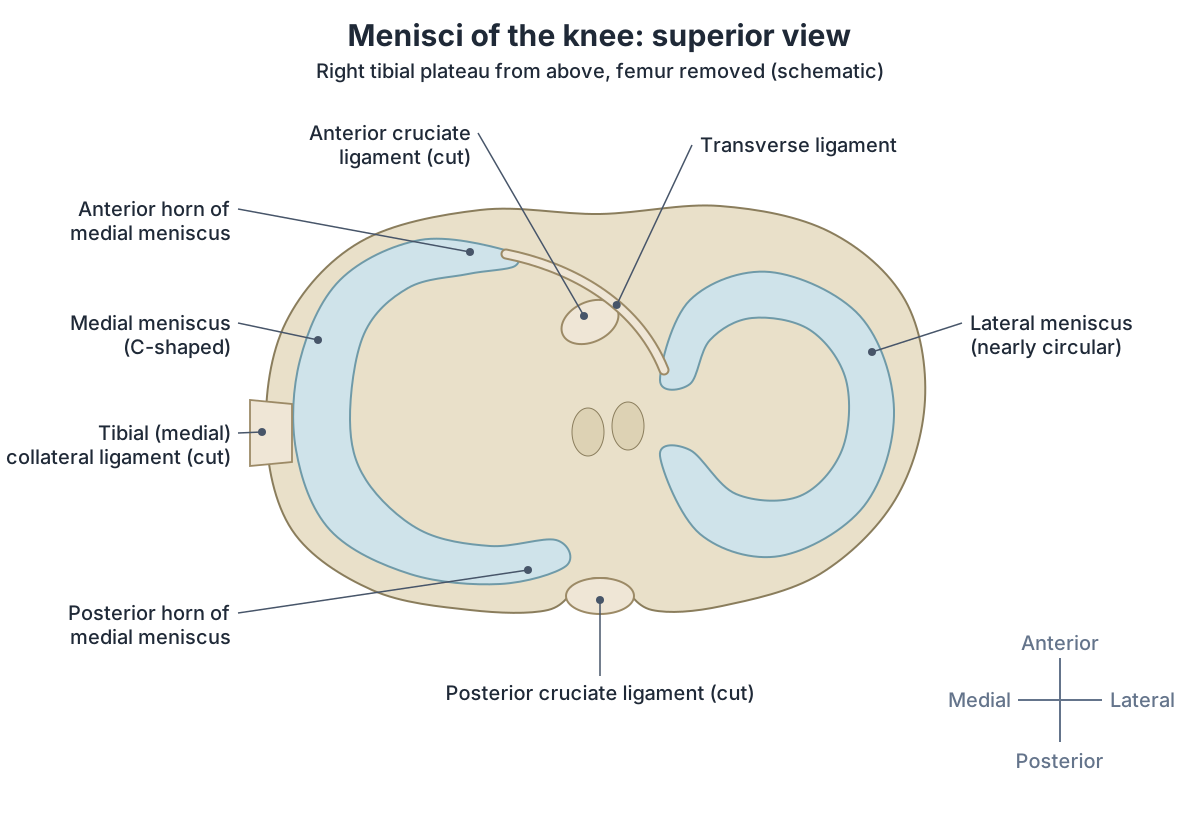
Joint capsule and bursae of the knee
The knee's capsule is relatively weak and is made largely of the tendons of the surrounding muscles and their expansions. It has an outer fibrous layer and an inner synovial membrane that makes synovial fluid to lubricate the joint and nourish the cartilage. (Kenhub: Knee joint.)
Bursae are fluid-filled sacs that reduce friction. The suprapatellar bursa lies between quadriceps femoris and the femur, the prepatellar bursa between the patella and the skin, and the deep and superficial infrapatellar bursae behind and in front of the patellar ligament. Behind the knee, the semimembranosus bursa lies between semimembranosus and the medial head of gastrocnemius. (TeachMeAnatomy: The Knee Joint.)
Inflammation of the prepatellar bursa is known as housemaid's knee and inflammation of the infrapatellar bursa as clergyman's knee. (TeachMeAnatomy: The Knee Joint.)
Knee flexion vs extension: movements and muscles
Flexion bends the knee and extension straightens it. The knee can flex to about 140 degrees with the hip flexed and about 120 degrees with the hip extended, and up to about 160 degrees when pushed passively; contact between calf and thigh is the main limit. (Kenhub: Knee joint.)
Extension is produced by quadriceps femoris. Flexion is produced by the hamstrings, helped by gracilis, sartorius and popliteus. Biceps femoris rotates the bent knee laterally, and semimembranosus, semitendinosus, gracilis, sartorius and popliteus rotate it medially. Rotation is only possible when the knee is bent. (TeachMeAnatomy: The Knee Joint.)
In the last part of extension the femur rotates slightly medially on the tibia. This 'locks' the knee. To bend the knee again, popliteus rotates the femur laterally to 'unlock' it. (OpenStax Anatomy and Physiology 2e, 9.6: Anatomy of Selected Synovial Joints.)
Kenhub calls this the screw-home mechanism: in full extension the knee is in its close-packed position, which allows it to bear weight for long periods without muscle activity. (Kenhub: Knee joint.)
Muscles that move the knee
| Movement | Main muscles |
|---|---|
| Extension | Quadriceps femoris (rectus femoris, vastus lateralis, vastus medialis, vastus intermedius) |
| Flexion | Hamstrings (biceps femoris, semitendinosus, semimembranosus), assisted by gracilis, sartorius and popliteus |
| Medial rotation (knee bent) | Semimembranosus, semitendinosus, gracilis, sartorius, popliteus |
| Lateral rotation (knee bent) | Biceps femoris |
| Unlocking from full extension | Popliteus |
From TeachMeAnatomy and Kenhub.
Articularis genus muscle
Articularis genus (articularis genu) is a small muscle on the front of the lower femur, deep to vastus intermedius. It arises from the front of the distal third of the femur and its bundles insert into the wall of the suprapatellar bursa and the synovial lining of the knee capsule. (Grob K, et al. The Anatomy of the Articularis Genus Muscle and Its Relation to the Extensor Apparatus of the Knee. JBJS Open Access. 2017.)
Its fibres are continuous with the deep fibres of vastus intermedius, and it is supplied by the femoral nerve (the medial deep division, which also supplies vastus medialis). It has long been thought to pull the suprapatellar bursa upward as the knee straightens. A 2017 dissection study suggests it is too small to do this alone and works together with vastus intermedius and vastus medialis. (Grob K, et al. The Anatomy of the Articularis Genus Muscle and Its Relation to the Extensor Apparatus of the Knee. JBJS Open Access. 2017.)
Blood and nerve supply of the knee
The knee is supplied by the genicular anastomosis, a network of genicular branches from the femoral and popliteal arteries. By Hilton's law, the nerves that supply the muscles crossing the knee also supply the joint: the femoral, tibial and common fibular nerves. (TeachMeAnatomy: The Knee Joint.)
Kenhub adds the posterior division of the obturator nerve to the knee's nerve supply, and notes that a 2013 dissection study found much more variation in the genicular network than textbook diagrams suggest. (Kenhub: Knee joint.)
Common knee ligament injuries
ACL tears usually happen with a sudden change of direction that twists and hyperextends the knee, or a forceful blow from the front. PCL injuries typically happen when the bent knee is struck and the tibia is driven backward, as when the knee hits the dashboard in a car crash. (OpenStax Anatomy and Physiology 2e, 9.6: Anatomy of Selected Synovial Joints.)
In the anterior drawer test the examiner pulls the tibia forward; extra forward movement suggests an ACL tear. In the posterior drawer test, extra backward movement suggests a PCL tear. (TeachMeAnatomy: The Knee Joint.)
A blow to the outer side of the knee injures the medial (tibial) collateral ligament, and a blow to the inner side injures the lateral (fibular) collateral ligament. (Kenhub: Knee joint.)
A hard blow to the lateral side of the straight knee can cause the 'unhappy' or 'terrible' triad: tears of the tibial collateral ligament, the medial meniscus and the ACL. (OpenStax Anatomy and Physiology 2e, 9.6: Anatomy of Selected Synovial Joints.)
Knee joint in the 3D model
Frequently asked questions
What are the four main ligaments of the knee?
The four main ligaments are the anterior cruciate ligament (ACL), the posterior cruciate ligament (PCL), the medial (tibial) collateral ligament (MCL) and the lateral (fibular) collateral ligament (LCL). The cruciates cross inside the joint and stop the tibia sliding forward or backward. The collaterals run down the sides and stop the knee bending sideways.
What is the difference between the ACL and PCL?
The ACL starts at the front of the tibia's intercondylar area and runs to the lateral femoral condyle; it stops the tibia moving forward and resists hyperextension. The PCL starts at the back of the tibia and runs to the medial femoral condyle; it stops the tibia moving backward. The PCL is the stronger of the two and supports the bent, weight-bearing knee.
Is the meniscus a ligament?
No. The menisci are C-shaped pads of fibrocartilage that sit on the tibia between the tibia and femur. They improve the fit of the joint, spread load and absorb shock. They are held in place by ligaments, such as the transverse and meniscofemoral ligaments, and the medial meniscus is also attached to the MCL.
Why is the medial meniscus injured more often than the lateral?
The medial meniscus is anchored to the joint capsule and the tibial collateral ligament, so it cannot move out of the way when the knee twists. The lateral meniscus is not attached to the lateral collateral ligament and moves more freely. OpenStax states the medial meniscus is about 20 times more likely to be injured.
What is the unhappy triad of the knee?
The unhappy (or terrible) triad is a combined injury of the medial collateral ligament, the medial meniscus and the anterior cruciate ligament. It happens when a strong force hits the outer side of the knee while the leg is straight and the foot is planted, as in a rugby or football tackle.
Why can't you see the knee ligaments in the 3D model?
The 3D model is built from the BodyParts3D data set, which includes the femur, tibia, fibula and patella but not the knee ligaments or menisci. The diagrams on this page show those structures instead, from the front, from behind and from above, so you can match them to the bones in the model.
References
- OpenStax. Anatomy and Physiology 2e, §9.6 Anatomy of Selected Synovial Joints. Rice University, 2022.
- OpenStax. Anatomy and Physiology 2e, §8.4 Bones of the Lower Limb. Rice University, 2022.
- TeachMeAnatomy. The Knee Joint.
- TeachMeAnatomy. The Femur.
- Kenhub. Knee joint.
- Kenhub. Anterior cruciate ligament.
- Fox AJS, Bedi A, Rodeo SA. The Basic Science of Human Knee Menisci: Structure, Composition, and Function. Sports Health. 2012;4(4):340–351. doi:10.1177/1941738111429419
- Grob K, Gilbey H, Manestar M, Ackland T, Kuster MS. The Anatomy of the Articularis Genus Muscle and Its Relation to the Extensor Apparatus of the Knee. JBJS Open Access. 2017;2(4):e0034. doi:10.2106/JBJS.OA.17.00034
- Mitsuhashi N. et al. (2009). BodyParts3D: 3D structure database for anatomical concepts. Nucleic Acids Research 37, D782–D785. Data © The Database Center for Life Science, CC BY 4.0.
Related anatomy
- FemurFemur bone labelled: head, neck, trochanters, linea aspera, condyles and epicondyles, with muscle attachments, blood…
- PatellaWhat is the patella? The kneecap explained: base, apex, facets, quadriceps tendon and patellar ligament, why it…
- TibiaTibia anatomy explained: where the shinbone is, the proximal tibia, tibial plateau, shaft and medial malleolus, its…
- FibulaFibula anatomy: the head of fibula, shaft and lateral malleolus, the proximal and distal tibiofibular joints and…
- Thigh musclesThigh muscles explained: quadriceps and sartorius at the front, adductor muscles of the inner thigh, hamstrings at the…
- Popliteal arteryPopliteal artery anatomy: course through the popliteal fossa, genicular branches, how to feel the popliteal pulse…
Explore the whole body in the interactive 3D human anatomy model, or browse the A–Z list of human structures.