

Popliteal artery at a glance
- What it is
- The continuation of the femoral artery behind the knee.
- Location
- It begins at the adductor hiatus, runs through the popliteal fossa and divides below the knee into anterior and posterior tibial arteries; branches include genicular and sural arteries.
- Function
- It supplies the knee joint and muscles of the posterior thigh and leg.
- Body system
- Cardiovascular · paired (left and right)
- FMA ID
- FMA77381 (Foundational Model of Anatomy)
Key facts
- Its pulse can be felt deep in the popliteal fossa.
- It is the deepest structure in the popliteal fossa, lying directly on the femur and knee joint capsule, so the pulse needs firm pressure with the knee flexed.
Clinical relevance
Popliteal aneurysm and knee dislocation can threaten limb circulation.
Where is the popliteal artery?
The popliteal artery begins where the femoral artery leaves the adductor canal through the adductor hiatus, at the junction of the middle and lower thirds of the thigh. It runs backwards into the popliteal fossa and down its floor, lying just behind (superficial to) the popliteus muscle. It ends at the lower border of popliteus by dividing into the anterior tibial artery and the tibioperoneal trunk. (Radiopaedia: Popliteal artery.)
The adductor hiatus is a gap in adductor magnus between its adductor part and its hamstring part. The femoral vein passes through the same gap and becomes the popliteal vein. (TeachMeAnatomy: The Adductor Canal.)
On its way down the back of the thigh and knee the artery gives genicular branches to the knee joint. It then leaves the popliteal fossa between gastrocnemius and popliteus to reach the back of the leg. (TeachMeAnatomy: Arterial Supply to the Lower Limb.)
For surgery and angiography the artery is divided into three segments: P1 from the adductor hiatus to the upper edge of the patella, P2 from there to the centre of the knee joint, and P3 from the centre of the joint to the origin of the anterior tibial artery. (Radiopaedia: Popliteal artery.)
In the 3D model, rotate to the posterior view to see the artery behind the knee with the popliteal vein and the five genicular arteries. The model is from BodyParts3D, an adult male data set, and does not include the nerves of the fossa, which are shown in the diagram instead.
The popliteal fossa: borders and contents
The popliteal fossa is a diamond-shaped space at the back of the knee and the main route for vessels and nerves between the thigh and the leg. Its upper medial border is semimembranosus, its upper lateral border is biceps femoris, its lower medial border is the medial head of gastrocnemius and its lower lateral border is the lateral head of gastrocnemius with plantaris. (TeachMeAnatomy: The Popliteal Fossa.)
The floor is the back of the femur, the posterior capsule of the knee joint and popliteus. The roof is the popliteal fascia, a continuation of the fascia lata, covered by skin. The fascia is tough and does not stretch, which matters when a mass grows inside the fossa. (TeachMeAnatomy: The Popliteal Fossa.)
The fossa contains the popliteal artery, the popliteal vein, the tibial nerve and the common fibular (peroneal) nerve. The two nerves are the most superficial contents and the popliteal artery is the deepest. The common fibular nerve follows the biceps femoris tendon along the lateral edge, and the small saphenous vein pierces the roof to join the popliteal vein. (TeachMeAnatomy: The Popliteal Fossa.)
The capsule, ligaments and menisci that the popliteal artery supplies are described on the Knee joint.
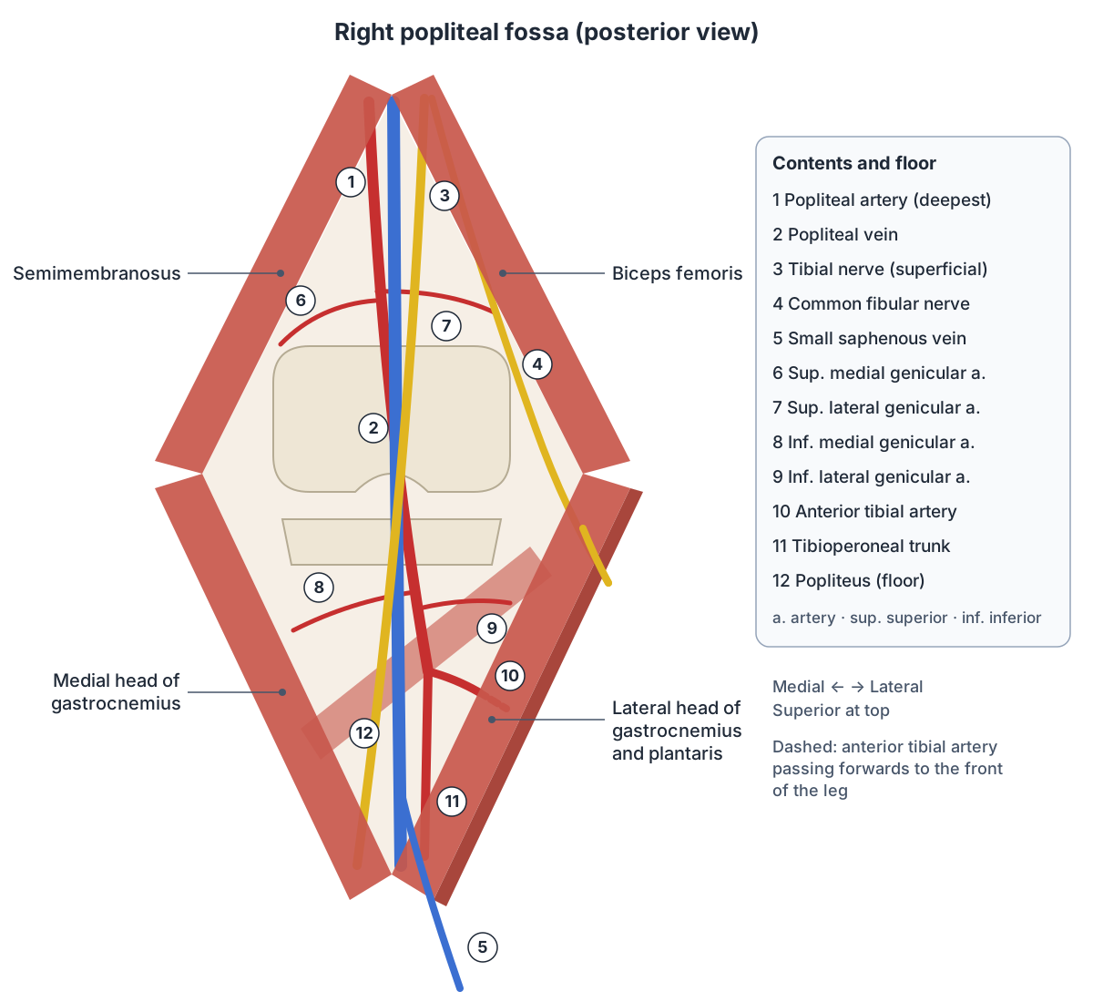
Relations: artery, vein and tibial nerve
From deep to superficial, the three main structures are stacked as artery, vein and tibial nerve. In front of (deep to) the artery lie the popliteal surface of the femur, separated by fat, then the knee joint capsule and popliteus. Behind it lie the popliteal vein and, most superficial, the tibial nerve. (Radiopaedia: Popliteal artery.)
Seen from side to side, the artery starts medial to the tibial nerve and ends lateral to it, so the nerve crosses behind the artery from lateral to medial as they descend. The popliteal vein always lies between the two. Above, semimembranosus and the medial femoral condyle lie medial to the artery and biceps femoris and the lateral femoral condyle lie lateral to it. (Radiopaedia: Popliteal artery.)
Branches of the popliteal artery: genicular and sural arteries
The popliteal artery gives five genicular (knee) arteries: superior lateral, superior medial, middle, inferior lateral and inferior medial. The superior pair wrap around the femoral condyles and supply the bone of the condyles, the synovium and the upper patella. The inferior pair wrap around the tibial condyles, deep to the collateral ligaments. (Kenhub: Popliteal artery.)
The single middle genicular artery pierces the back of the knee capsule (the oblique popliteal ligament) and supplies the posterior cruciate ligament, part of the anterior cruciate ligament and the back of the menisci. (Kenhub: Popliteal artery.)
Together the genicular arteries form the genicular anastomosis, a network around the knee that supplies the capsule and ligaments. From below, the anterior tibial recurrent artery climbs up through tibialis anterior to join the lateral genicular branches. (Radiopaedia: Anterior tibial artery.)
Two large sural arteries, one on each side, supply gastrocnemius, soleus and plantaris. Other muscular branches supply the hamstrings and adductor magnus, and small cutaneous branches supply the skin over the back of the knee. (Kenhub: Popliteal artery.)
Branches of the popliteal artery
| Branch | Course | Supply |
|---|---|---|
| Superior medial and superior lateral genicular | Wind around the femoral condyles | Condyles, knee synovium, upper patella |
| Middle genicular | Pierces the oblique popliteal ligament | Cruciate ligaments, back of the menisci |
| Inferior medial and inferior lateral genicular | Wind around the tibial condyles, deep to the collateral ligaments | Capsule, collateral ligaments, lower patella |
| Sural arteries (two) | One on each side | Gastrocnemius, soleus, plantaris |
| Muscular and cutaneous branches | Upper fossa and skin | Hamstrings, adductor magnus, skin over the knee |
| Anterior tibial artery (terminal) | Through the gap above the interosseous membrane | Anterior compartment of the leg, dorsum of the foot |
| Tibioperoneal trunk (terminal) | Divides into posterior tibial and fibular arteries | Posterior and lateral compartments of the leg, sole |
Based on Kenhub, Radiopaedia and TeachMeAnatomy (see references).
How the popliteal artery divides: normal pattern and variants
In about 90% of people the popliteal artery divides at the lower border of popliteus into the anterior tibial artery and the tibioperoneal trunk; the trunk then divides into the posterior tibial and fibular (peroneal) arteries. The other 10% show a variant. (Radiopaedia: Popliteal artery.)
Radiopaedia lists hypoplastic leg arteries in about 4%, a trifurcation into all three leg arteries with no tibioperoneal trunk in about 2%, a high origin of the anterior tibial artery in about 2.5% and a high origin of the posterior tibial artery in about 1%. Rarely the fibular artery arises from the anterior tibial artery, or the popliteal artery continues from a persistent sciatic artery. (Radiopaedia: Popliteal artery.)
A high-origin anterior tibial artery may run between the back of the tibia and popliteus. Knowing about this matters in knee replacement, high tibial osteotomy, meniscal repair and posterior cruciate ligament reconstruction, where it could be injured. (Radiopaedia: Anterior tibial artery.)
How to feel the popliteal pulse
The popliteal pulse is the hardest lower limb pulse to feel because the artery is the deepest structure in the fossa. It needs firm, deep pressure, and bending the knee slightly helps by relaxing the popliteal fascia. (TeachMeAnatomy: Arterial Supply to the Lower Limb.)
Kenhub describes the technique with the patient lying down, the knee raised and flexed to relax the popliteal fascia and the hamstrings, and the fingers pressing into the lower part of the fossa. A weak or absent popliteal pulse may point to a blockage higher up, for example in the femoral artery. (Kenhub: Popliteal artery.)
Popliteal artery aneurysm
A popliteal artery aneurysm is an abnormal widening of the artery behind the knee. It is the most common aneurysm of a peripheral artery and the second most common aneurysm overall after abdominal aortic aneurysm. It occurs almost only in men (up to 97% of cases), becomes more common with age and is bilateral in 50 to 70% of cases. (Radiopaedia: Popliteal artery aneurysm.)
About 40% of people with a popliteal aneurysm also have an abdominal aortic aneurysm, so finding one leads doctors to check for the other. The most common complication is a clot breaking off and blocking smaller arteries downstream (distal thromboembolism), in 18 to 31% of untreated cases; rupture is rare. Asymptomatic aneurysms larger than 2 cm are considered for elective repair. (Radiopaedia: Popliteal artery aneurysm.)
TeachMeAnatomy defines an aneurysm as a widening of an artery to more than 50% above its normal diameter. Because the popliteal fascia does not stretch, a large popliteal aneurysm presses on its neighbours, especially the tibial nerve. This can weaken plantarflexion and cause tingling in the foot and back of the leg. The aneurysm may be felt as a pulsating swelling behind the knee, sometimes with a bruit. A Baker's cyst, a swollen semimembranosus bursa, is the other common cause of a lump in the fossa. (TeachMeAnatomy: The Popliteal Fossa.)
Popliteal artery entrapment syndrome
Popliteal artery entrapment syndrome happens when the artery is squeezed by a muscle it develops in an abnormal relation to, most often the medial head of gastrocnemius, less often popliteus or a fibrous band. The anatomical anomaly is found in up to about 3 to 3.5% of people, about two thirds of them on both sides, but most never have symptoms. (Radiopaedia: Popliteal artery entrapment syndrome.)
Symptoms are most common in young, muscular athletes: about 60% of cases are under 30 and men outnumber women by about 15 to 1. The typical complaint is cramping calf pain on exercise (claudication), and pulses may weaken when the ankle is pointed down (plantarflexed). Repeated compression can lead to thrombosis and acute limb ischaemia. (Radiopaedia: Popliteal artery entrapment syndrome.)
The usual classification has six types. In type I the artery takes an abnormal course around the medial head of gastrocnemius; in type II the medial head attaches too far laterally; in type III an extra slip of the medial head wraps the artery; in type IV popliteus or a fibrous band traps it; type V involves the vein as well; and type VI is a functional form caused by an enlarged gastrocnemius. Treatment is surgical release, sometimes with a saphenous vein bypass. (Radiopaedia: Popliteal artery entrapment syndrome.)
Popliteal artery injury in knee dislocation
Because the popliteal artery lies directly on the back of the knee joint, a knee dislocation, usually from high-energy trauma, can stretch, bruise, tear or sever it. The popliteal vein and calf muscles may be damaged too, and untreated arterial injury can lead to loss of the limb. (Kenhub: Popliteal artery.)
Structures next to the popliteal artery
The nearest named structures in the 3D model, measured from the centre of each part:
- Middle genicular artery · cardiovascular
- Popliteal vein · cardiovascular
- Inferior lateral genicular artery · cardiovascular
- Medial superior genicular artery · cardiovascular
- Lateral superior genicular artery · cardiovascular
- Great saphenous vein · cardiovascular
- Anterior tibial recurrent artery · cardiovascular
- Patella · skeletal
- Inferior medial genicular artery · cardiovascular
- Popliteus · muscular
Frequently asked questions
Where is the popliteal artery?
The popliteal artery lies behind the knee, in the diamond-shaped popliteal fossa. It continues the femoral artery from the adductor hiatus, just above the knee, and runs down on the back of the femur and knee joint capsule, deeper than the popliteal vein and tibial nerve. It ends just below the knee at the lower border of the popliteus muscle.
Why is the popliteal pulse hard to feel?
The popliteal artery is the deepest structure in the popliteal fossa, under the tibial nerve, popliteal vein, fat and the tough popliteal fascia. To feel it, the knee is bent slightly to relax the fascia and hamstrings, and the fingers press firmly into the lower part of the fossa against the back of the knee.
What are the branches of the popliteal artery?
It gives five genicular arteries to the knee (superior medial and lateral, middle, and inferior medial and lateral), two sural arteries to the calf muscles, and muscular and skin branches. It ends by dividing into the anterior tibial artery and the tibioperoneal trunk, which then splits into the posterior tibial and fibular (peroneal) arteries.
What is a popliteal aneurysm?
A popliteal aneurysm is an abnormal ballooning of the artery behind the knee. It is the most common peripheral artery aneurysm and affects mostly older men, often on both sides. About 40% of patients also have an abdominal aortic aneurysm. Its main danger is clots breaking off and blocking leg arteries, rather than rupture.
What is popliteal artery entrapment syndrome?
It is compression of the popliteal artery by a muscle or band it has developed in an abnormal relation to, most often the medial head of gastrocnemius. It mainly affects young, muscular athletes, usually men, and causes cramping calf pain on exercise. It is treated surgically by releasing the artery, sometimes with a vein bypass.
What is the popliteal fossa?
The popliteal fossa is the diamond-shaped hollow at the back of the knee. Semimembranosus and biceps femoris form its upper borders and the two heads of gastrocnemius its lower borders. It contains the popliteal artery and vein, the tibial and common fibular nerves, and the end of the small saphenous vein.
References
- Popliteal artery. Radiopaedia reference article.
- Popliteal artery aneurysm. Radiopaedia reference article.
- Popliteal artery entrapment syndrome. Radiopaedia reference article.
- Anterior tibial artery. Radiopaedia reference article.
- Jones O. The Popliteal Fossa. TeachMeAnatomy.
- Jones O. Arterial Supply to the Lower Limb. TeachMeAnatomy.
- Jones O. The Adductor Canal. TeachMeAnatomy.
- Popliteal artery. Kenhub anatomy library.
- Foundational Model of Anatomy: Popliteal artery (FMA77381). EMBL-EBI Ontology Lookup Service.
- Mitsuhashi N. et al. (2009). BodyParts3D: 3D structure database for anatomical concepts. Nucleic Acids Research 37, D782–D785. Data © The Database Center for Life Science, CC BY 4.0.
Related anatomy
- Lower limb arteriesArteries of the leg explained: common, internal and external iliac, femoral, deep femoral, popliteal, tibial, fibular…
- Femoral arteryFemoral artery anatomy: where it is, the femoral triangle and sheath (NAVEL), adductor canal, branches, deep femoral…
- Knee jointLigaments of the knee explained with labelled diagrams: ACL, PCL, MCL, LCL, patellar ligament and menisci, plus knee…
- Popliteal veinPopliteal vein: A deep vein behind the knee formed by the joining of the anterior and posterior tibial veins. See…
- Leg musclesLower leg muscles by compartment: tibialis anterior, fibularis (peroneus) longus and brevis, gastrocnemius, soleus…
- Great saphenous veinSaphenous vein anatomy: course of the great and small saphenous veins, saphenofemoral junction, valves, saphenous…
Explore the whole body in the interactive 3D human anatomy model, or browse the A–Z list of human structures.