

What is the rib cage and the thorax?
The thoracic cage, or rib cage, forms the thorax (chest) part of the body. It consists of 12 pairs of ribs with their costal cartilages and the sternum. At the back, the ribs are anchored to the 12 thoracic vertebrae, T1 to T12. The cage protects the heart and lungs. (OpenStax Anatomy and Physiology 2e, 7.4: The Thoracic Cage.)
The ribs also take part in breathing. They are not rigid hoops: they move at their joints with the spine and sternum, so the cage can expand and shrink as air is drawn in and pushed out. (TeachMeAnatomy: The Ribs.)
The 3D model above shows all 24 ribs, the manubrium, body and xiphoid process of the sternum, the thoracic vertebrae and the costal cartilages of ribs 1 to 7. It is based on BodyParts3D, an adult male data set.
How many ribs does a human have?
Humans normally have 12 pairs of ribs, 24 in total, numbered 1 to 12 from the top to match the thoracic vertebra each one joins. Each rib is a curved, flattened bone that forms part of the wall of the thorax. (OpenStax Anatomy and Physiology 2e, 7.4: The Thoracic Cage.)
The ribs lengthen from rib 1 to rib 7 and then shorten from rib 8 to rib 12. The spaces between neighbouring ribs are the intercostal spaces. (Radiopaedia: Ribs.)
Variation is not rare. About 5 to 8% of people have only 11 pairs as a normal variant. Extra ribs can arise from C7 (cervical ribs) or L1 (lumbar ribs), and forked (bifid) ribs occur in about 0.2% of people. (Radiopaedia: Ribs.)
A cervical rib is found in about 0.5% of the population, more often in females, and is usually on both sides. Most are harmless, but a cervical rib can compress the brachial plexus or the subclavian vessels and cause thoracic outlet syndrome. (Radiopaedia: Cervical rib.)
True, false and floating ribs
Ribs 1 to 7 are true ribs (vertebrosternal ribs): each one's costal cartilage attaches directly to the sternum. Ribs 8 to 12 are false ribs, because their cartilages do not reach the sternum directly. The cartilages of ribs 8 to 10 join the cartilage of the rib above (10 to 9, 9 to 8, 8 to 7), so they reach the sternum only indirectly (vertebrochondral ribs). (OpenStax Anatomy and Physiology 2e, 7.4: The Thoracic Cage.)
Ribs 11 and 12 are floating ribs (vertebral ribs). They are short and do not attach to the sternum at all; their cartilages end in the muscles of the lateral abdominal wall. In OpenStax's scheme the floating ribs are a subgroup of the false ribs. (OpenStax Anatomy and Physiology 2e, 7.4: The Thoracic Cage.)
Some sources, including Radiopaedia, use 'false ribs' only for ribs 8 to 10 and list ribs 11 and 12 separately as floating ribs. Both conventions describe the same anatomy; only the label differs. (Radiopaedia: Ribs.)
The costal margin (costal arch) is the curved lower edge of the front of the rib cage. It is formed by the costal cartilage of rib 7 and the joined costal cartilages of ribs 8 to 10, which are linked to each other by small interchondral synovial joints. Some sources count only cartilages 8 to 10, because cartilage 7 also reaches the sternum. (Radiopaedia: Costal cartilage.)
Classification of the ribs
| Ribs | Group | Anterior attachment | Notes |
|---|---|---|---|
| 1 to 7 | True (vertebrosternal) | Own costal cartilage joins the sternum directly | Rib 1 joins the manubrium; rib 2 the sternal angle; rib 7 near the xiphisternal joint |
| 8 to 10 | False (vertebrochondral) | Cartilage joins the cartilage of the rib above | Their cartilages, with cartilage 7, form the costal margin |
| 11 and 12 | Floating (vertebral); counted as false ribs by OpenStax | None; end in the abdominal wall muscles | Short, single head facet, no neck or tubercle |
Based on OpenStax and Radiopaedia, which differ only in whether floating ribs are counted as false ribs.
Parts of a typical rib
Ribs 3 to 9 are typical ribs. A typical rib has a head, a neck, a tubercle and a long, curved body (shaft). The head is wedge-shaped, with two facets separated by a ridge of bone: one facet joins the vertebra of the same number and the other joins the vertebra above. (TeachMeAnatomy: The Ribs.)
The neck is a short narrowed part just lateral to the head. The tubercle is a small bump on the back of the rib where the neck meets the body; it carries a facet that joins the transverse process of the vertebra of the same number. (OpenStax Anatomy and Physiology 2e, 7.4: The Thoracic Cage.)
Just lateral to the tubercle is the angle of the rib, where the rib curves most sharply. The angles of the ribs form the back limit of the cage. The rest of the rib is the body or shaft. (OpenStax Anatomy and Physiology 2e, 7.4: The Thoracic Cage.)
Along the lower inner edge of the shaft runs the costal groove, which shelters the intercostal neurovascular bundle. From top to bottom the order is vein, artery, nerve (often remembered as VAN). (TeachMeAnatomy: The Ribs.)
Atypical ribs: 1, 2, 10, 11 and 12
Rib 1 is short, broad and sharply curved. Its head has a single facet, because there is no vertebra above T1 for it to touch. Its upper surface has two grooves for the subclavian vessels. (TeachMeAnatomy: The Ribs.)
Rib 2 is thinner and longer than rib 1 and has the usual two facets on its head. Its upper surface has a roughened area where serratus anterior arises. (TeachMeAnatomy: The Ribs.)
Rib 10 usually has a single facet on its head for its own vertebra and may lack a tubercle. Ribs 11 and 12 are short, have a single facet, and have no neck or tubercle; they do not join the sternum. (Radiopaedia: Ribs.)
Joints of the ribs
Each rib makes two joints at the back. At the costovertebral joint, the head of the rib meets the superior costal facet of its own vertebra and the inferior costal facet of the vertebra above. At the costotransverse joint, the tubercle meets the transverse costal facet of its own vertebra. (TeachMeAnatomy: The Ribs.)
On a typical thoracic vertebra such as T5, the superior costal facet (demifacet) on the body takes rib 5 and the inferior demifacet takes rib 6, while the facet on the transverse process takes the tubercle of rib 5. (Radiopaedia: Typical thoracic vertebrae.)
At the front, each rib joins its costal cartilage at a costochondral joint. The first costal cartilage joins the manubrium by a cartilaginous joint (synchondrosis), while costal cartilages 2 to 7 join the sternum by small synovial joints. (Radiopaedia: Sternum.)
Sternum anatomy: manubrium, body and xiphoid process
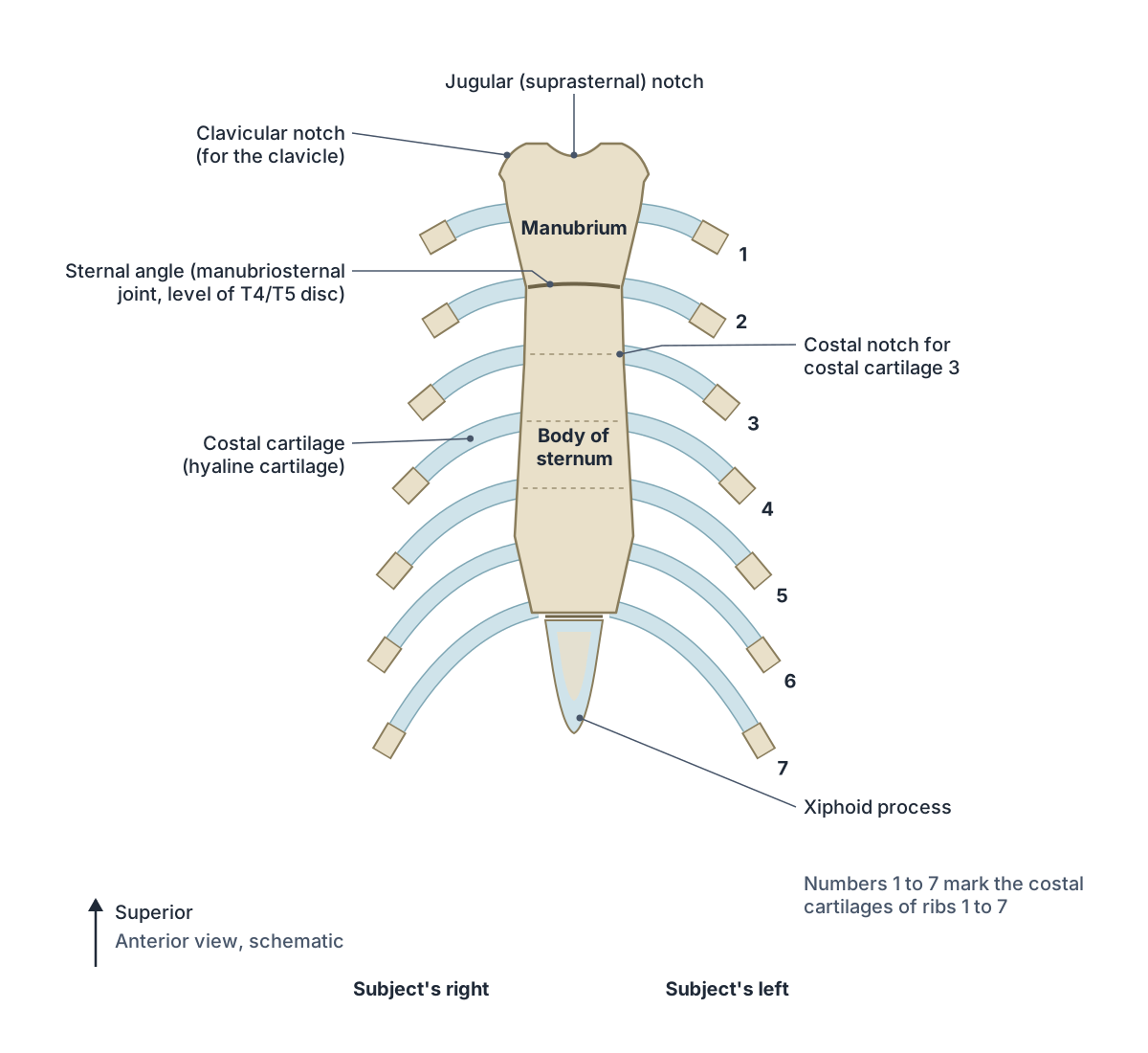
The sternum (breastbone) is the flat bone in the midline of the front of the chest. It has three parts, from top to bottom: the manubrium, the body and the xiphoid process. In children the three parts are joined by cartilage, which ossifies in adulthood. (TeachMeAnatomy: The Sternum.)
The manubrium is the wide upper part. Its upper border dips into the jugular (suprasternal) notch, which you can feel at the base of the neck between the inner ends of the clavicles. On each side of the notch is a clavicular notch, where the clavicle forms the sternoclavicular joint. The first costal cartilage attaches to the side of the manubrium. (OpenStax Anatomy and Physiology 2e, 7.4: The Thoracic Cage.)
The manubrium meets the body at the sternal angle (angle of Louis), a slight bend you can feel as a horizontal ridge. The second costal cartilage attaches here. Because rib 1 is hidden behind the clavicle, rib 2 is the highest rib you can feel, so the sternal angle is the starting point for counting ribs. (OpenStax Anatomy and Physiology 2e, 7.4: The Thoracic Cage.)
The joint at the sternal angle is the manubriosternal joint, a secondary cartilaginous joint (symphysis). It lies at the level of the second costal cartilage at the front and the intervertebral disc between T4 and T5 at the back. (Radiopaedia: Manubriosternal joint.)
The body of the sternum is the largest part. It develops from four segments called sternebrae and carries facets for costal cartilages 3 to 6, plus half-facets for cartilages 2 and 7. It meets the xiphoid process at the xiphisternal joint. (TeachMeAnatomy: The Sternum.)
The xiphoid process is the smallest, lowest part, and its shape varies a lot. It stays largely cartilaginous for many years and ossifies completely at around 40 years of age. (TeachMeAnatomy: The Sternum.)
Superior thoracic aperture (thoracic inlet)
The superior thoracic aperture is the opening at the top of the rib cage that connects the root of the neck with the thorax. It is bounded by the body of T1 at the back, the first ribs and their cartilages at the sides, and the upper border of the manubrium at the front. It is kidney-shaped, about 10 cm wide and 5 cm from front to back, and slopes downwards and forwards. (Radiopaedia: Thoracic inlet.)
The trachea, oesophagus (esophagus), thoracic duct, the great vessels to the head and upper limbs, the vagus and phrenic nerves, and the suprapleural membrane (Sibson's fascia) all pass through or lie in this opening. Radiopaedia treats 'thoracic inlet' and 'thoracic outlet' as synonyms for it; 'thoracic outlet' is also part of the name of thoracic outlet syndrome. (Radiopaedia: Thoracic inlet.)
Costal cartilages
There are ten pairs of costal cartilages, one for each of ribs 1 to 10. Cartilages 1 to 7 join the sternum, and cartilages 8 to 10 join each other to form the costal margin. Calcification of the cartilages is common on X-rays and CT; in males it typically lies along the margins of the cartilage, while in females it has a central, tongue-like pattern. (Radiopaedia: Costal cartilage.)
The costal cartilages are made of hyaline cartilage and can extend for several inches, linking the bony ribs to the sternum at the front of the cage. (OpenStax Anatomy and Physiology 2e, 7.4: The Thoracic Cage.)
Muscles of the thoracic wall: intercostals and others
Three layers of intercostal muscles fill each intercostal space. The external intercostals run from the lower border of one rib to the upper border of the rib below, with fibres running downwards and forwards. They steady the ribs in quiet breathing and lift them in forced inspiration. (TeachMeAnatomy: Muscles of the Thoracic Cage.)
The internal intercostals lie deep to the externals and their fibres run the opposite way, downwards and backwards. The innermost intercostals lie deepest, separated from the internal intercostals by the intercostal nerve and vessels. Both deep layers help depress the ribs in forced expiration. All three layers are supplied by the intercostal nerves. (TeachMeAnatomy: Muscles of the Thoracic Cage.)
In quiet breathing, the diaphragm and external intercostals contract during inspiration; the external intercostals move the ribs upwards and outwards so the cage expands. Quiet expiration is passive. In forced expiration, accessory muscles, mainly the internal intercostals, compress the rib cage. (OpenStax Anatomy and Physiology 2e, 22.3: The Process of Breathing.)
The transversus thoracis lies on the inside of the front of the chest wall. It arises from the back of the lower sternum and lower costal cartilages, inserts on the inner surfaces of costal cartilages 2 to 6, and acts as a weak depressor of the ribs. It is supplied by intercostal nerves T2 to T6. The subcostales run from the inner surface of lower ribs to the second or third rib below. (TeachMeAnatomy: Muscles of the Thoracic Cage.)
The levatores costarum are twelve small muscles on each side. They arise from the tips of the transverse processes of C7 to T11 and insert on the rib below, between its tubercle and angle; in the lower ones some fibres skip a rib. They lift the ribs, and they are the only thoracic wall muscles supplied by dorsal rami (C8 to T11). (Radiopaedia: Levatores costarum.)
Muscles of the thoracic wall
| Muscle | Attachments | Nerve supply | Main action |
|---|---|---|---|
| External intercostals | Lower border of a rib to upper border of the rib below | Intercostal nerves (T1 to T11) | Steady ribs; elevate them in forced inspiration |
| Internal intercostals | Costal groove (lateral edge) to upper border of the rib below | Intercostal nerves (T1 to T11) | Depress ribs in forced expiration |
| Innermost intercostals | Costal groove (medial edge) to upper border of the rib below | Intercostal nerves (T1 to T11) | As internal intercostals |
| Subcostales | Inner surface of lower ribs to the second or third rib below | Intercostal nerves | Depress ribs in forced expiration |
| Transversus thoracis | Back of lower sternum and lower costal cartilages to costal cartilages 2 to 6 | Intercostal nerves (T2 to T6) | Weak depressor of the ribs |
| Levatores costarum | Transverse processes of C7 to T11 to the rib below (between tubercle and angle) | Dorsal rami of C8 to T11 | Elevate the ribs |
From TeachMeAnatomy (intercostals, subcostales, transversus thoracis) and Radiopaedia (levatores costarum).
Clinical relevance: rib and sternal fractures
Rib fractures are most common in the middle ribs and usually follow crushing injuries or direct blows. Broken ends can injure the lungs, spleen or diaphragm. When two or more adjacent ribs are each broken in two or more places, the segment becomes a flail chest: it moves inwards when the patient breathes in, which impairs ventilation. (TeachMeAnatomy: The Ribs.)
Sternal fractures are uncommon and follow severe blunt chest trauma such as car accidents. The commonest site is the manubriosternal joint. Their high mortality (25 to 45%) comes mainly from injuries to the heart and lungs beneath, so patients are checked for those injuries. (TeachMeAnatomy: The Sternum.)
Rib cage in the 3D model
- Body of sternum
- The long middle part of the sternum.
- Eighth rib
- The eighth rib. A typical rib and the first of the false ribs.
- Eighth thoracic vertebra
- The eighth of the twelve thoracic vertebrae, labelled T8, which articulate with ribs.
- Eleventh rib
- The eleventh rib. A short floating rib with a pointed end and no neck or tubercle.
- Eleventh thoracic vertebra
- The eleventh of the twelve thoracic vertebrae, labelled T11, which articulate with ribs.
- Fifth costal cartilage
- The bar of hyaline cartilage that connects the fifth rib to the sternum.
- Fifth rib
- The fifth rib. A typical rib.
- Fifth thoracic vertebra
- The fifth of the twelve thoracic vertebrae, labelled T5, which articulate with ribs.
- First costal cartilage
- The bar of hyaline cartilage that connects the first rib to the sternum.
- First rib
- The first rib. Usually the shortest, and the broadest and most strongly curved rib, flat from top to bottom.
- First thoracic vertebra
- The first of the twelve thoracic vertebrae, labelled T1, which articulate with ribs.
- Fourth costal cartilage
- The bar of hyaline cartilage that connects the fourth rib to the sternum.
- Fourth rib
- The fourth rib. A typical rib.
- Fourth thoracic vertebra
- The fourth of the twelve thoracic vertebrae, labelled T4, which articulate with ribs.
- Manubrium
- The upper part of the sternum.
- Ninth rib
- The ninth rib. A typical rib in the false group.
- Ninth thoracic vertebra
- The ninth of the twelve thoracic vertebrae, labelled T9, which articulate with ribs.
- Second costal cartilage
- The bar of hyaline cartilage that connects the second rib to the sternum.
- Second rib
- The second rib. A thinner, longer rib than the first, with a flattened upper surface.
- Second thoracic vertebra
- The second of the twelve thoracic vertebrae, labelled T2, which articulate with ribs.
- Seventh costal cartilage
- The bar of hyaline cartilage that connects the seventh rib to the sternum.
- Seventh rib
- The seventh rib. A typical rib and the last of the true ribs.
- Seventh thoracic vertebra
- The seventh of the twelve thoracic vertebrae, labelled T7, which articulate with ribs.
- Sixth costal cartilage
- The bar of hyaline cartilage that connects the sixth rib to the sternum.
- Sixth rib
- The sixth rib. A typical rib.
- Sixth thoracic vertebra
- The sixth of the twelve thoracic vertebrae, labelled T6, which articulate with ribs.
- Tenth rib
- The tenth rib. The last of the false ribs, with a single articular facet on its head.
- Tenth thoracic vertebra
- The tenth of the twelve thoracic vertebrae, labelled T10, which articulate with ribs.
- Third costal cartilage
- The bar of hyaline cartilage that connects the third rib to the sternum.
- Third rib
- The third rib. A typical rib.
- Third thoracic vertebra
- The third of the twelve thoracic vertebrae, labelled T3, which articulate with ribs.
- Twelfth rib
- The twelfth rib. The lowest floating rib, often very short, with no neck or tubercle.
- Twelfth thoracic vertebra
- The twelfth of the twelve thoracic vertebrae, labelled T12, which articulate with ribs.
- Xiphoid process
- The small lowest part of the sternum.
Frequently asked questions
How many ribs does a human have?
A human normally has 24 ribs in 12 pairs, and the number is the same in men and women. Ribs 1 to 7 are true ribs, 8 to 10 are false ribs and 11 and 12 are floating ribs. A few percent of people have 11 pairs, and about 0.5% have an extra cervical rib arising from C7.
What are false ribs?
False ribs are ribs whose costal cartilages do not attach directly to the sternum. Ribs 8 to 10 join the cartilage of the rib above and so reach the sternum indirectly. Many textbooks also count ribs 11 and 12 as false ribs, while others list them separately as floating ribs.
What are floating ribs?
Floating ribs are ribs 11 and 12. They are short, have no attachment to the sternum and end in the muscles of the side of the abdominal wall. Each has a single facet on its head and no neck or tubercle. They still join the vertebrae T11 and T12 at the back.
Where is the jugular notch?
The jugular (suprasternal) notch is the U-shaped dip at the top of the manubrium of the sternum. You can feel it at the base of the front of the neck, between the inner ends of the two clavicles. It is a useful surface landmark in clinical examination.
What is the sternal angle and why is it important?
The sternal angle (angle of Louis) is the ridge where the manubrium meets the body of the sternum. The second costal cartilage attaches here, so it is the reference point for counting ribs and intercostal spaces. It lies level with the disc between the T4 and T5 vertebrae.
What is the xiphoid process?
The xiphoid process is the small, lowest part of the sternum. It varies in shape, is largely cartilaginous in young people and ossifies fully at around 40 years. It joins the body of the sternum at the xiphisternal joint and can sometimes articulate with part of the seventh costal cartilage.
What forms the costal margin?
The costal margin is the curved lower border of the front of the rib cage on each side. Most textbooks say it is formed by the costal cartilages of ribs 7 to 10: cartilages 8 to 10 join each other by small interchondral joints and connect to the cartilage of rib 7, which runs up to the sternum.
References
- OpenStax. Anatomy and Physiology 2e, §7.4 The Thoracic Cage. Rice University, 2022.
- OpenStax. Anatomy and Physiology 2e, §22.3 The Process of Breathing. Rice University, 2022.
- Jones O. The Ribs. TeachMeAnatomy.
- Jones O. The Sternum. TeachMeAnatomy.
- Jones O. Muscles of the Thoracic Cage. TeachMeAnatomy.
- Ribs. Radiopaedia reference article.
- Sternum. Radiopaedia reference article.
- Manubriosternal joint. Radiopaedia reference article.
- Costal cartilage. Radiopaedia reference article.
- Thoracic inlet. Radiopaedia reference article.
- Levatores costarum. Radiopaedia reference article.
- Cervical rib. Radiopaedia reference article.
- Typical thoracic vertebrae. Radiopaedia reference article.
- Mitsuhashi N. et al. (2009). BodyParts3D: 3D structure database for anatomical concepts. Nucleic Acids Research 37, D782–D785. Data © The Database Center for Life Science, CC BY 4.0.
Related anatomy
- Vertebral columnHuman spine anatomy: cervical, thoracic and lumbar vertebrae, curvatures, discs, facet joints and spinal ligaments…
- ManubriumManubrium: The upper part of the sternum. See where it is in an interactive 3D human body, what it does and which…
- Body of sternumBody of sternum: The long middle part of the sternum. See where it is in an interactive 3D human body, what it does…
- Xiphoid processXiphoid process: The small lowest part of the sternum. See where it is in an interactive 3D human body, what it does…
- First ribFirst rib: The first rib. Usually the shortest, and the broadest and most strongly curved rib, flat from top to…
- Skeletal systemExplore a realistic 3D human skeleton: the 206 bones, axial vs appendicular skeleton, bone types, bony landmarks and…
Explore the whole body in the interactive 3D human anatomy model, or browse the A–Z list of human structures.