

What is the vertebral column?
The vertebral column forms the central axis of the body. It protects the spinal cord, carries the weight of the body above the pelvis, and contributes to posture and movement. (TeachMeAnatomy: The Vertebral Column.)
The spine belongs to the axial skeleton, the vertical, central part of the skeleton. The adult axial skeleton has 80 bones: the skull and associated bones (including the hyoid and the ear ossicles), the vertebral column, and the thoracic cage of 12 pairs of ribs and the sternum. The limbs and their girdles make up the appendicular skeleton. (OpenStax Anatomy and Physiology 2e, 7.1: Divisions of the Skeletal System.)
In the 3D model above every vertebra from C1 to L5, the sacrum and the intervertebral discs are highlighted. The model comes from BodyParts3D, an adult male data set; it has no coccyx and no spinal ligaments, so these are shown in the diagrams instead.
Regions of the spine: how many vertebrae are there?
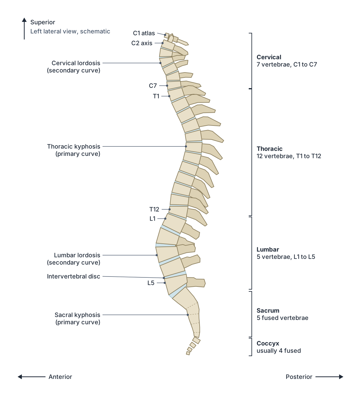
The column starts as 33 vertebrae in the embryo. In the adult there are 24 separate vertebrae plus the sacrum and coccyx: 7 cervical (C1 to C7), 12 thoracic (T1 to T12) and 5 lumbar (L1 to L5). The sacrum forms from 5 fused sacral vertebrae and the coccyx from fused coccygeal vertebrae. (OpenStax Anatomy and Physiology 2e, 7.3: The Vertebral Column.)
The sacrum and coccyx are sometimes called false vertebrae because their segments are fused. The vertebral bodies get bigger and thicker from top to bottom, matching the increasing weight they carry. (TeachMeAnatomy: The Vertebral Column.)
Counts vary a little between people. The lumbar spine may have 4 or 6 vertebrae, and transitional vertebrae at the lumbosacral junction (a sacralised L5 or lumbarised S1) are common. (Radiopaedia: Lumbar vertebrae.)
Regions of the vertebral column
| Region | Vertebrae | Curve | Distinctive features |
|---|---|---|---|
| Cervical | 7 (C1 to C7) | Lordosis (secondary) | Small bodies, transverse foramina, bifid spinous processes (C3 to C6), atlas and axis |
| Thoracic | 12 (T1 to T12) | Kyphosis (primary) | Heart-shaped bodies, costal facets for ribs, long downward-sloping spinous processes |
| Lumbar | 5 (L1 to L5) | Lordosis (secondary) | Largest kidney-shaped bodies, short hatchet-shaped spinous processes, no costal facets |
| Sacral | 5 fused (sacrum) | Kyphosis (primary) | Triangular bone, sacral foramina, joins the hip bones |
| Coccygeal | Usually 4 fused (coccyx) | Part of the sacrococcygeal curve | Small tailbone with no vertebral canal |
Based on OpenStax and TeachMeAnatomy (see references).
Curvatures of the spine
Seen from the side, the adult spine has four curves. The thoracic and sacrococcygeal curves are primary curves, kept from the C-shaped spine of the fetus. The cervical and lumbar curves are secondary curves that develop after birth, and they are concave towards the back. (OpenStax Anatomy and Physiology 2e, 7.3: The Vertebral Column.)
The cervical curve forms when a baby begins to hold its head upright, and the lumbar curve forms when the child begins to stand and walk. The curves deepen when the spine is loaded and spring back when the load is removed, which helps the column absorb shock. The adult lumbar curve is generally deeper in females. (OpenStax Anatomy and Physiology 2e, 7.3: The Vertebral Column.)
Cervical and lumbar intervertebral discs are thicker at the front than at the back, and this wedge shape contributes to the normal forward curves (lordoses) of the neck and lower back. (Radiopaedia: Intervertebral disc.)
Abnormal curves have their own names. Kyphosis is an exaggerated thoracic curve (hunchback), for example after osteoporotic collapse of upper thoracic vertebrae. Lordosis is an exaggerated lumbar curve (swayback), linked with obesity and late pregnancy. Scoliosis is a sideways curve with twisting; its cause is usually unknown and it is the most common vertebral abnormality in girls. (OpenStax Anatomy and Physiology 2e, 7.3: The Vertebral Column.)
Parts of a vertebra: body, arch, spinous and transverse processes
A typical vertebra has a body at the front, which bears weight, and a vertebral arch at the back. The arch is made of two pedicles, which attach to the body, and two laminae, which meet in the midline. Between the body and the arch is the vertebral foramen. (OpenStax Anatomy and Physiology 2e, 7.3: The Vertebral Column.)
Seven processes project from the arch: one spinous process pointing backwards in the midline, two transverse processes pointing out to the sides, and two superior and two inferior articular processes that form the facet joints with the vertebrae above and below. (OpenStax Anatomy and Physiology 2e, 7.3: The Vertebral Column.)
The spinous process and transverse processes are levers for muscles and attachment points for ligaments. Pedicles join the body to the transverse processes, and laminae join the transverse processes to the spinous process. The articular processes arise where the pedicles and laminae meet. (TeachMeAnatomy: The Vertebral Column.)
Notches above and below the pedicles of neighbouring vertebrae line up to make an intervertebral foramen on each side. A spinal nerve leaves the vertebral canal through each of these foramina. (OpenStax Anatomy and Physiology 2e, 7.3: The Vertebral Column.)
Thoracic vertebrae (T1 to T12) and costal facets
The twelve thoracic vertebrae are the ones that carry ribs. Their bodies are medium-sized and heart-shaped, their vertebral foramina are round, and their long spinous processes slope steeply downwards, overlapping the vertebra below. (Radiopaedia: Typical thoracic vertebrae.)
A typical thoracic vertebra has two costal facets (demifacets) on each side of its body. The superior costal facet takes part of the head of the rib with the same number, and the inferior costal facet takes part of the head of the rib below. On T5, for example, the superior demifacet joins rib 5 and the inferior demifacet joins rib 6. Each transverse process also has a transverse costal facet for the tubercle of its own rib (the costotransverse joint). (Radiopaedia: Typical thoracic vertebrae.)
T1 and T9 to T12 are atypical. T1 has a full facet for the first rib and a demifacet for the second, and looks a little like a lower cervical vertebra. T9 may lack an inferior demifacet. T10 usually has a single full facet for rib 10 and sometimes no transverse costal facet. (Radiopaedia: Atypical thoracic vertebrae.)
T11 and T12 each have one full facet for the head of their own rib, and their small transverse processes have no transverse costal facets. The inferior articular processes of T12 face outwards like those of the lumbar vertebrae, so T12 is a transition between the thoracic and lumbar spine. (Radiopaedia: Atypical thoracic vertebrae.)
Lumbar vertebrae (L1 to L5), including L4 and L5
The five lumbar vertebrae are the largest vertebrae and carry the most weight. They have large, kidney-shaped bodies, a triangular vertebral foramen, and no transverse foramina or costal facets. Their spinous processes are short, thick and hatchet-shaped, and point almost straight backwards. (Kenhub: Lumbar vertebrae.)
The superior articular processes have a small knob, the mammillary process, where multifidus attaches, and there is an accessory process at the root of each transverse process. L3 usually has the longest transverse processes, which helps when counting levels on X-rays. The lumbar facet joints allow flexion, extension and side bending, but rotation is greatly limited. (Radiopaedia: Lumbar vertebrae.)
L4 is a typical lumbar vertebra; its transverse processes arise from the pedicles and the back of the body and point slightly upwards. L5 is atypical: its body is deeper in front than behind, which creates the lumbosacral angle, and its thick transverse processes arise from both the body and the pedicles. (Kenhub: Lumbar vertebrae.)
L5 is the most common site of spondylolysis (a defect in the pars interarticularis) and spondylolisthesis, where a vertebra slips forwards on the one below. (Kenhub: Lumbar vertebrae.)
The L4/L5 and L5/S1 discs are the most common sites of disc herniation, and pressure on the nerve roots there can cause sciatica. In the neck the commonest levels are C5/C6 and C6/C7. (OpenStax Anatomy and Physiology 2e, 7.3: The Vertebral Column.)
Vertebral canal and vertebral cavity
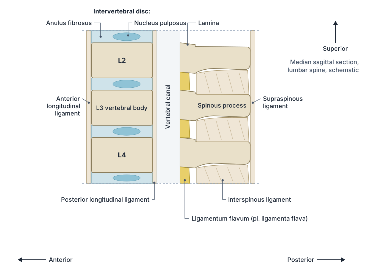
The vertebral foramina of all the vertebrae line up to form the vertebral (spinal) canal. It is bounded in front by the vertebral bodies, discs and posterior longitudinal ligament, at the sides by the pedicles, and behind by the laminae and ligamenta flava. It runs from the foramen magnum of the skull to the sacral hiatus. (Radiopaedia: Vertebral canal.)
The canal contains the spinal cord with its meninges and cerebrospinal fluid, nerve roots, blood vessels and the epidural space. The spinal cord fills only the upper two-thirds of the canal and ends at about the middle of the L1 vertebral body in adults. The canal is small and triangular in the neck, small and round in the thorax, and large and triangular in the lumbar region. (Radiopaedia: Vertebral canal.)
In the language of body cavities, the vertebral (spinal) cavity is the part of the dorsal body cavity that encloses the spinal cord. It is continuous with the cranial cavity, which houses the brain. (OpenStax Anatomy and Physiology 2e, 1.6: Anatomical Terminology.)
Intervertebral discs
An intervertebral disc is a pad of fibrocartilage between two vertebral bodies. There are 23, from C2/C3 down to L5/S1; there is no disc between the skull and C1 or between C1 and C2. The discs are secondary cartilaginous joints and make up about a quarter of the height of the column. (Radiopaedia: Intervertebral disc.)
Each disc has a tough outer ring, the anulus (annulus) fibrosus, and a soft, gel-like centre with a high water content, the nucleus pulposus, which resists compression. Discs are thinnest in the neck and thickest in the lumbar region. With age the nucleus loses water, so discs become thinner and less flexible. (OpenStax Anatomy and Physiology 2e, 7.3: The Vertebral Column.)
Adult discs have almost no blood supply. Only the outer anulus has vessels and nerves; the inner anulus and nucleus are fed by diffusion through the vertebral end plates. The outer anulus is supplied by sinuvertebral nerves. (Radiopaedia: Intervertebral disc.)
In a herniated (slipped) disc, the nucleus pulposus pushes through a weak point in the anulus, usually backwards and to one side, and can press on a spinal nerve. (TeachMeAnatomy: The Vertebral Column.)
Facet joints (zygapophyseal joints)
The facet joints, also called zygapophyseal or apophyseal joints, are paired synovial joints between the inferior articular process of one vertebra and the superior articular process of the vertebra below. Each has a thin, loose capsule that merges with the ligamentum flavum. (Radiopaedia: Facet joint.)
Their orientation changes down the spine and controls which movements are possible. Cervical facets slope downwards and backwards and have loose capsules, allowing a wide range of movement. Thoracic facets are nearly vertical. Lumbar facets are curved and lie mainly in the sagittal plane, resisting rotation. (Radiopaedia: Facet joint.)
Pain fibres from each facet joint travel in the medial branch of a dorsal ramus. Each medial branch supplies the joint at its own level and the joint below; for example, the L1 medial branch supplies the L1/2 and L2/3 joints. Every facet joint therefore has two nerve supplies, which matters for medial branch blocks. (Radiopaedia: Facet joint.)
Ligaments of the vertebral column
The anterior longitudinal ligament is a long band down the front of the vertebral bodies and discs, from the front of the sacrum up to the anterior arch of the atlas, where it continues as the anterior atlanto-occipital membrane. (Radiopaedia: Anterior longitudinal ligament.)
The posterior longitudinal ligament runs inside the vertebral canal on the back of the vertebral bodies, in front of the spinal cord. It is firmly attached to the discs but only loosely to the bodies. It broadens as it ascends, and at C2 it continues as the tectorial membrane to the base of the skull. (Radiopaedia: Posterior longitudinal ligament.)
The ligamenta flava (singular ligamentum flavum) join the laminae of neighbouring vertebrae from C2/C3 down to the sacrum and form part of the back wall of the vertebral canal. They are yellowish because they contain many elastic fibres; they are thin in the neck and thickest in the lumbar spine. (Radiopaedia: Ligamentum flavum.)
The interspinous ligaments are relatively weak sheets between adjacent spinous processes, and they blend with the stronger supraspinous ligament, which runs along the tips of the spinous processes. Above C7 the supraspinous ligament continues as the nuchal ligament. (Radiopaedia: Interspinous ligament.)
The nuchal ligament (ligamentum nuchae) is a thick median ligament from the spinous process of C7 to the external occipital protuberance of the skull. It limits flexion of the neck and anchors the trapezius, splenius capitis and rhomboid minor. (Radiopaedia: Nuchal ligament.)
Intertransverse ligaments join neighbouring transverse processes. Overall, the anterior longitudinal ligament limits hyperextension, and the posterior longitudinal and interspinous ligaments resist hyperflexion. (TeachMeAnatomy: The Vertebral Column.)
Ligaments of the vertebral column
| Ligament | Runs between | Position | Main role |
|---|---|---|---|
| Anterior longitudinal | Front of vertebral bodies and discs, sacrum to atlas | In front of the bodies | Limits hyperextension |
| Posterior longitudinal | Back of vertebral bodies and discs, sacrum to C2 (then tectorial membrane) | Inside the canal, in front of the cord | Limits hyperflexion |
| Ligamentum flavum | Laminae of adjacent vertebrae, C2/C3 to sacrum | Back wall of the canal | Elastic; part of the posterior ligament complex |
| Interspinous | Adjacent spinous processes | Between the spines | Resists hyperflexion |
| Supraspinous | Tips of the spinous processes | Behind the spines | Continues as the nuchal ligament above C7 |
| Nuchal | C7 spinous process to external occipital protuberance | Back of the neck | Limits neck flexion; anchors neck muscles |
| Intertransverse | Adjacent transverse processes | Between transverse processes | Links transverse processes |
Compiled from Radiopaedia, OpenStax and TeachMeAnatomy (see references).
Vertebral column in the 3D model
- Atlas
- The first cervical vertebra (C1), a ring-shaped bone that supports the skull.
- Axis
- The second cervical vertebra (C2), distinguished by a tooth-like upward projection called the dens.
- Eighth thoracic vertebra
- The eighth of the twelve thoracic vertebrae, labelled T8, which articulate with ribs.
- Eleventh thoracic vertebra
- The eleventh of the twelve thoracic vertebrae, labelled T11, which articulate with ribs.
- Fifth cervical vertebra
- The fifth of the seven cervical (neck) vertebrae, labelled C5.
- Fifth lumbar vertebra
- The fifth of the five lumbar vertebrae, labelled L5, in the lower back.
- Fifth thoracic vertebra
- The fifth of the twelve thoracic vertebrae, labelled T5, which articulate with ribs.
- First lumbar vertebra
- The first of the five lumbar vertebrae, labelled L1, in the lower back.
- First thoracic vertebra
- The first of the twelve thoracic vertebrae, labelled T1, which articulate with ribs.
- Fourth cervical vertebra
- The fourth of the seven cervical (neck) vertebrae, labelled C4.
- Fourth lumbar vertebra
- The fourth of the five lumbar vertebrae, labelled L4, in the lower back.
- Fourth thoracic vertebra
- The fourth of the twelve thoracic vertebrae, labelled T4, which articulate with ribs.
- Intervertebral disk
- A fibrocartilage cushion between two adjacent vertebral bodies, consisting of a gel-like nucleus pulposus inside a ring-like annulus fibrosus.
- Intervertebral disk of axis
- The intervertebral disc between C2 and C3 (the C2–C3 disc), a fibrocartilage pad with a gel-like nucleus pulposus inside a ring-like anulus fibrosus.
- Intervertebral disk of eighth thoracic vertebra
- The intervertebral disc between T8 and T9 (the T8–T9 disc), a fibrocartilage pad with a gel-like nucleus pulposus inside a ring-like anulus fibrosus.
- Intervertebral disk of eleventh thoracic vertebra
- The intervertebral disc between T11 and T12 (the T11–T12 disc), a fibrocartilage pad with a gel-like nucleus pulposus inside a ring-like anulus fibrosus.
- Intervertebral disk of fifth cervical vertebra
- The intervertebral disc between C5 and C6 (the C5–C6 disc), a fibrocartilage pad with a gel-like nucleus pulposus inside a ring-like anulus fibrosus.
- Intervertebral disk of fifth lumbar vertebra
- The intervertebral disc between L5 and S1 (the L5–S1 disc), a fibrocartilage pad with a gel-like nucleus pulposus inside a ring-like anulus fibrosus.
- Intervertebral disk of fifth thoracic vertebra
- The intervertebral disc between T5 and T6 (the T5–T6 disc), a fibrocartilage pad with a gel-like nucleus pulposus inside a ring-like anulus fibrosus.
- Intervertebral disk of first lumbar vertebra
- The intervertebral disc between L1 and L2 (the L1–L2 disc), a fibrocartilage pad with a gel-like nucleus pulposus inside a ring-like anulus fibrosus.
- Intervertebral disk of first thoracic vertebra
- The intervertebral disc between T1 and T2 (the T1–T2 disc), a fibrocartilage pad with a gel-like nucleus pulposus inside a ring-like anulus fibrosus.
- Intervertebral disk of fourth cervical vertebra
- The intervertebral disc between C4 and C5 (the C4–C5 disc), a fibrocartilage pad with a gel-like nucleus pulposus inside a ring-like anulus fibrosus.
- Intervertebral disk of fourth lumbar vertebra
- The intervertebral disc between L4 and L5 (the L4–L5 disc), a fibrocartilage pad with a gel-like nucleus pulposus inside a ring-like anulus fibrosus.
- Intervertebral disk of fourth thoracic vertebra
- The intervertebral disc between T4 and T5 (the T4–T5 disc), a fibrocartilage pad with a gel-like nucleus pulposus inside a ring-like anulus fibrosus.
- Intervertebral disk of ninth thoracic vertebra
- The intervertebral disc between T9 and T10 (the T9–T10 disc), a fibrocartilage pad with a gel-like nucleus pulposus inside a ring-like anulus fibrosus.
- Intervertebral disk of second lumbar vertebra
- The intervertebral disc between L2 and L3 (the L2–L3 disc), a fibrocartilage pad with a gel-like nucleus pulposus inside a ring-like anulus fibrosus.
- Intervertebral disk of second thoracic vertebra
- The intervertebral disc between T2 and T3 (the T2–T3 disc), a fibrocartilage pad with a gel-like nucleus pulposus inside a ring-like anulus fibrosus.
- Intervertebral disk of seventh cervical vertebra
- The intervertebral disc between C7 and T1 (the C7–T1 disc), a fibrocartilage pad with a gel-like nucleus pulposus inside a ring-like anulus fibrosus.
- Intervertebral disk of seventh thoracic vertebra
- The intervertebral disc between T7 and T8 (the T7–T8 disc), a fibrocartilage pad with a gel-like nucleus pulposus inside a ring-like anulus fibrosus.
- Intervertebral disk of sixth cervical vertebra
- The intervertebral disc between C6 and C7 (the C6–C7 disc), a fibrocartilage pad with a gel-like nucleus pulposus inside a ring-like anulus fibrosus.
- Intervertebral disk of sixth thoracic vertebra
- The intervertebral disc between T6 and T7 (the T6–T7 disc), a fibrocartilage pad with a gel-like nucleus pulposus inside a ring-like anulus fibrosus.
- Intervertebral disk of tenth thoracic vertebra
- The intervertebral disc between T10 and T11 (the T10–T11 disc), a fibrocartilage pad with a gel-like nucleus pulposus inside a ring-like anulus fibrosus.
- Intervertebral disk of third cervical vertebra
- The intervertebral disc between C3 and C4 (the C3–C4 disc), a fibrocartilage pad with a gel-like nucleus pulposus inside a ring-like anulus fibrosus.
- Intervertebral disk of third lumbar vertebra
- The intervertebral disc between L3 and L4 (the L3–L4 disc), a fibrocartilage pad with a gel-like nucleus pulposus inside a ring-like anulus fibrosus.
- Intervertebral disk of third thoracic vertebra
- The intervertebral disc between T3 and T4 (the T3–T4 disc), a fibrocartilage pad with a gel-like nucleus pulposus inside a ring-like anulus fibrosus.
- Ninth thoracic vertebra
- The ninth of the twelve thoracic vertebrae, labelled T9, which articulate with ribs.
- Sacrum
- A triangular bone of five fused sacral vertebrae.
- Second lumbar vertebra
- The second of the five lumbar vertebrae, labelled L2, in the lower back.
- Second thoracic vertebra
- The second of the twelve thoracic vertebrae, labelled T2, which articulate with ribs.
- Seventh cervical vertebra
- The seventh of the seven cervical (neck) vertebrae, labelled C7.
- Seventh thoracic vertebra
- The seventh of the twelve thoracic vertebrae, labelled T7, which articulate with ribs.
- Sixth cervical vertebra
- The sixth of the seven cervical (neck) vertebrae, labelled C6.
- Sixth thoracic vertebra
- The sixth of the twelve thoracic vertebrae, labelled T6, which articulate with ribs.
- Tenth thoracic vertebra
- The tenth of the twelve thoracic vertebrae, labelled T10, which articulate with ribs.
- Third cervical vertebra
- The third of the seven cervical (neck) vertebrae, labelled C3.
- Third lumbar vertebra
- The third of the five lumbar vertebrae, labelled L3, in the lower back.
- Third thoracic vertebra
- The third of the twelve thoracic vertebrae, labelled T3, which articulate with ribs.
- Twelfth thoracic vertebra
- The twelfth of the twelve thoracic vertebrae, labelled T12, which articulate with ribs.
Frequently asked questions
How many vertebrae are in the human spine?
An adult has 24 separate vertebrae: 7 cervical, 12 thoracic and 5 lumbar. Below them, 5 fused sacral vertebrae form the sacrum and usually 4 fused coccygeal vertebrae form the coccyx, giving 33 vertebral segments in total. Small variations, such as a sixth lumbar vertebra, are fairly common.
What are the four curves of the spine?
The cervical and lumbar curves bend forwards (lordoses) and the thoracic and sacrococcygeal curves bend backwards (kyphoses). The thoracic and sacral curves are primary curves present before birth. The cervical curve forms when a baby holds up its head and the lumbar curve when the child stands and walks.
What is the difference between the vertebral canal and the vertebral foramen?
The vertebral foramen is the hole in a single vertebra, between its body and arch. When all the vertebrae are stacked, their foramina line up to form one long tube, the vertebral canal, which runs from the foramen magnum to the sacral hiatus and contains the spinal cord, its meninges and the nerve roots.
What are costal facets?
Costal facets are small joint surfaces on the thoracic vertebrae where the ribs attach. Each typical thoracic vertebra has superior and inferior costal facets (demifacets) on its body for rib heads, and a transverse costal facet on each transverse process for the rib tubercle. T1, T10, T11 and T12 have full facets instead.
What does the ligamentum flavum do?
The ligamenta flava are elastic yellow ligaments that join the laminae of neighbouring vertebrae from C2 down to the sacrum. They form part of the back wall of the vertebral canal. Their elastic fibres help the spine return to an upright position after bending forwards.
Where does the spinal cord end?
In adults the spinal cord ends at about the level of the L1 vertebra, so it fills only the upper two-thirds of the vertebral canal. Below this the canal contains nerve roots (the cauda equina) in a sac of cerebrospinal fluid. This is why lumbar punctures are done between lower lumbar vertebrae.
References
- OpenStax. Anatomy and Physiology 2e, §7.3 The Vertebral Column. Rice University, 2022.
- OpenStax. Anatomy and Physiology 2e, §7.1 Divisions of the Skeletal System. Rice University, 2022.
- OpenStax. Anatomy and Physiology 2e, §1.6 Anatomical Terminology. Rice University, 2022.
- Jones O. The Vertebral Column. TeachMeAnatomy.
- Intervertebral disc. Radiopaedia reference article.
- Anterior longitudinal ligament. Radiopaedia reference article.
- Posterior longitudinal ligament. Radiopaedia reference article.
- Ligamentum flavum. Radiopaedia reference article.
- Interspinous ligament. Radiopaedia reference article.
- Supraspinous ligament. Radiopaedia reference article.
- Nuchal ligament. Radiopaedia reference article.
- Facet joint. Radiopaedia reference article.
- Vertebral canal. Radiopaedia reference article.
- Typical thoracic vertebrae. Radiopaedia reference article.
- Atypical thoracic vertebrae. Radiopaedia reference article.
- Lumbar vertebrae. Radiopaedia reference article.
- Lumbar vertebrae. Kenhub anatomy library.
- Mitsuhashi N. et al. (2009). BodyParts3D: 3D structure database for anatomical concepts. Nucleic Acids Research 37, D782–D785. Data © The Database Center for Life Science, CC BY 4.0.
Related anatomy
- Cervical vertebraeCervical vertebrae explained: typical C3 to C6 features, the atlas (C1), axis and dens (C2), C7 vertebra prominens, AO…
- SacrumSacrum anatomy explained: promontory, ala, sacral foramina, crests and hiatus, joints and ligaments, sacrum vs coccyx…
- Rib cageRib cage anatomy: 12 pairs of ribs, true, false and floating ribs, parts of a rib, sternum and jugular notch, costal…
- Spinal cordSpinal cord anatomy with labelled diagrams: cross-section, grey matter horns, spinothalamic and dorsal column tracts…
- Back musclesBack muscles explained layer by layer: trapezius, rhomboids, serratus posterior, erector spinae, multifidus and…
- Skeletal systemExplore a realistic 3D human skeleton: the 206 bones, axial vs appendicular skeleton, bone types, bony landmarks and…
Explore the whole body in the interactive 3D human anatomy model, or browse the A–Z list of human structures.