

What is the shoulder joint?
The glenohumeral joint is a multiaxial ball-and-socket joint. It has the largest range of motion of any joint in the body, and that mobility comes at the cost of stability. The hip and the glenohumeral joint are the only ball-and-socket joints in the body. (OpenStax Anatomy and Physiology 2e, 9.6: Anatomy of Selected Synovial Joints.)
The two surfaces are badly matched in size. The glenoid cavity is small and shallow, about one-third the size of the humeral head. A rim of fibrocartilage, the glenoid labrum, runs around its margin and deepens the socket slightly. (OpenStax Anatomy and Physiology 2e, 9.6: Anatomy of Selected Synovial Joints.)
Kenhub gives the humeral head about three to four times the surface area of the glenoid, so only about a third of the head touches the socket and labrum at any moment. TeachMeAnatomy compares the arrangement to a golf ball sitting on a tee. (Kenhub: Glenohumeral (shoulder) joint.)
Both articular surfaces are covered with hyaline cartilage, as in most synovial joints. The labrum also forms a seal around the humeral head, which helps resist dislocation. (TeachMeAnatomy: The Shoulder (Glenohumeral) Joint.)
The three joints of the shoulder
The shoulder (pectoral) girdle is made of the clavicle and the scapula. The clavicle meets the sternum at the sternoclavicular joint and the acromion of the scapula at the acromioclavicular joint. The sternoclavicular joint is the only bony articulation between the upper limb and the axial skeleton. (OpenStax Anatomy and Physiology 2e, 8.1: The Pectoral Girdle.)
The scapula itself has no joint with the ribs. It is held against the back of the chest by muscles and slides over it during elevation, depression, protraction and retraction of the shoulder. (OpenStax Anatomy and Physiology 2e, 8.1: The Pectoral Girdle.)
In OpenStax's classification, the glenohumeral joint is a ball-and-socket joint, the acromioclavicular joint is a plane joint and the sternoclavicular joint is a saddle joint. (OpenStax Anatomy and Physiology 2e, 9.4: Synovial Joints.)
Joint capsule and bursae
The fibrous capsule of the glenohumeral joint runs from the rim of the glenoid fossa to the anatomical neck of the humerus. It is lax, which allows a wide range of movement, especially abduction. A synovial membrane lines it and produces synovial fluid. (TeachMeAnatomy: The Shoulder (Glenohumeral) Joint.)
The capsule has two openings: one between the greater and lesser tubercles for the tendon of the long head of biceps, and one between the superior and middle glenohumeral ligaments where the subscapular bursa communicates with the joint. Its weakest part is the inferior capsule. (Kenhub: Glenohumeral (shoulder) joint.)
A bursa is a thin connective tissue sac filled with lubricating fluid that reduces friction. The subacromial bursa lies under the deltoid and acromion and above the supraspinatus tendon, letting the rotator cuff glide under the deltoid. The subscapular bursa lies between the subscapularis tendon and the scapula. (TeachMeAnatomy: The Shoulder (Glenohumeral) Joint.)
Shoulder ligaments
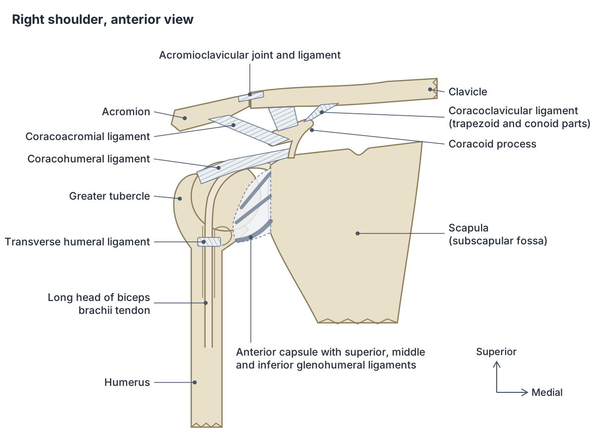
TeachMeAnatomy lists the superior, middle and inferior glenohumeral ligaments, the coracohumeral ligament and the transverse humeral ligament as the ligaments of the glenohumeral joint, together with the coracoacromial ligament, which runs between two parts of the scapula and forms a roof above the joint. (TeachMeAnatomy: The Shoulder (Glenohumeral) Joint.)
The coracohumeral ligament runs from the base of the coracoid process to the humerus in two bands that reach the greater and lesser tubercles. It reinforces the top of the capsule, forms a tunnel for the long head of biceps tendon and blends with the superior glenohumeral ligament. It tightens in external rotation, limiting excessive external rotation and downward slipping of the humerus. (Kenhub: Coracohumeral ligament.)
The transverse humeral ligament bridges the intertubercular (bicipital) groove between the two tubercles and holds the long head of biceps tendon in the groove. (TeachMeAnatomy: The Shoulder (Glenohumeral) Joint.)
The coracoacromial ligament is a strong triangular band between the lateral border of the coracoid process and the apex of the acromion. With these two processes it forms the coracoacromial arch over the head of the humerus, which stops the head being pushed upward out of the glenoid cavity. (Kenhub: Coracoacromial ligament.)
Glenohumeral ligaments: superior, middle and inferior
The three glenohumeral ligaments are thickenings on the front of the capsule that strengthen its superior and anterior walls. (OpenStax Anatomy and Physiology 2e, 9.6: Anatomy of Selected Synovial Joints.)
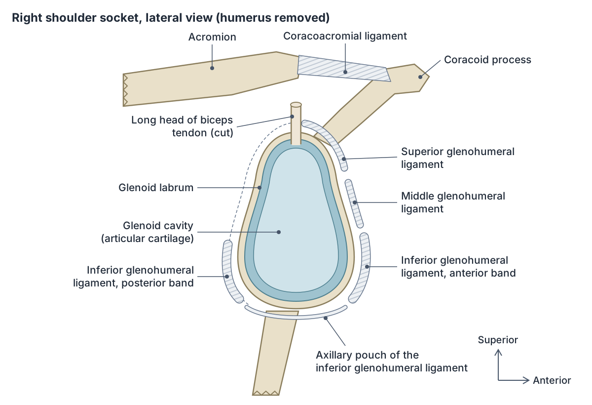
The superior glenohumeral ligament runs from near the supraglenoid tubercle to the top of the lesser tubercle and limits downward slipping of the humerus, especially with the arm at the side. The middle ligament runs from the front of the glenoid margin to the lesser tubercle, blends with the subscapularis tendon and limits external rotation, most of all with the arm partly raised (about 45 to 60 degrees of abduction). (Kenhub: Glenohumeral (shoulder) joint.)
The inferior glenohumeral ligament is the strongest. It hangs like a sling from the labrum to the lower humeral neck, with an anterior band, a posterior band and a loose axillary pouch between them. The anterior band limits external rotation, the posterior band limits internal rotation, and the whole ligament steadies the humeral head when the arm is raised above 90 degrees. (Kenhub: Glenohumeral (shoulder) joint.)
Ligaments of the shoulder region
| Ligament | Joint | Attachments | Main role |
|---|---|---|---|
| Superior glenohumeral | Glenohumeral | Near supraglenoid tubercle to top of lesser tubercle | Limits downward slip of the humerus with the arm at the side |
| Middle glenohumeral | Glenohumeral | Anterior glenoid margin to lesser tubercle | Limits external rotation in mid-abduction |
| Inferior glenohumeral | Glenohumeral | Labrum to lower humeral neck (anterior band, axillary pouch, posterior band) | Main stabiliser of the raised arm |
| Coracohumeral | Glenohumeral | Base of coracoid to greater and lesser tubercles | Supports the top of the capsule; limits external rotation |
| Transverse humeral | Glenohumeral | Across the bicipital groove between the tubercles | Holds the long head of biceps tendon in its groove |
| Coracoacromial | None (forms an arch) | Coracoid process to acromion | Roof over the humeral head; blocks upward displacement |
| Acromioclavicular | AC | Acromion to lateral clavicle (over the joint) | Reinforces the top of the AC capsule |
| Coracoclavicular (conoid + trapezoid) | AC (accessory) | Coracoid to conoid tubercle and trapezoid line of clavicle | Main support of the AC joint; suspends the arm from the clavicle |
| Sternoclavicular (anterior, posterior) | SC | Clavicle to manubrium, front and back | Reinforce the SC capsule |
| Interclavicular | SC | Sternal end of one clavicle to the other, over the manubrium | Reinforces the capsule from above |
| Costoclavicular | SC (accessory) | First rib and costal cartilage to the underside of the clavicle | Main stabiliser of the SC joint |
Compiled from Kenhub, TeachMeAnatomy and OpenStax (see references).
Acromioclavicular (AC) joint and the coracoclavicular ligament
The acromioclavicular joint (articulatio acromioclavicularis) joins the lateral end of the clavicle to the acromion. It is a plane synovial joint whose surfaces are covered with fibrocartilage rather than hyaline cartilage. (Kenhub: Acromioclavicular (AC) joint.)
Sources differ on the articular disc. TeachMeAnatomy describes a wedge of fibrocartilage hanging from the upper capsule into the joint. Kenhub says the disc is often absent or incomplete and that a complete disc is very rare. (TeachMeAnatomy: The Acromioclavicular Joint.)
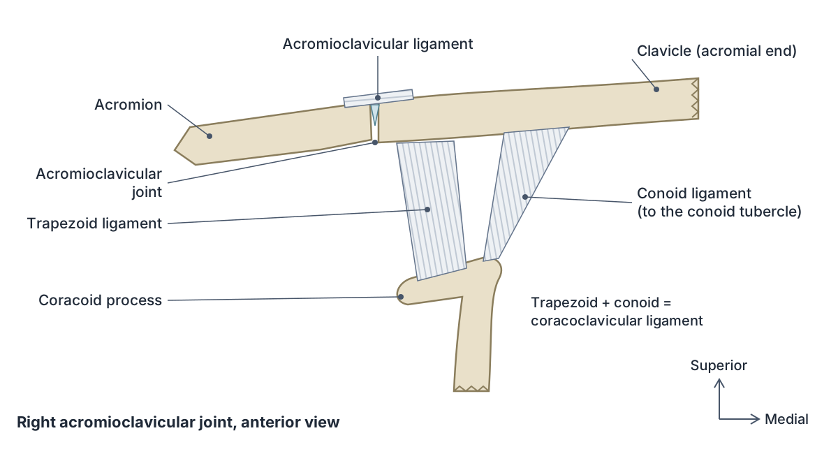
The acromioclavicular ligament runs horizontally over the top of the joint from the acromion to the clavicle. The coracoclavicular (CC) ligament lies medial to the joint and has two parts: the conoid ligament, which runs up from the coracoid process to the conoid tubercle of the clavicle, and the trapezoid ligament, which runs to the trapezoid line just lateral to it. (TeachMeAnatomy: The Acromioclavicular Joint.)
The ligaments of the AC joint itself are relatively weak. The coracoclavicular ligament is very strong and gives the joint its main support, effectively suspending the weight of the arm from the clavicle. (OpenStax Anatomy and Physiology 2e, 8.1: The Pectoral Girdle.)
No muscle acts directly on the AC joint. It glides and rotates slightly as the scapula moves. It is supplied by the suprascapular and thoracoacromial arteries and innervated by branches of the suprascapular and lateral pectoral nerves. (TeachMeAnatomy: The Acromioclavicular Joint.)
Sternoclavicular joint (articulatio sternoclavicularis)
The sternoclavicular joint (articulatio sternoclavicularis) is the joint between the sternal end of the clavicle, the clavicular notch of the manubrium and the upper surface of the first costal cartilage. It is a saddle-type synovial joint, but it behaves almost like a ball-and-socket joint with three degrees of freedom. (Kenhub: Sternoclavicular joint.)
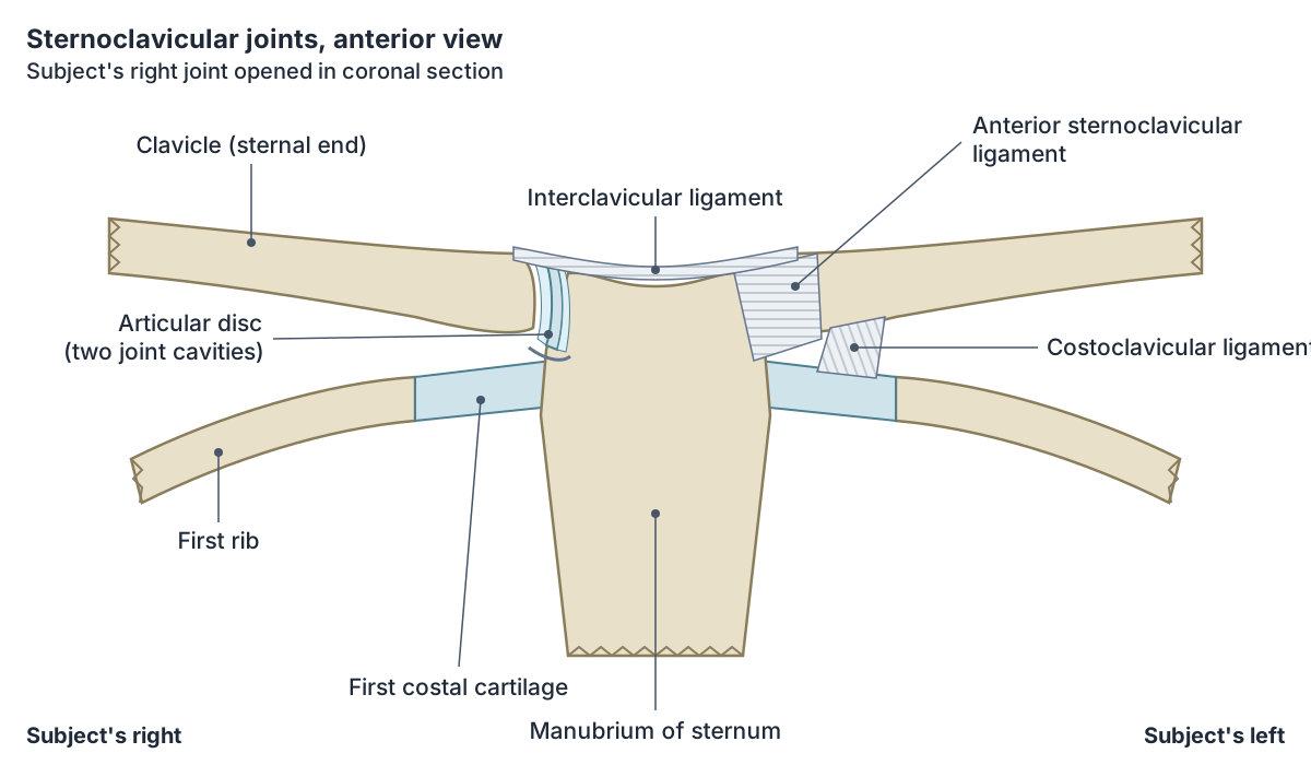
Like the AC joint, its surfaces are covered with fibrocartilage. A fibrocartilage articular disc divides it into two compartments, which lets it move in an extra axis. The capsule is thick and strong. (TeachMeAnatomy: The Sternoclavicular Joint.)
Four ligaments support it. The anterior and posterior sternoclavicular ligaments reinforce the capsule in front and behind, and the interclavicular ligament joins the sternal ends of both clavicles over the top of the manubrium. The costoclavicular ligament, from the first rib and its cartilage to the underside of the clavicle, is the main stabiliser of the joint. (TeachMeAnatomy: The Sternoclavicular Joint.)
The joint allows elevation (shrugging), depression, protraction and retraction of the shoulder, and the clavicle rotates passively on its long axis when the arm is raised overhead. Kenhub gives average active ranges of about 40 degrees for elevation and depression together and about 35 degrees for protraction and retraction. (Kenhub: Sternoclavicular joint.)
Sternoclavicular dislocation is rare and needs a large force. Anterior dislocation, from a blow that drives the shoulder backward, is more common than posterior dislocation, which follows a force driving the shoulder forward or a direct blow to the joint. In adolescents the growth plate at the sternal end of the clavicle has not yet closed, so a dislocation usually comes with a fracture through the plate. (TeachMeAnatomy: The Sternoclavicular Joint.)
Kenhub notes that large blood vessels lie directly behind the sternoclavicular joint, an important relation for any injury that pushes the clavicle backward. (Kenhub: Sternoclavicular joint.)
Shoulder movements: flexion vs extension and more
In the upper limb, every forward movement at the shoulder is flexion and every backward movement is extension. Flexion moves the arm forward and up in front of the body; extension moves it back behind the body. (OpenStax Anatomy and Physiology 2e, 9.5: Types of Body Movements.)
Abduction raises the arm out to the side, away from the body, and adduction brings it back down to the side. Medial (internal) rotation turns the front of the arm toward the midline, and lateral (external) rotation turns it away. Circumduction moves the arm in a circle; it is a combination of the other movements and is possible because the shoulder is a ball-and-socket joint. (OpenStax Anatomy and Physiology 2e, 9.5: Types of Body Movements.)
Kenhub gives the active range of the glenohumeral joint alone as about 110 degrees of flexion, 60 degrees of extension, 120 degrees of abduction and 90 degrees each of internal and external rotation. When movement of the scapula is added, flexion and abduction each reach about 180 degrees. (Kenhub: Glenohumeral (shoulder) joint.)
Abduction happens in stages. Supraspinatus starts the movement over the first 15 degrees, the middle fibres of deltoid continue it to about 90 degrees, and beyond that trapezius and serratus anterior rotate the scapula upward to lift the arm overhead. (TeachMeAnatomy: The Shoulder (Glenohumeral) Joint.)
The main shoulder extensors are latissimus dorsi, teres major and the posterior fibres of deltoid. The main flexors are pectoralis major and the anterior fibres of deltoid, helped by coracobrachialis, with biceps brachii assisting weakly. (TeachMeAnatomy: The Shoulder (Glenohumeral) Joint.)
Shoulder movements and the main muscles
| Movement | Glenohumeral range (Kenhub) | Main muscles |
|---|---|---|
| Flexion | about 110° | Pectoralis major (clavicular part), anterior deltoid; coracobrachialis and biceps assist |
| Extension | about 60° | Latissimus dorsi, teres major, posterior deltoid |
| Abduction | about 120° | Supraspinatus (first 15°), deltoid; trapezius and serratus anterior rotate the scapula beyond 90° |
| Adduction | back to the side | Pectoralis major, latissimus dorsi, teres major |
| Medial (internal) rotation | about 90° | Subscapularis, teres major, pectoralis major, latissimus dorsi, anterior deltoid |
| Lateral (external) rotation | about 90° | Infraspinatus, teres minor, posterior deltoid |
| Circumduction | combination | Sequential action of all the above |
Ranges are for the glenohumeral joint alone; adding scapular movement takes flexion and abduction to about 180°. Muscle groupings from TeachMeAnatomy and Kenhub.
What keeps the shoulder stable?
The rotator cuff muscles, supraspinatus, infraspinatus, teres minor and subscapularis, arise from the scapula and attach to the tubercles of the humerus. Their tendons fuse with the capsule. Because they constantly adjust their tension, they act as dynamic ligaments and are the main source of the joint's structural support. (OpenStax Anatomy and Physiology 2e, 9.6: Anatomy of Selected Synovial Joints.)
Their resting tone presses the humeral head into the glenoid cavity. The labrum deepens the socket, the ligaments reinforce the capsule and form the coracoacromial arch, and the long head of biceps acts as a minor depressor of the humeral head. (TeachMeAnatomy: The Shoulder (Glenohumeral) Joint.)
When deltoid contracts it tends to pull the humerus upward. The rotator cuff counters this pull, which helps prevent upward displacement of the humeral head. (Kenhub: Glenohumeral (shoulder) joint.)
Nerve and blood supply of the shoulder joint
The glenohumeral joint receives blood from the anterior and posterior circumflex humeral arteries, branches of the axillary artery, and from the suprascapular artery, a branch of the thyrocervical trunk. (TeachMeAnatomy: The Shoulder (Glenohumeral) Joint.)
Its capsule is supplied by the suprascapular nerve (posterior and superior parts), the axillary nerve (anteroinferior part) and the lateral pectoral nerve (anterosuperior part). Kenhub also lists the subscapular nerve. (Kenhub: Glenohumeral (shoulder) joint.)
Shoulder dislocation, AC separation and rotator cuff problems
TeachMeAnatomy classifies shoulder dislocations by where the humeral head ends up: anterior in about 95% of cases, posterior in 4% and inferior in 1%. Anterior dislocation usually follows forced extension and lateral rotation, which drives the head forward and downward through the weakest part of the capsule. (TeachMeAnatomy: The Shoulder (Glenohumeral) Joint.)
OpenStax describes most dislocations as inferior, typically when force is applied with the arm fully abducted. TeachMeAnatomy's anterior dislocation also forces the head forward and downward, so the two descriptions overlap: they name the same common injury from different directions. (OpenStax Anatomy and Physiology 2e, 9.6: Anatomy of Selected Synovial Joints.)
A dislocation can leave a dent on the back of the humeral head (Hill-Sachs lesion) and tear the front lower labrum (Bankart lesion). The axillary nerve runs close to the joint and can be injured, weakening deltoid and numbing the skin over the lower deltoid. (TeachMeAnatomy: The Shoulder (Glenohumeral) Joint.)
An AC separation ("separated shoulder") is a different injury from a shoulder dislocation. A strong blow to the side of the shoulder can tear the acromioclavicular and coracoclavicular ligaments, and the weight of the arm then pulls the scapula downward. It is common in ice hockey, football and martial arts. (OpenStax Anatomy and Physiology 2e, 8.1: The Pectoral Girdle.)
Kenhub describes the Tossy grading: grade I, ligaments stretched but intact; grade II, partial rupture of the AC ligament with subluxation; grade III, rupture of all the ligaments with dislocation. A clavicle that springs back up after being pressed down is called the piano key sign. (Kenhub: Acromioclavicular (AC) joint.)
Repetitive overhead use, as in throwing, swimming and racquet sports, can inflame the bursae and rotator cuff tendons, tear the glenoid labrum and wear down the cuff. Scar tissue after inflammation can cause adhesive capsulitis (frozen shoulder), which stiffens the joint. (OpenStax Anatomy and Physiology 2e, 9.6: Anatomy of Selected Synovial Joints.)
In rotator cuff impingement, pain typically occurs between about 60 and 120 degrees of abduction, when the inflamed tissue passes under the acromion. This is called the painful arc. (TeachMeAnatomy: The Shoulder (Glenohumeral) Joint.)
Shoulder joint in the 3D model
Frequently asked questions
What are the main ligaments of the shoulder?
The glenohumeral joint is reinforced by the superior, middle and inferior glenohumeral ligaments, the coracohumeral ligament and the transverse humeral ligament. The coracoacromial ligament forms an arch above the joint. Nearby, the acromioclavicular and coracoclavicular ligaments hold the AC joint, and the sternoclavicular, interclavicular and costoclavicular ligaments hold the SC joint.
What is the CC ligament?
The CC ligament is the coracoclavicular ligament. It joins the coracoid process of the scapula to the underside of the clavicle and has two parts: the conoid ligament medially and the trapezoid ligament laterally. It is the main support of the acromioclavicular joint and is torn, together with the AC ligament, in a complete AC separation.
What is the difference between shoulder flexion and extension?
Shoulder flexion moves the arm forward and upward in front of the body, as when reaching for a shelf. Shoulder extension moves it backward behind the body, as when reaching into a back pocket. Flexion is driven mainly by pectoralis major and anterior deltoid; extension by latissimus dorsi, teres major and posterior deltoid.
What is circumduction of the shoulder?
Circumduction is moving the arm in a circle, so that the hand traces a circle while the shoulder stays at the centre. It is not a separate movement but a sequence of flexion, abduction, extension and adduction. The shoulder can do it because it is a ball-and-socket joint that moves in several planes.
What type of joint is the sternoclavicular joint?
The sternoclavicular joint is a saddle-type synovial joint between the clavicle, the manubrium and the first costal cartilage. An articular disc divides it into two compartments, so in practice it moves almost like a ball-and-socket joint. It is the only bony joint linking the upper limb to the axial skeleton.
Why does the shoulder dislocate so easily?
The humeral head is much larger than the shallow glenoid cavity and the capsule is loose, so bony fit gives little stability. The joint depends on the labrum, ligaments and rotator cuff muscles. The inferior capsule is the weakest part, and most dislocations send the head forward and downward out of the socket.
References
- OpenStax. Anatomy and Physiology 2e, §8.1 The Pectoral Girdle. Rice University, 2022.
- OpenStax. Anatomy and Physiology 2e, §9.4 Synovial Joints. Rice University, 2022.
- OpenStax. Anatomy and Physiology 2e, §9.5 Types of Body Movements. Rice University, 2022.
- OpenStax. Anatomy and Physiology 2e, §9.6 Anatomy of Selected Synovial Joints. Rice University, 2022.
- TeachMeAnatomy. The Shoulder (Glenohumeral) Joint.
- TeachMeAnatomy. The Acromioclavicular Joint.
- TeachMeAnatomy. The Sternoclavicular Joint.
- Kenhub. Glenohumeral (shoulder) joint: bones, movements, muscles.
- Kenhub. Acromioclavicular (AC) joint: anatomy, function.
- Kenhub. Sternoclavicular joint: bones, ligaments, movements.
- Kenhub. Coracoacromial ligament: anatomy and function.
- Kenhub. Coracohumeral ligament: anatomy and function.
- Mitsuhashi N. et al. (2009). BodyParts3D: 3D structure database for anatomical concepts. Nucleic Acids Research 37, D782–D785. Data © The Database Center for Life Science, CC BY 4.0.
Related anatomy
- Rotator cuffThe rotator cuff muscles explained: supraspinatus, infraspinatus, teres minor and subscapularis, with attachments…
- ScapulaScapula anatomy explained: spine, acromion, coracoid process, glenoid cavity, supraspinous fossa, borders and angles…
- ClavicleWhere is your clavicle? The collarbone's sternal end, shaft and acromial end, its joints, ligaments and muscles, why…
- HumerusHumerus bone labeled: head, anatomical and surgical neck, tubercles, deltoid tuberosity, radial groove, epicondyles…
- Arm musclesArm muscles labeled in 3D: biceps brachii, brachialis, coracobrachialis, triceps brachii, deltoid and anconeus, with…
- Types of jointsTypes of joints explained: fibrous, cartilaginous and synovial (diarthrosis) joints, a labelled synovial joint diagram…
Explore the whole body in the interactive 3D human anatomy model, or browse the A–Z list of human structures.