

Categories of joints: structural and functional
A joint, also called an articulation, is any place where adjacent bones, or bone and cartilage, come together to form a connection. Joints are classified in two ways: by their structure (what joins the bones) and by their function (how much movement they allow). (OpenStax Anatomy and Physiology 2e, 9.1: Classification of Joints.)
Structurally there are three classes. In fibrous joints the bones are united by fibrous connective tissue. In cartilaginous joints they are joined by hyaline cartilage or fibrocartilage. In synovial joints the bone ends are not directly connected but meet inside a fluid-filled joint cavity. Synovial joints are the most common type of joint in the body. (OpenStax Anatomy and Physiology 2e, 9.1: Classification of Joints.)
Functionally there are also three classes. A synarthrosis is an immobile or nearly immobile joint, such as a suture of the skull. An amphiarthrosis has limited mobility, such as an intervertebral disc or the pubic symphysis. A diarthrosis is freely mobile, such as the elbow, the knuckles, the shoulder and the hip. All diarthroses are synovial joints, which is why synovial joints are often called diarthroses or synovial diarthroses. (OpenStax Anatomy and Physiology 2e, 9.1: Classification of Joints.)
Structural and functional classification of joints
| Structural type | What joins the bones | Usual functional type | Examples |
|---|---|---|---|
| Fibrous: suture | Fibrous connective tissue | Synarthrosis | Sutures of the skull |
| Fibrous: syndesmosis | Ligament or interosseous membrane | Amphiarthrosis | Distal tibiofibular joint; radius and ulna |
| Fibrous: gomphosis | Periodontal ligament | Synarthrosis | Tooth in its socket |
| Cartilaginous: synchondrosis | Hyaline cartilage | Synarthrosis | Epiphyseal plate; first rib with manubrium |
| Cartilaginous: symphysis | Fibrocartilage | Amphiarthrosis | Pubic symphysis; intervertebral discs |
| Synovial | Articular capsule around a fluid-filled cavity | Diarthrosis | Shoulder, elbow, hip, knee |
Fibrous and cartilaginous joints
Fibrous joints have no joint cavity. There are three kinds. A suture is the narrow fibrous joint between most bones of the skull. A syndesmosis unites two parallel bones by fibrous tissue, as the interosseous membranes join the shafts of the radius and ulna and of the tibia and fibula. A gomphosis is the peg-in-socket joint that anchors the root of a tooth in its bony socket by the periodontal ligament. (OpenStax Anatomy and Physiology 2e, 9.2: Fibrous Joints.)
The distal tibiofibular joint is a syndesmosis in which ligaments hold the lower ends of the tibia and fibula together. This firmly locks the talus in place at the ankle, and the joint is classed as an amphiarthrosis. (OpenStax Anatomy and Physiology 2e, 9.2: Fibrous Joints.)
Cartilaginous joints come in two forms. In a synchondrosis the bones are joined by hyaline cartilage: the epiphyseal (growth) plate of a growing long bone is a temporary example, and the joint between the first rib's costal cartilage and the manubrium is a permanent one. In a symphysis the bones are joined by fibrocartilage, as at the pubic symphysis and between the bodies of adjacent vertebrae, where the thick intervertebral discs allow small movements and cushion the spine. (OpenStax Anatomy and Physiology 2e, 9.3: Cartilaginous Joints.)
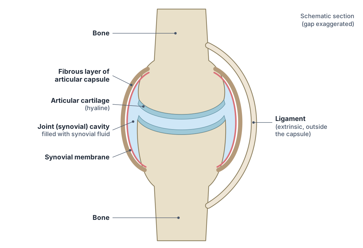
Synovial joint labelled: structure and parts
At a synovial joint the articulating surfaces of the bones meet within a joint cavity, whose walls are formed by the articular capsule. Each articulating surface is covered by a thin layer of hyaline cartilage, the articular cartilage, and the cartilages of the two bones are not continuous with each other. (OpenStax Anatomy and Physiology 2e, 9.4: Synovial Joints.)
The articular capsule has an outer fibrous layer and an inner lining, the synovial membrane. The synovial membrane secretes synovial fluid, which lubricates the joint, reduces friction and supplies nutrients to the articular cartilage, which has no blood vessels of its own. (OpenStax Anatomy and Physiology 2e, 9.4: Synovial Joints.)
Ligaments hold the bones together while limiting the range of movement. They are described as extrinsic (outside the capsule), intrinsic (fused to the capsule wall) or intracapsular (inside it). Muscles and their tendons crossing the joint act as dynamic ligaments, which is especially important at the shoulder, where the ligaments are relatively weak. (OpenStax Anatomy and Physiology 2e, 9.4: Synovial Joints.)
Some synovial joints have extra structures. Articular discs and menisci are fibrocartilage pads between the bones, as in the knee and the temporomandibular joint. Fat pads cushion some joints. Bursae are fluid-filled sacs that reduce friction between bone and skin, muscle or tendon, and tendon sheaths are similar sacs wrapped around tendons where they cross a joint. (OpenStax Anatomy and Physiology 2e, 9.4: Synovial Joints.)
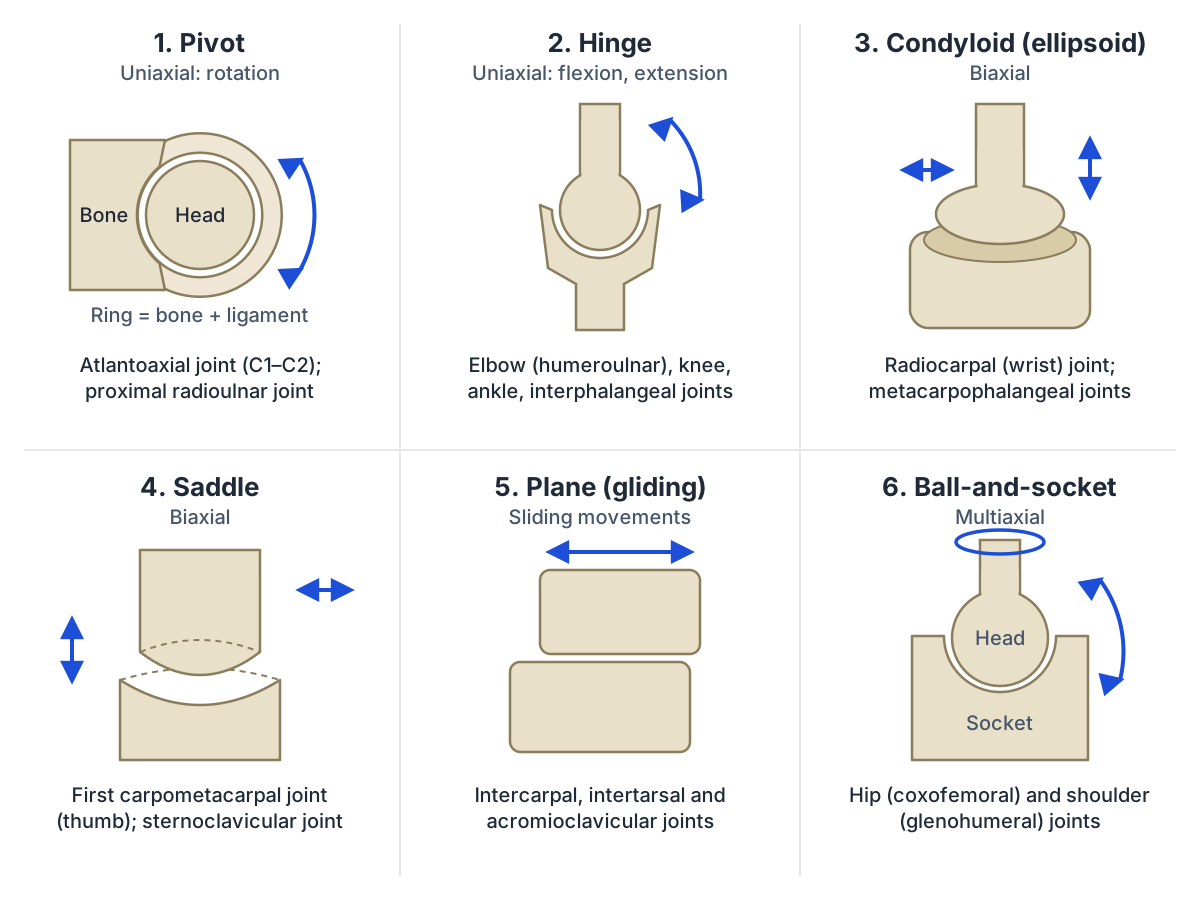
The six types of synovial joints with examples
Synovial joints are subdivided by the shapes of their articulating surfaces, which decide how many axes they can move around. A pivot joint is uniaxial: a rounded bone rotates within a ring formed by another bone and a ligament, as at the atlantoaxial joint between the first and second cervical vertebrae and the proximal radioulnar joint. (OpenStax Anatomy and Physiology 2e, 9.4: Synovial Joints.)
A hinge joint is also uniaxial: a convex end fits into a concave end so the joint bends and straightens, as at the elbow, knee, ankle and the interphalangeal joints of the fingers and toes. A condyloid joint, also called an ellipsoid joint, is biaxial: an oval, rounded surface sits in a shallow depression, allowing movement in two planes. Examples are the metacarpophalangeal (knuckle) joints and the radiocarpal joint of the wrist. (OpenStax Anatomy and Physiology 2e, 9.4: Synovial Joints.)
A saddle joint is biaxial too, but both surfaces are concave in one direction and convex in the other, like a rider in a saddle. The first carpometacarpal joint at the base of the thumb is the classic example, and OpenStax also classes the sternoclavicular joint as a saddle joint. (OpenStax Anatomy and Physiology 2e, 9.4: Synovial Joints.)
In a plane (gliding) joint, flat or slightly curved surfaces of similar size slide against each other, as between the carpal bones, between the tarsal bones, at the acromioclavicular joint and at the facet joints of the vertebrae. A ball-and-socket joint has a rounded head in a cup-like socket and is multiaxial, with the widest range of motion of any joint: the hip and the shoulder (glenohumeral) joints are the two examples. (OpenStax Anatomy and Physiology 2e, 9.4: Synovial Joints.)
The six types of synovial joints
| Type | Shape of the surfaces | Axes | Examples |
|---|---|---|---|
| Pivot | Rounded bone rotates in a ring of bone and ligament | Uniaxial | Atlantoaxial (C1–C2); proximal radioulnar |
| Hinge | Convex end in a concave end | Uniaxial | Elbow, knee, ankle, interphalangeal joints |
| Condyloid (ellipsoid) | Oval convex surface in a shallow oval depression | Biaxial | Radiocarpal (wrist); metacarpophalangeal |
| Saddle | Each surface concave one way, convex the other | Biaxial | First carpometacarpal (thumb); sternoclavicular |
| Plane (gliding) | Flat or slightly curved surfaces | Multiaxial gliding, often limited by ligaments | Intercarpal, intertarsal, acromioclavicular, facet joints |
| Ball-and-socket | Rounded head in a cup-shaped socket | Multiaxial | Hip (coxofemoral); shoulder (glenohumeral) |
Movements at synovial joints
In the limbs, flexion decreases the angle between bones and extension increases it. Abduction moves a limb laterally away from the midline and adduction brings it back toward or across it. Circumduction moves a body region in a circle, and rotation turns a bone around its own long axis, medially (internally) or laterally (externally). (OpenStax Anatomy and Physiology 2e, 9.5: Types of Body Movements.)
Some movements have special names. Pronation moves the forearm out of the supinated (anatomical) position, so the distal radius crosses over the ulna; supination rotates the radius back until the two bones are parallel again. Dorsiflexion lifts the front of the foot and plantar flexion points the toes down. Inversion turns the sole toward the midline and eversion turns it away. Opposition brings the tip of the thumb to the tip of a finger. (OpenStax Anatomy and Physiology 2e, 9.5: Types of Body Movements.)
Joint type limits the movements available. A uniaxial hinge joint allows only bending and straightening along a single axis. A biaxial condyloid joint allows movement in two planes, forward and backward and side to side. In a multiaxial ball-and-socket joint the femur or humerus can also rotate around its own long axis. (OpenStax Anatomy and Physiology 2e, 9.4: Synovial Joints.)
Elbow joint, elbow ligaments and the radioulnar joints
The elbow is a hinge-type synovial joint that flexes and extends the forearm. It has two articulations inside a single joint capsule: the trochlea of the humerus with the trochlear notch of the ulna (humeroulnar joint), and the capitulum of the humerus with the head of the radius (humeroradial joint). Flexion is produced by brachialis, biceps brachii and brachioradialis; extension by triceps brachii and anconeus. (TeachMeAnatomy: The Elbow Joint.)
The main elbow ligaments are collateral ligaments on each side. The ulnar collateral ligament arises from the medial epicondyle of the humerus and attaches to the medial side of the proximal ulna; it can be injured by frequent, forceful extensions of the forearm, as in baseball pitchers. The radial collateral ligament arises from the lateral epicondyle of the humerus and blends with the annular ligament. (OpenStax Anatomy and Physiology 2e, 9.6: Anatomy of Selected Synovial Joints.)
The annular ligament of the elbow encircles the head of the radius and holds it against the radial notch of the ulna. It attaches to the front and back of that notch, forming a tight loop around the radial head, and together with the interosseous membrane it stabilises the proximal radioulnar joint. Its anterior part is where the radial collateral ligament inserts. Radiopaedia lists pulled elbow (nursemaid's elbow) among the conditions involving this ligament. (Radiopaedia: Annular ligament of the elbow.)
There are two radioulnar joints, and both are pivot joints that turn the forearm into pronation and supination. At the proximal joint the head of the radius turns in the radial notch of the ulna, inside the annular ligament; this joint shares the elbow's capsule. At the distal joint the ulnar notch of the radius moves around the head of the ulna, and an articular disc separates it from the wrist joint. Between them, the interosseous membrane binds the shafts together and transfers force from the radius to the ulna. (TeachMeAnatomy: The Radioulnar Joints.)
Radiocarpal (wrist) joint and wrist ligaments
The wrist (radiocarpal) joint is a condyloid, or ellipsoid, synovial joint. Its concave proximal surface is formed by the distal end of the radius and the articular disc; its convex distal surface is formed by the proximal row of carpal bones, excluding the pisiform. Note that the ulna does not articulate directly with the carpal bones here: the articular disc lies in between. (TeachMeAnatomy: The Wrist Joint.)
Four main ligaments support the wrist. The palmar radiocarpal ligament on the front and the dorsal radiocarpal ligament on the back run from the radius to both rows of carpal bones and make the hand follow the forearm in supination and pronation. The ulnar collateral ligament of the wrist runs from the ulnar styloid process to the triquetrum and pisiform and limits radial deviation; the radial collateral ligament runs from the radial styloid process to the scaphoid and trapezium and limits ulnar deviation. (TeachMeAnatomy: The Wrist Joint.)
Being biaxial, the wrist allows flexion and extension and also abduction (radial deviation) and adduction (ulnar deviation). These movements are produced by the muscles of the forearm, whose tendons cross the joint. (TeachMeAnatomy: The Wrist Joint.)
Proximal interphalangeal joints and the thumb IP joint
The interphalangeal (IP) joints of the hand are synovial hinge joints and uniaxial, allowing only flexion and extension. The index, middle, ring and little fingers each have a proximal interphalangeal (PIP) joint and a distal interphalangeal (DIP) joint. The thumb has only two phalanges, so it has a single joint, the interphalangeal joint of the thumb. (Kenhub: Interphalangeal joints of the hand.)
At the PIP joint the head of the proximal phalanx articulates with the base of the middle phalanx. Each IP joint is reinforced by medial and lateral collateral ligaments and by a palmar ligament, also called the palmar or volar plate. (Kenhub: Interphalangeal joints of the hand.)
Flexor digitorum superficialis and flexor digitorum profundus flex the PIP joints of the fingers, while flexor digitorum profundus alone flexes the DIP joints. At the thumb IP joint, flexor pollicis longus is the flexor and extensor pollicis longus is the extensor. (Kenhub: Interphalangeal joints of the hand.)
Hip (coxofemoral) joint
The hip joint, also called the coxofemoral joint, is a ball-and-socket (spheroidal) joint in which the femoral head is largely contained within the acetabulum of the hip bone. Embryologically, about two fifths of the acetabulum come from the ilium, two fifths from the ischium and one fifth from the pubis. (Navarro-Zarza JE et al. Clinical anatomy of the pelvis and hip. Reumatol Clin. 2012.)
A fibrocartilaginous collar, the acetabular labrum, deepens the socket. Three capsular ligaments reinforce the joint: the iliofemoral ligament, the strongest of the three; the pubofemoral ligament, from the superior pubic ramus to the intertrochanteric line of the femur; and the ischiofemoral ligament, from the ischium to the greater trochanter. Inside the capsule, the ligament of the head of the femur runs from the acetabular fossa to the fovea of the femoral head. The hip allows flexion, extension, abduction, adduction and medial and lateral rotation. (TeachMeAnatomy: The Hip Joint.)
OpenStax notes that the three capsular ligaments spiral around the head and neck of the femur, and that the ligament of the head of the femur carries an artery to the femoral head. Because the hip is built for stability and weight bearing, its range of motion is more limited than at the shoulder, which has the largest range of motion of any joint in the body. (OpenStax Anatomy and Physiology 2e, 9.6: Anatomy of Selected Synovial Joints.)
Joints of the ankle: articulations and ligaments
The ankle (talocrural) joint is a synovial hinge joint between the bones of the leg, the tibia and fibula, and the talus of the foot. The distal tibia and fibula form a bracket-shaped socket, the mortise, which grips the talus. The ankle allows dorsiflexion and plantarflexion. (TeachMeAnatomy: The Ankle Joint.)
On the medial side the strong deltoid (medial) ligament has four parts that fan out from the medial malleolus to the talus, calcaneus and navicular, and it resists over-eversion. On the lateral side three separate ligaments arise from the lateral malleolus: the anterior talofibular, posterior talofibular and calcaneofibular ligaments, which resist over-inversion. (TeachMeAnatomy: The Ankle Joint.)
The joint is most stable in dorsiflexion, when the wider front part of the talus is held in the mortise. Most ankle sprains happen through excessive inversion of a plantarflexed, weight-bearing foot, and the anterior talofibular ligament is the ligament most often injured. (TeachMeAnatomy: The Ankle Joint.)
Just above the talocrural joint, the distal tibiofibular joint is a fibrous syndesmosis: ligaments hold the lower tibia and fibula tightly together so that they firmly lock the talus in place. (OpenStax Anatomy and Physiology 2e, 9.2: Fibrous Joints.)
Inversion and eversion of the foot are often blamed on the ankle, but they are not movements of the ankle joint. They take place at the multiple plane joints between the tarsal bones of the posterior foot, the intertarsal joints. (OpenStax Anatomy and Physiology 2e, 9.5: Types of Body Movements.)
Selected limb joints and their key ligaments
| Joint | Bones | Type | Key ligaments |
|---|---|---|---|
| Elbow | Humerus, ulna, radius | Hinge | Ulnar collateral, radial collateral |
| Proximal radioulnar | Head of radius, radial notch of ulna | Pivot | Annular ligament |
| Radiocarpal (wrist) | Radius and articular disc; scaphoid, lunate, triquetrum | Condyloid | Palmar and dorsal radiocarpal, ulnar and radial collateral |
| Interphalangeal (PIP, DIP, thumb IP) | Adjacent phalanges | Hinge | Collateral ligaments, palmar plate |
| Hip (coxofemoral) | Femoral head, acetabulum | Ball-and-socket | Iliofemoral, pubofemoral, ischiofemoral, ligament of head of femur |
| Ankle (talocrural) | Tibia, fibula, talus | Hinge | Deltoid; anterior and posterior talofibular, calcaneofibular |
Frequently asked questions
What are the six types of synovial joints?
The six types are pivot, hinge, condyloid (ellipsoid), saddle, plane (gliding) and ball-and-socket. Pivot and hinge joints are uniaxial, condyloid and saddle joints are biaxial, and ball-and-socket joints are multiaxial. Examples are the atlantoaxial joint, the elbow, the wrist, the base of the thumb, the intercarpal joints and the hip and shoulder.
Are synovial joints the same as diarthroses?
Nearly. Diarthrosis is a functional term meaning a freely movable joint, and synovial is a structural term meaning a joint with a fluid-filled cavity inside an articular capsule. All diarthroses are synovial joints, so the terms are often used together as synovial diarthrosis. A few synovial joints, such as some plane joints held tight by ligaments, move only slightly.
What is an ellipsoid joint?
An ellipsoid joint, also called a condyloid joint, is a synovial joint in which an oval, convex surface fits into a shallow oval depression. It is biaxial, allowing movement in two planes: flexion and extension, and abduction and adduction. The radiocarpal (wrist) joint and the metacarpophalangeal (knuckle) joints are examples.
What does the annular ligament of the elbow do?
The annular ligament forms a ring around the head of the radius and attaches to the front and back of the radial notch of the ulna. It holds the radial head against the ulna while it rotates during pronation and supination, so it stabilises the proximal radioulnar joint. The radial collateral ligament blends into it on the lateral side.
What type of joint is the hip?
The hip, or coxofemoral joint, is a synovial ball-and-socket joint between the head of the femur and the acetabulum of the hip bone. It is multiaxial, allowing flexion, extension, abduction, adduction and rotation. Its deep socket, deepened further by the acetabular labrum, and its strong iliofemoral ligament make it more stable but less mobile than the shoulder.
How many interphalangeal joints does the thumb have?
One. The thumb has only two phalanges, proximal and distal, so it has a single interphalangeal joint, while each finger has a proximal and a distal interphalangeal joint. The thumb IP joint is a hinge joint flexed by flexor pollicis longus and extended by extensor pollicis longus.
Which bones form the ankle joint?
The ankle (talocrural) joint is formed by the distal ends of the tibia and fibula, which make a socket called the mortise, and the talus, which sits inside it. It is a hinge joint for dorsiflexion and plantarflexion. The tibia and fibula are also bound to each other just above the joint by the distal tibiofibular syndesmosis.
References
- OpenStax. Anatomy and Physiology 2e, §9.1 Classification of Joints. Rice University, 2022.
- OpenStax. Anatomy and Physiology 2e, §9.2 Fibrous Joints. Rice University, 2022.
- OpenStax. Anatomy and Physiology 2e, §9.3 Cartilaginous Joints. Rice University, 2022.
- OpenStax. Anatomy and Physiology 2e, §9.4 Synovial Joints. Rice University, 2022.
- OpenStax. Anatomy and Physiology 2e, §9.5 Types of Body Movements. Rice University, 2022.
- OpenStax. Anatomy and Physiology 2e, §9.6 Anatomy of Selected Synovial Joints. Rice University, 2022.
- TeachMeAnatomy. The Elbow Joint.
- TeachMeAnatomy. The Radioulnar Joints.
- TeachMeAnatomy. The Wrist Joint.
- TeachMeAnatomy. The Hip Joint.
- TeachMeAnatomy. The Ankle Joint.
- Beger A. Interphalangeal joints of the hand. Kenhub.
- Feger J, Knipe H, et al. Annular ligament of the elbow. Radiopaedia.org.
- Navarro-Zarza JE, Villaseñor-Ovies P, Vargas A, et al. Clinical anatomy of the pelvis and hip. Reumatol Clin. 2012;8(S2):33–38.
Related anatomy
- Knee jointLigaments of the knee explained with labelled diagrams: ACL, PCL, MCL, LCL, patellar ligament and menisci, plus knee…
- Shoulder jointShoulder ligaments explained: glenohumeral, coracohumeral, coracoacromial and coracoclavicular (CC) ligaments, AC and…
- Skeletal systemExplore a realistic 3D human skeleton: the 206 bones, axial vs appendicular skeleton, bone types, bony landmarks and…
- Anatomical planesAnatomical planes explained: midsagittal vs parasagittal, coronal (frontal) and transverse (axial) planes, axes of…
- Hand bonesHow many bones are in the human hand? Learn the 8 carpal bones, 5 metacarpals and 14 phalanges by name, plus the thumb…
- Foot bonesFoot bone anatomy with a labeled diagram and 3D model: all 26 bones of the foot, the 7 tarsals, 5 metatarsals and 14…
Explore the whole body in the interactive 3D human anatomy model, or browse the A–Z list of human structures.