

How many bones are in the skull?
The adult skull has 22 bones. Twenty-one of them are fused into one immobile unit, and the mandible (lower jaw) is the only bone that moves. The bones fall into two groups: the brain case, which surrounds and protects the brain, and the facial skeleton, which supports the face and forms the orbits, nasal cavity and mouth. (OpenStax Anatomy and Physiology 2e, 7.2: The Skull.)
Some books give a larger number because they count other small bones of the head. If the six ear ossicles (malleus, incus and stapes on each side) and the hyoid bone in the neck are added, the total becomes 29. The hyoid is not usually counted as part of the skull because it does not touch any other bone. (Kenhub: The skull.)
Anatomists name the two groups the neurocranium (cranium or brain case) and the viscerocranium (facial skeleton). The roof of the neurocranium is called the calvaria (skullcap) and is formed by the frontal bone, the two parietal bones and the occipital bone. The floor, or cranial base, is formed mainly by the frontal, sphenoid, ethmoid, occipital, parietal and temporal bones. (TeachMeAnatomy: The Skull.)
The 8 cranial bones (neurocranium)
The neurocranium contains eight bones. Two are paired (the parietal and temporal bones) and four are single (the frontal, occipital, sphenoid and ethmoid bones). The flat bones of the top and sides of the brain case are often called the flat bones of the skull, and together they make the rounded calvaria. (OpenStax Anatomy and Physiology 2e, 7.2: The Skull.)
The frontal bone forms the forehead and the roof of each orbit. Each parietal bone forms most of the upper side of the skull and is bordered by the frontal bone in front, the temporal bone below and the occipital bone behind. The temporal bone forms the lower side of the skull and houses the middle and inner ear in its petrous part. The occipital bone forms the back of the skull and the back of the cranial base, and the spinal cord passes through its foramen magnum. (OpenStax Anatomy and Physiology 2e, 7.2: The Skull.)
The sphenoid bone is a single central bone that joins almost every other skull bone, which is why it is called the keystone of the cranial base. Its body carries the sella turcica, a saddle-shaped area whose hypophyseal fossa holds the pituitary gland. The ethmoid bone sits in the midline between the orbits. Its cribriform plates let olfactory nerve fibres reach the nasal cavity, and its perpendicular plate forms the upper part of the nasal septum. (OpenStax Anatomy and Physiology 2e, 7.2: The Skull.)
The 8 cranial bones
| Bone | Number | Where it is | Key features |
|---|---|---|---|
| Frontal bone | 1 | Forehead and roof of the orbits | Supraorbital margins, glabella, frontal sinuses |
| Parietal bone | 2 | Upper sides and roof of the skull | Meets its partner at the sagittal suture; parietal eminence |
| Temporal bone | 2 | Lower side of the skull, around the ear | Squamous, petrous, tympanic and mastoid parts; zygomatic and styloid processes; external acoustic meatus |
| Occipital bone | 1 | Back and base of the skull | Foramen magnum, occipital condyles, external occipital protuberance |
| Sphenoid bone | 1 | Middle of the skull base | Body with sella turcica, greater and lesser wings, pterygoid processes |
| Ethmoid bone | 1 | Between the orbits, roof of the nasal cavity | Cribriform plate, crista galli, perpendicular plate, ethmoid air cells |
Paired bones are counted twice, giving 8 cranial bones in total (OpenStax 7.2).
The 14 facial bones (viscerocranium)
The facial skeleton has 14 bones. Six are paired: the maxilla, palatine, zygomatic, nasal and lacrimal bones and the inferior nasal concha. Two are single: the vomer and the mandible. The maxillae form the upper jaw and most of the hard palate, the palatine bones form the back quarter of the hard palate, and the mandible forms the lower jaw. (OpenStax Anatomy and Physiology 2e, 7.2: The Skull.)
The facial bones hold the soft tissues of the face and surround the orbits, the nasal cavity, the mouth and the paranasal sinuses. Some textbooks also count the frontal bone as a facial bone because it forms the forehead and the upper rim of each orbit, so do not be surprised to see it listed in both groups. (TeachMeAnatomy: The Skull.)
Anterior view of the skull: what you see from the front
From the front, the frontal bone forms the forehead at the top. Below it are the two orbits, the openings for the eyes. The two nasal bones form the bridge of the nose, the maxillae fill the middle of the face, and the mandible forms the chin and lower jaw at the bottom. (Kenhub: The skull.)
Seven bones build the walls of each orbit: the frontal bone (roof), the zygomatic bone (lateral wall and floor), the maxilla (most of the floor), a small part of the palatine bone, the ethmoid and lacrimal bones (most of the medial wall) and the sphenoid bone at the back. Small openings near the orbital rim, the supraorbital and infraorbital foramina, carry sensory nerves to the forehead and cheek. (OpenStax Anatomy and Physiology 2e, 7.2: The Skull.)
Inside the nose, the bony nasal septum is made of the perpendicular plate of the ethmoid above and the vomer below. On each lateral wall there are three conchae: the superior and middle conchae belong to the ethmoid bone, while the inferior nasal concha, the largest, is a separate bone. (OpenStax Anatomy and Physiology 2e, 7.2: The Skull.)
Sutures of the skull: coronal, sagittal, lambdoid and squamous
A suture is a fibrous joint between skull bones. In adults the bone edges sit very close together with a thin layer of dense connective tissue between them. The edges are often wavy and interlock, which makes a tight union that prevents most movement. Because they allow little or no movement, sutures are classed as synarthroses. Late in life the sagittal, coronal and lambdoid sutures begin to ossify and their lines gradually disappear; bony fusion of a joint like this is called a synostosis. (OpenStax Anatomy and Physiology 2e, 9.2: Fibrous Joints.)
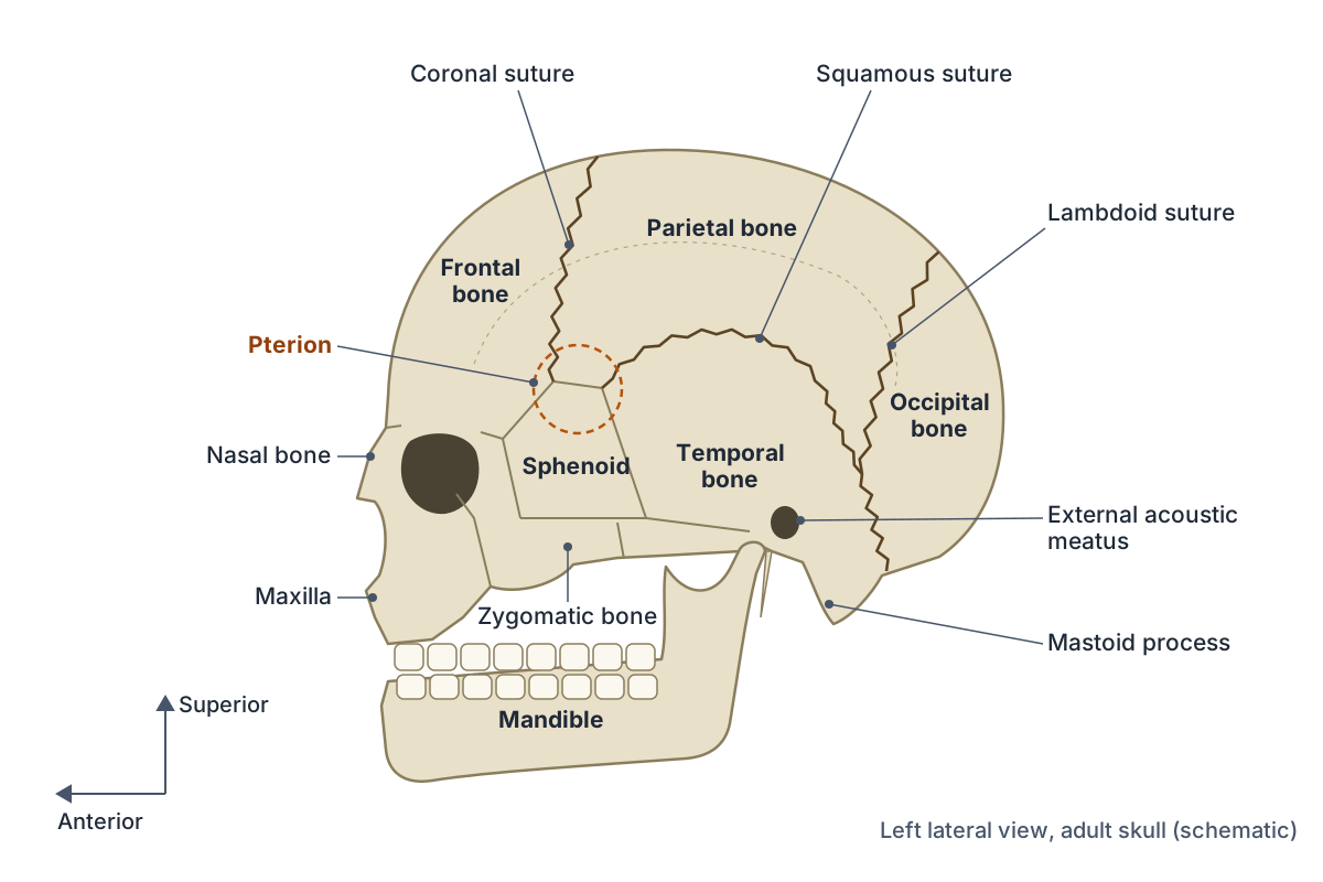
The coronal suture runs from side to side across the top of the skull and joins the frontal bone to the two parietal bones. The sagittal suture runs along the midline of the top of the skull and joins the two parietal bones. The lambdoid suture, shaped like an upside-down letter V (the Greek letter lambda), joins the occipital bone to the parietal and temporal bones. The squamous suture, on the side of the skull, joins the squamous part of the temporal bone to the parietal bone. (OpenStax Anatomy and Physiology 2e, 7.2: The Skull.)
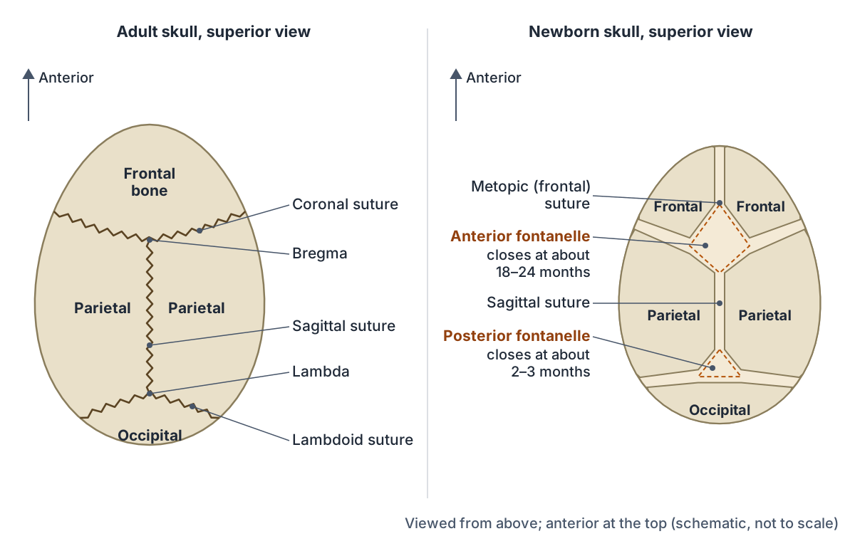
The points where the main sutures meet have names used in surgery and anthropology. Bregma is where the coronal and sagittal sutures meet, and lambda is where the sagittal and lambdoid sutures meet. Kenhub notes that the skull bones form 33 sutures in total, but the coronal, sagittal and lambdoid sutures are the most important to know. (Kenhub: The skull.)
Sutures do not all fuse at the same time. Radiopaedia lists approximate fusion ages of about 22 years for the sagittal suture, 24 years for the coronal and 26 years for the lambdoid, while the squamous suture may not close until about 60. These ages are approximate. (Radiopaedia: Craniosynostosis.)
Skull sutures and when they usually fuse
| Suture | Bones it joins | Approximate fusion age |
|---|---|---|
| Metopic (frontal) | Two halves of the frontal bone | 3 to 9 months |
| Sagittal | Right and left parietal bones | About 22 years |
| Coronal | Frontal bone and both parietal bones | About 24 years |
| Lambdoid | Occipital bone and both parietal bones | About 26 years |
| Squamous (squamosal) | Temporal and parietal bones | About 60 years, or may stay open |
Fusion ages from Radiopaedia's craniosynostosis article; individual skulls vary widely.
Squamous suture
The squamous (squamosal) suture is the curved suture between the temporal bone and the parietal bone on each side of the skull. It starts at the pterion, runs backwards in an arch over the temporal bone, then curves downwards and continues as the parietomastoid (parietotemporal) suture towards the back of the ear. (Radiopaedia: Squamosal suture.)
It is usually the last major suture to fuse and may not close completely until about 60 years of age. Growth at this suture, together with growth around the pterion, the asterion and the frontozygomatic suture, adds to the height of the cranium as the skeleton matures. (Radiopaedia: Squamosal suture.)
The pterion: the thinnest part of the skull
The pterion is a small H-shaped junction on the side of the skull where four bones meet: the frontal bone, the parietal bone, the squamous part of the temporal bone and the greater wing of the sphenoid. It lies about two finger widths above the zygomatic arch and is described as the weakest part of the skull. (OpenStax Anatomy and Physiology 2e, 7.2: The Skull.)
The middle meningeal artery runs just deep to the pterion. A blow here can fracture the thin bone and injure the artery, so blood collects between the skull and the dura mater (an extradural, or epidural, haematoma) and raises the pressure inside the skull. (TeachMeAnatomy: The Skull.)
The pattern of sutures at the pterion varies. A 2025 meta-analysis of 79 studies (18,694 skull sides) found the usual sphenoparietal type in about 79% of sides, with the epipteric type (with extra small bones) in about 8%, the frontotemporal type in about 6% and the stellate type in about 4%. On average the pterion lay about 39 mm above the midpoint of the zygomatic arch. (Triantafyllou G et al. Unveiling pterion variability: a meta-analytic approach. Surg Radiol Anat 2025.)
Fontanelles: the soft spots of a baby's skull
In a fetus and newborn, the bones of the brain case are separated by areas of dense connective tissue called fontanelles, the soft spots on a baby's head. They let the skull change shape as it passes through the birth canal and allow the skull to grow quickly as the brain enlarges after birth. The bones of the top and sides of the brain case form by intramembranous ossification, in which mesenchymal cells turn directly into bone-forming cells without a cartilage model. (OpenStax Anatomy and Physiology 2e, 7.5: Embryonic Development of the Axial Skeleton.)
The anterior fontanelle is the largest. It is diamond-shaped and lies where the coronal and sagittal sutures meet, and it usually persists until about 18 to 24 months, when its site becomes bregma. The posterior fontanelle is triangular, lies where the lambdoid and sagittal sutures meet, and closes at about 2 to 3 months, when its site becomes lambda. (Radiopaedia: Anterior fontanelle.)
Closure times vary between children. A CT study summarised by Lottering and colleagues reported complete closure of the anterior fontanelle at 14 to 19 months in boys and 16 to 22.5 months in girls, while another study found that half of New Zealand European infants aged 19 to 24 months still had remnants of it. (Lottering N et al. Temporal mapping of the closure of the anterior fontanelle and contiguous sutures. J Anat 2020.)
At birth the frontal bone is in two halves joined by the metopic suture. This suture normally fuses in infancy: Radiopaedia gives fusion starting as early as 3 months and complete in most children by about 9 months. In a small number of adults it persists as a vertical line in the middle of the forehead, which can be mistaken for a fracture on an X-ray. (Radiopaedia: Metopic suture.)
Female skull vs male skull: do women have smaller skulls?
On average, yes. A 2023 study of 228 crania found that female skulls are, on average, smaller and more gracile than male skulls, and that much of the shape difference is linked to size. The sexes overlap widely, though, so a small skull is not necessarily female and a large skull is not necessarily male. (Del Bove A et al. Mapping sexual dimorphism signal in the human cranium. Sci Rep 2023.)
The same study found that the most sexually dimorphic areas are the glabella and brow (supraciliary) region, the mastoid process and the nasal region. Using these regions, sex was estimated correctly in about 73 to 77% of skulls. The external occipital protuberance varied mainly with size rather than with sex. (Del Bove A et al. Mapping sexual dimorphism signal in the human cranium. Sci Rep 2023.)
Forensic anthropologists often use Walker's method, which scores five skull traits from 1 (gracile) to 5 (robust): the nuchal crest, the mastoid process, the supraorbital margin, the supraorbital ridge (glabella region) and the mental eminence of the chin. Its accuracy depends on the population. In a South Asian sample it classed only 63% of individuals correctly because many males had gracile skulls. (Rogers LM et al. Comparing Walker's (2008) skull trait sex estimation standard to proteomic sex estimation. Forensic Sci Int Synerg 2024.)
Variations and clinical notes
Craniosynostosis is the premature fusion of one or more sutures, which changes the shape of the growing head. It occurs in about 1 in 2,000 births. The sagittal suture is involved in about half of cases, giving a long, narrow head (scaphocephaly). Treatment is usually surgical, and earlier surgery gives better results. (OpenStax Anatomy and Physiology 2e, 7.5: Embryonic Development of the Axial Skeleton.)
Early fusion of other sutures gives other head shapes: both coronal sutures produce a short, wide head (brachycephaly), one coronal or lambdoid suture produces an asymmetrical head (plagiocephaly), and the metopic suture produces a triangular forehead (trigonocephaly). (Radiopaedia: Craniosynostosis.)
Wormian (intrasutural) bones are small extra bones within the sutures, most often in the lambdoid suture. A few are a normal variant, but many of them, especially more than five, are linked to conditions such as osteogenesis imperfecta and cleidocranial dysostosis. (Radiopaedia: Wormian bones.)
Skull fractures are classed as linear (the most common, with lines radiating from the point of impact), depressed (bone pushed inwards), comminuted (broken into pieces) or contrecoup (on the side opposite the impact). A fracture of the cranial base can damage the internal carotid artery in the carotid canal. (OpenStax Anatomy and Physiology 2e, 7.2: The Skull.)
Exploring the skull in 3D
The 3D skull model on this page highlights all six types of cranial bone and every facial bone from the BodyParts3D dataset, which is based on an adult male. Rotate it to see the pterion and the squamous suture from the side, then turn to the base to find the sphenoid and the foramen magnum of the occipital bone. The model does not include the ear ossicles, and its sutures are not modelled as separate structures, so use the diagrams above to study the suture lines. (Mitsuhashi N et al. BodyParts3D: 3D structure database for anatomical concepts. Nucleic Acids Res 2009.)
Skull bones in the 3D model
- Ethmoid
- A delicate cube-shaped bone between the orbits.
- Frontal bone
- The bone of the forehead and the roof of the orbits.
- Inferior nasal concha
- A thin curved bone on the lateral wall of the nose.
- Lacrimal bone
- A small, delicate bone of the medial orbit wall.
- Mandible
- The lower jaw bone.
- Maxilla
- The upper jaw bone, paired.
- Nasal bone
- A small, paired rectangular bone forming the bridge of the nose.
- Occipital bone
- The bone forming the back and base of the skull.
- Palatine bone
- An L-shaped bone behind the maxilla.
- Parietal bone
- A paired, curved bone forming the roof and sides of the skull.
- Sphenoid bone
- A butterfly-shaped bone at the base of the skull.
- Temporal bone
- A paired bone forming part of the side and base of the skull.
- Vomer
- A thin, flat bone of the nasal septum.
- Zygomatic bone
- The cheekbone.
Frequently asked questions
How many cranial bones are there?
There are 8 cranial bones: the frontal bone, two parietal bones, two temporal bones, the occipital bone, the sphenoid bone and the ethmoid bone. Together they form the neurocranium, the brain case that surrounds and protects the brain. The other 14 bones of the skull are facial bones, giving 22 skull bones in total.
What are the 4 main sutures of the skull?
The four main sutures are the coronal suture (frontal bone to both parietal bones), the sagittal suture (between the two parietal bones), the lambdoid suture (occipital bone to the parietal bones) and the squamous suture (temporal bone to parietal bone, on each side). All are fibrous joints that allow little or no movement.
What is the difference between the skull and the cranium?
The skull is the whole bony framework of the head, including the facial bones and the mandible. The cranium, or neurocranium, is only the brain case: the eight bones that enclose the brain. In everyday use the two words are often used interchangeably, so check which meaning a textbook uses.
Is a suture a joint?
Yes. A suture is a fibrous joint in which neighbouring skull bones are joined by a thin layer of dense connective tissue. The bone edges often interlock like a zip. Sutures are classed as synarthroses because they allow little or no movement, and with age many of them ossify and fuse completely.
When do the soft spots on a baby's head close?
The posterior fontanelle at the back of the head usually closes at about 2 to 3 months after birth. The larger anterior fontanelle on top of the head usually closes between about 18 and 24 months, though studies report a wide normal range, and it tends to close slightly later in girls than in boys.
Do women have smaller skulls than men?
On average, yes. Female skulls tend to be smaller and more gracile, with a smoother brow, a smaller mastoid process and less marked muscle ridges. The ranges overlap widely, so skull size alone cannot reliably tell sex. Forensic methods combine several features and still classify only about 70 to 80% of skulls correctly.
Why is the pterion dangerous?
The pterion is the thinnest part of the side of the skull, where the frontal, parietal, temporal and sphenoid bones meet. The anterior branch of the middle meningeal artery runs just beneath it, so a fracture here can tear the artery and cause an extradural haematoma that compresses the brain and needs urgent surgery.
References
- Betts JG, et al. Anatomy and Physiology 2e, §7.2 The Skull. OpenStax, Rice University, 2022.
- Betts JG, et al. Anatomy and Physiology 2e, §7.5 Embryonic Development of the Axial Skeleton. OpenStax, Rice University, 2022.
- Betts JG, et al. Anatomy and Physiology 2e, §9.2 Fibrous Joints. OpenStax, Rice University, 2022.
- TeachMeAnatomy. The Skull. Accessed 2026-10-19.
- Kenhub. The skull. Accessed 2026-10-19.
- Radiopaedia. Squamosal suture. Accessed 2026-10-19.
- Radiopaedia. Craniosynostosis. Accessed 2026-10-19.
- Radiopaedia. Metopic suture. Accessed 2026-10-19.
- Radiopaedia. Wormian bones. Accessed 2026-10-19.
- Radiopaedia. Anterior fontanelle. Accessed 2026-10-19.
- Radiopaedia. Posterior fontanelle. Accessed 2026-10-19.
- Lottering N, Alston CL, Barry MD, MacGregor DM, Gregory LS. Temporal mapping of the closure of the anterior fontanelle and contiguous sutures using computed tomography, in silico models of modern infants. J Anat. 2020;237(2):379-390.
- Triantafyllou G, Karangeli N, Duparc F, et al. Unveiling pterion variability: a meta-analytic approach to enhance neurosurgical precision. Surg Radiol Anat. 2025;47(1):66.
- Del Bove A, Menéndez L, Manzi G, et al. Mapping sexual dimorphism signal in the human cranium. Sci Rep. 2023;13(1):16847.
- Mitsuhashi N, et al. BodyParts3D: 3D structure database for anatomical concepts. Nucleic Acids Res. 2009;37:D782-D785.
- Rogers LM, Halcrow SE, Kleffmann T, King CL. Comparing Walker's (2008) skull trait sex estimation standard to proteomic sex estimation for a group of South Asian individuals. Forensic Sci Int Synerg. 2024;8:100450.
- Mitsuhashi N. et al. (2009). BodyParts3D: 3D structure database for anatomical concepts. Nucleic Acids Research 37, D782–D785. Data © The Database Center for Life Science, CC BY 4.0.
Related anatomy
- Frontal boneThe frontal bone (os frontale) explained: squamous, orbital and nasal parts, supraorbital margin and foramen, frontal…
- Parietal boneThe parietal bones (os parietale) explained: four borders, four angles, the sutures and fontanelles at their corners…
- Temporal boneThe temporal bone (os temporale) explained: petrous part and inner ear, mastoid process, zygomatic and styloid…
- Occipital boneThe occipital bone (os occipitale) explained: what passes through the foramen magnum, the external occipital…
- Sphenoid boneThe sphenoid bone (os sphenoidale): sella turcica and hypophyseal fossa, greater and lesser wings, pterygoid process…
- Ethmoid boneThe ethmoid bone (os ethmoidale) explained: cribriform plate, crista galli, perpendicular plate, ethmoid sinus air…
Explore the whole body in the interactive 3D human anatomy model, or browse the A–Z list of human structures.