

Mandible at a glance
- Also called
- Jaw
- What it is
- The lower jaw bone.
- Location
- Articulates with the temporal bones at the temporomandibular joints and holds the lower teeth.
- Function
- Allows chewing and speaking.
- Body system
- Skeletal · single, midline structure
- FMA ID
- FMA52748 (Foundational Model of Anatomy)
Key facts
- The only movable bone of the skull apart from the ear ossicles. Its two halves fuse at the midline symphysis during the first year of life.
- Has a body, ramus, condylar and coronoid processes, and mental and mandibular foramina.
Clinical relevance
Because the mandible is a ring with the skull base, fractures often occur in two places.
Where is the mandible?
The mandible is the single midline bone of the lower jaw and one of the facial bones. It articulates with both temporal bones at the mandibular fossae, forming the two temporomandibular joints, and carries the lower teeth in its alveolar process. (Radiopaedia: Mandible.)
Its condyle articulates with the mandibular fossa and articular tubercle of the temporal bone, just in front of each ear. (OpenStax Anatomy and Physiology 2e, 7.2: The Skull.)
To find it on yourself, run a finger along the lower border of the jaw from the chin back to the angle, then up the back edge of the ramus. Placing a fingertip just in front of the ear canal and opening the mouth wide, you can feel the head of the mandible glide forward.
Parts of the mandible
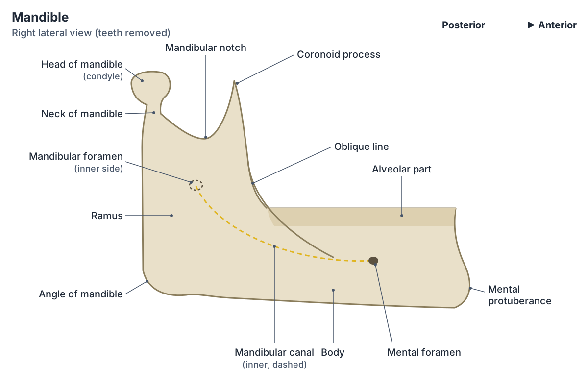
Each side of the mandible has a horizontal body and a vertical ramus. The outside corner where the body and ramus meet is the angle of the mandible. At the top of the ramus are two processes: the flattened coronoid process in front, which anchors a biting muscle, and the condylar process behind, which forms the jaw joint. The broad U-shaped curve between them is the mandibular notch. The upper border of the body, which holds the lower teeth, is the alveolar process. (OpenStax Anatomy and Physiology 2e, 7.2: The Skull.)
The body is horseshoe-shaped. In the midline, the mandibular symphysis marks where the two halves fused. The condylar process has a head (the articular condyle) on a narrower neck. The coronoid process is a thin triangular plate, separated from the condylar process by the mandibular (sigmoid) notch. (Radiopaedia: Mandible.)
Landmarks on the outer surface
The mandibular symphysis encloses the mental protuberance, which forms the chin. The mental foramen lies on the outer surface below the second premolar tooth. The alveolar border has 16 tooth sockets in an adult. (TeachMeAnatomy: The Mandible.)
The position of the mental foramen varies: it is usually below the second premolar but may lie anywhere from below the canine to below the first molar. The buccinator attaches along the alveolar border, and the lower border may be grooved by the facial artery near the front of the ramus. (Radiopaedia: Mandible.)
The oblique line is a ridge that runs from the front of the ramus down onto the outer surface of the body. Depressor anguli oris attaches along it. Two small mental tubercles lie on either side of the chin. (Kenhub: The mandible.)
Landmarks on the inner surface
On the inner surface of the body, near the midline, are small mental spines (genial tubercles) that anchor genioglossus and geniohyoid. The mylohyoid line runs back along the inner surface and gives origin to mylohyoid. Above the line is a fossa for the sublingual gland and below it a fossa for the submandibular gland. (Radiopaedia: Mandible.)
On the inner surface of the ramus is the mandibular foramen, the entrance to the mandibular canal. In front of it is a small tongue of bone, the lingula, which gives attachment to the sphenomandibular ligament. (Radiopaedia: Mandible.)
The digastric fossa is a small depression on the lower border of the body on each side of the midline, where the digastric muscle attaches. The mylohyoid groove (sulcus) on the inner surface of the ramus carries the mylohyoid nerve and artery. (Kenhub: The mandible.)
Ramus of the mandible and its muscles
The ramus is the flat, roughly four-sided upright part of the mandible on each side. Its outer surface is covered by the masseter. Medial pterygoid inserts on the inner side of the angle. Temporalis inserts on both surfaces of the coronoid process. The posterior border of the ramus is covered by the parotid gland. (Radiopaedia: Mandible.)
Lateral pterygoid attaches to the neck of the condylar process. TeachMeAnatomy lists four chewing muscles attached to the rami: masseter, temporalis, medial pterygoid and lateral pterygoid. The body gives attachment to muscles of the face (mentalis, buccinator, platysma, depressor labii inferioris, depressor anguli oris) on its outer surface and to genioglossus, geniohyoid, mylohyoid and digastric on its inner surface. (TeachMeAnatomy: The Mandible.)
The masseteric nerve and vessels cross the mandibular notch to reach the masseter from its deep side. (Kenhub: The mandible.)
Nerves and blood supply
The inferior alveolar nerve and artery enter the mandibular foramen and run forward in the mandibular canal to the mental foramen. There the nerve gives off the mental nerve, which supplies the skin of the lower lip and chin. (TeachMeAnatomy: The Mandible.)
The mandibular canal runs obliquely down and forward through the ramus, then horizontally through the body. Branches of the inferior alveolar nerve, including the incisive nerve, supply the lower teeth. The mandible receives blood from the inferior alveolar artery (a branch of the maxillary artery) and from the facial and lingual arteries (branches of the external carotid). (Radiopaedia: Mandible.)
The inferior alveolar nerve is a branch of the mandibular nerve (V3), which enters the infratemporal fossa through the foramen ovale and runs in the infratemporal fossa, deep to the ramus, before entering the mandibular foramen. Its other branches there include the lingual, buccal and auriculotemporal nerves. (TeachMeAnatomy: The Infratemporal Fossa.)
Jaw anatomy: the temporomandibular joint
The temporomandibular joint (TMJ) is a synovial joint between the head of the mandible and the mandibular fossa and articular tubercle of the temporal bone. An articular disc divides it into upper and lower cavities, so the bones never touch directly. The joint surfaces are covered by fibrocartilage rather than hyaline cartilage. (TeachMeAnatomy: The Temporomandibular Joint.)
Elevation (closing) is powered by temporalis, masseter and medial pterygoid. Depression (opening) is mainly due to gravity, helped by digastric, geniohyoid and mylohyoid. Lateral pterygoid protrudes the jaw, and the back fibres of temporalis retract it. Alternating protrusion and retraction on each side produce the side-to-side grinding of chewing. (TeachMeAnatomy: The Temporomandibular Joint.)
The TMJ usually dislocates forward, after a blow to the side of the face, a big yawn or a large bite. The head of the mandible slips out of the mandibular fossa in front of the articular tubercle and the person cannot close the mouth. (TeachMeAnatomy: The Temporomandibular Joint.)
Development and age changes
The mandible begins as two halves. The midline symphysis between them ossifies by the end of the first year of life, leaving one bone. The angle between body and ramus (the gonial angle) is usually 110 to 130 degrees and varies with age, sex and ethnic background. (Kenhub: The mandible.)
After teeth are lost, the alveolar bone that held them is resorbed, which changes the shape of the body in older, edentulous (toothless) people. (Kenhub: The mandible.)
Mandible fractures
The mandible and the skull base are traditionally described as a bony ring, broken only at the TMJs, so a single break is thought to be unusual. In practice about 40% of mandibular fractures are single and about 60% are multiple. Common sites are the angle (20 to 33%), the condyle or condylar neck (15 to 36%), the body (15 to 25%) and the parasymphysis (14 to 15%); the ramus, coronoid process and alveolar ridge are less often affected. (Radiopaedia: Mandibular fracture.)
Signs include pain, a bite that no longer meets properly (malocclusion), difficulty opening the mouth (trismus), damaged teeth, abnormal movement of the jaw and numbness of the chin from injury to the mental nerve. In a New Zealand trauma centre series, half were caused by assault and most patients were young men. (Radiopaedia: Mandibular fracture.)
Because the mandible rarely breaks in only one place, finding one fracture should prompt a search for another, often on the opposite side. Body fractures often pass through the region of the canine tooth, and angle fractures may involve the third molar socket. (TeachMeAnatomy: The Mandible.)
Structures next to the mandible
The nearest named structures in the 3D model, measured from the centre of each part:
- Tongue · digestive
- Gingiva of lower jaw · digestive
- Genioglossus · muscular
- Gingiva of upper jaw · digestive
- Mylohyoid · muscular
- Upper second secondary molar tooth · digestive
- Submandibular gland · digestive
- Superior pharyngeal constrictor · muscular
- Lower second secondary molar tooth · digestive
- Hyoglossus · muscular
Frequently asked questions
What are the parts of the mandible?
A horseshoe-shaped body that carries the lower teeth, and two vertical rami that rise from the angles. Each ramus ends in a coronoid process in front (for temporalis) and a condylar process behind (head and neck, forming the jaw joint), separated by the mandibular notch. The chin is the mental protuberance at the symphysis.
What is the ramus of the mandible?
The ramus is the flat, upright part of the mandible on each side, rising from the angle to the coronoid and condylar processes. Masseter covers its outer surface and medial pterygoid attaches to its inner surface near the angle. The mandibular foramen, where the inferior alveolar nerve enters, is on its inner surface.
Is the mandible the only movable bone of the skull?
Almost. The mandible is the only skull bone that moves against the rest of the skull, at the two temporomandibular joints. The tiny ear ossicles inside the temporal bone also move, but against each other. The other skull bones are joined by sutures that do not move in adults.
What nerve runs through the mandible?
The inferior alveolar nerve, a branch of the mandibular nerve (V3). It enters the mandibular foramen on the inner surface of the ramus, runs in the mandibular canal and supplies the lower teeth. At the mental foramen it gives off the mental nerve to the skin of the chin and lower lip.
Why does the mandible often break in two places?
The mandible and skull base form a ring broken only at the jaw joints, and a force applied to a ring tends to break it in more than one place. About 60% of mandibular fractures are multiple, so doctors check the opposite side whenever they find one fracture.
References
- Betts JG, et al. Anatomy and Physiology 2e, §7.2 The Skull. OpenStax, Rice University, 2022.
- Radiopaedia. Mandible (reference article).
- TeachMeAnatomy. The Mandible: Structure, Attachments, Fractures.
- Kenhub. The mandible: anatomy, structure, function.
- TeachMeAnatomy. The Temporomandibular Joint: Structure and Function.
- Radiopaedia. Mandibular fracture (reference article).
- TeachMeAnatomy. The Infratemporal Fossa: Borders and Contents.
- Foundational Model of Anatomy: Mandible (FMA52748). EMBL-EBI Ontology Lookup Service.
- Mitsuhashi N. et al. (2009). BodyParts3D: 3D structure database for anatomical concepts. Nucleic Acids Research 37, D782–D785. Data © The Database Center for Life Science, CC BY 4.0.
Related anatomy
- Muscles of masticationThe muscles of mastication explained: masseter, temporalis, medial and lateral pterygoid, with labelled diagrams, the…
- Skeletal systemExplore a realistic 3D human skeleton: the 206 bones, axial vs appendicular skeleton, bone types, bony landmarks and…
- Skull bonesLearn the 22 bones of the skull, the 8 cranial bones of the neurocranium and the coronal, sagittal, lambdoid and…
- Skull foraminaSkull foramina explained: the three cranial fossae, foramen magnum, jugular foramen and every other opening, with a…
- MaxillaMaxilla bone anatomy: the body and four processes, palatine and alveolar processes, maxillary sinus, articulations, Le…
- Temporal boneThe temporal bone (os temporale) explained: petrous part and inner ear, mastoid process, zygomatic and styloid…
- TeethHuman teeth explained: incisors, canine (cuspid) teeth, premolars and molars, Universal and FDI numbering chart, tooth…
Explore the whole body in the interactive 3D human anatomy model, or browse the A–Z list of human structures.