

What is the aorta? Parts of the aorta
The aorta is the largest artery in the body. It leaves the left ventricle of the heart and carries blood to the systemic circulation. Its parts are named by position: the ascending aorta, the aortic arch, and the descending aorta, which is called the thoracic aorta above the diaphragm and the abdominal aorta below it. (OpenStax Anatomy and Physiology 2e, 20.5: Circulatory Pathways.)
The descending aorta passes from the chest into the abdomen through an opening in the diaphragm called the aortic hiatus, at the level of T12. In the abdomen it ends by dividing (bifurcating) into the right and left common iliac arteries at L4. (OpenStax Anatomy and Physiology 2e, 20.5: Circulatory Pathways.)
The 3D model on this page comes from BodyParts3D, an adult male data set. It includes the aorta and most of its large branches, but not the median sacral artery, and as a male model it shows testicular rather than ovarian arteries.
Ascending aorta
The ascending aorta is the first part of the aorta. It rises upwards from the left ventricle for about 5 cm before curving into the arch. (OpenStax Anatomy and Physiology 2e, 20.5: Circulatory Pathways.)
The ascending aorta lies inside the pericardium, the fibrous sac around the heart, covered by its visceral layer. Beside it, the superior vena cava enters the middle mediastinum on its right. (TeachMeAnatomy: Great Vessels of the Heart.)
Its lowest part, the aortic root, runs from the aortic valve ring (annulus) to the sinotubular junction. Above that the ascending aorta continues to about the level of the fourth thoracic vertebra, where the brachiocephalic artery arises. (di Gioia CRT, Ascione A, Carletti R, Giordano C. Thoracic Aorta: Anatomy and Pathology. Diagnostics (Basel). 2023.)
The two coronary arteries arise from the first portion of the ascending aorta, just above the aortic valve, and supply the heart muscle. (OpenStax Anatomy and Physiology 2e, 19.1: Heart Anatomy.)
Aortic arch and its three branches
The aortic arch (arch of the aorta, sometimes searched as 'arc of aorta') continues the ascending aorta and forms an arc to the left. OpenStax places its end at the intervertebral disc between T4 and T5; Radiopaedia gives the lower border of T4. There it becomes the descending aorta. (OpenStax Anatomy and Physiology 2e, 20.5: Circulatory Pathways.)
In most people three large branches arise from the upper (convex) side of the arch. From right to left (proximal to distal) they are the brachiocephalic trunk (truncus brachiocephalicus), the left common carotid artery and the left subclavian artery. The arch lies in front of and to the left of the trachea. (Radiopaedia: Aortic arch.)
The brachiocephalic trunk divides into the right subclavian artery and the right common carotid artery. On the left, the common carotid and subclavian arteries come straight off the arch, so the two sides are not symmetrical. (Radiopaedia: Aortic arch.)
The arch is the part of the aorta from which the major arteries to the head, neck and upper limbs branch: the carotid arteries go mainly to the head and neck, and the subclavian arteries to the upper limbs. (TeachMeAnatomy: Great Vessels of the Heart.)
Just beyond the left subclavian artery is the aortic isthmus, a slight narrowing where the ligamentum arteriosum attaches. This ligament is the remnant of the fetal ductus arteriosus, and the isthmus is the usual site of aortic injury in sudden deceleration, such as a car crash. (Radiopaedia: Aortic arch.)
The branching pattern varies. The normal three-branch pattern is seen in about 75% of people. In about 15%, the brachiocephalic trunk and left common carotid share a common origin (the so-called bovine arch), and in about 10% the left common carotid arises from the brachiocephalic trunk itself. The left vertebral artery can also arise directly from the arch, usually between the left common carotid and left subclavian arteries. (Radiopaedia: Aortic arch.)
Descending thoracic aorta
The thoracic (descending) aorta begins at the lower edge of T4 and descends through the posterior mediastinum on the left side of the vertebral bodies, moving towards the midline as it goes down. At T12 it passes through the aortic hiatus of the diaphragm and becomes the abdominal aorta. (TeachMeAnatomy: The Posterior Mediastinum.)
Its branches are small but numerous. Nine pairs of posterior intercostal arteries leave its back to supply the intercostal spaces (the first two spaces are supplied from elsewhere). Usually one or two bronchial arteries supply the airways, usually two oesophageal (esophageal) arteries arise from its front, and superior phrenic arteries supply the upper surface of the diaphragm. (TeachMeAnatomy: The Posterior Mediastinum.)
OpenStax groups these as visceral branches, which supply organs (bronchial, pericardial, oesophageal and mediastinal arteries), and parietal or somatic branches, which supply the body wall (the intercostal and superior phrenic arteries). (OpenStax Anatomy and Physiology 2e, 20.5: Circulatory Pathways.)
Branches of the aortic arch and thoracic aorta
| Part | Branches | Supplies |
|---|---|---|
| Ascending aorta | Right and left coronary arteries | Heart muscle |
| Aortic arch | Brachiocephalic trunk (to right subclavian and right common carotid), left common carotid, left subclavian | Head, neck and upper limbs |
| Descending thoracic aorta (visceral) | Bronchial, oesophageal, pericardial and mediastinal arteries | Airways, oesophagus, pericardium, mediastinum |
| Descending thoracic aorta (parietal) | Posterior intercostal arteries (9 pairs), superior phrenic arteries | Chest wall, upper surface of diaphragm |
Abdominal aorta: where it is and what lies around it
The abdominal aorta begins at T12, immediately below the aortic hiatus of the diaphragm, and descends in front of the vertebral column to end at L4, where it divides into the right and left common iliac arteries. TeachMeAnatomy gives its average length as about 13 cm. (TeachMeAnatomy: The Abdominal Aorta.)
It lies on the front of the lumbar vertebral bodies, slightly to the left of the midline, with the inferior vena cava on its right. It enters the abdomen behind the median arcuate ligament and the crura of the diaphragm. Its diameter falls from about 28 mm in the chest to under 20 mm below the kidneys. (Radiopaedia: Abdominal aorta.)
Several structures cross in front of it. Between the origins of the coeliac trunk and the superior mesenteric artery lie the splenic vein and the body of the pancreas. Between the superior and inferior mesenteric arteries lie the left renal vein, the uncinate process of the pancreas and the third part of the duodenum. (Radiopaedia: Abdominal aorta.)
Branches of the abdominal aorta
The abdominal aorta has three groups of branches. Three unpaired anterior (visceral) branches go to the gut: the coeliac trunk at T12, the superior mesenteric artery at L1 and the inferior mesenteric artery at L3. (TeachMeAnatomy: The Abdominal Aorta.)
Three paired lateral (visceral) branches go to the glands and kidneys: the middle suprarenal arteries at L1, the renal arteries between L1 and L2, and the gonadal (testicular or ovarian) arteries at L2. (TeachMeAnatomy: The Abdominal Aorta.)
The posterior (parietal) branches supply the body wall: the inferior phrenic arteries at T12, four pairs of lumbar arteries between L1 and L4, and the median sacral artery at L4. A useful way to remember the unpaired gut arteries is that they arise roughly one vertebra apart: T12, L1, then a gap to L3. (TeachMeAnatomy: The Abdominal Aorta.)
Levels vary between sources and people. Radiopaedia, for example, places the renal arteries at L1 to L2 and the gonadal arteries between L2 and L3, and lists variants such as a shared coeliac and superior mesenteric origin (coeliacomesenteric trunk), multiple renal arteries and a variable bifurcation level. (Radiopaedia: Abdominal aorta.)
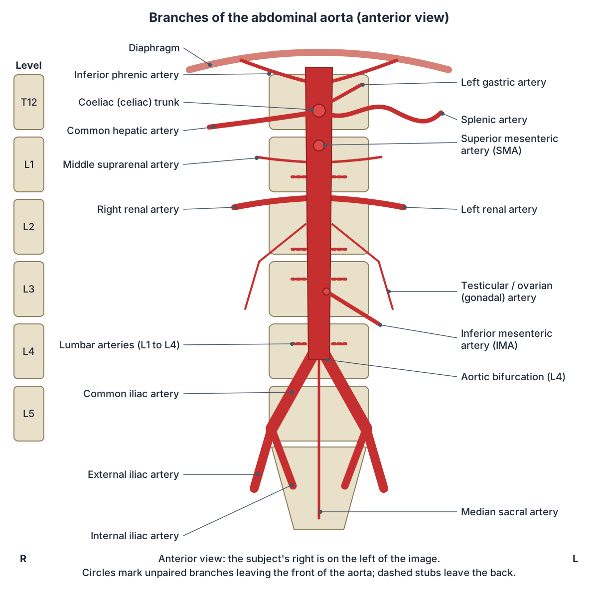
Abdominal aorta branches and their vertebral levels
| Branch | Type | Vertebral level | Main territory |
|---|---|---|---|
| Inferior phrenic arteries | Paired, posterior (parietal) | T12 | Underside of the diaphragm |
| Coeliac trunk | Unpaired, anterior (visceral) | T12 | Stomach, abdominal oesophagus, liver, gallbladder, spleen, pancreas, duodenum |
| Superior mesenteric artery | Unpaired, anterior (visceral) | L1 | Midgut: second part of duodenum to distal transverse colon |
| Middle suprarenal arteries | Paired, lateral (visceral) | L1 | Adrenal glands |
| Renal arteries | Paired, lateral (visceral) | L1 to L2 | Kidneys |
| Gonadal (testicular or ovarian) arteries | Paired, lateral (visceral) | L2 (L2 to L3 in some sources) | Testes or ovaries |
| Inferior mesenteric artery | Unpaired, anterior (visceral) | L3 | Hindgut: splenic flexure to upper two-thirds of rectum |
| Lumbar arteries (4 pairs) | Paired, posterior (parietal) | L1 to L4 | Posterior abdominal wall |
| Median sacral artery | Unpaired, posterior (parietal) | L4 | Sacral region (continues the aorta onto the sacrum) |
| Common iliac arteries (terminal) | Paired, terminal branches | L4 | Pelvis (internal iliac) and lower limb (external iliac) |
Levels from TeachMeAnatomy; territories from TeachMeAnatomy, Radiopaedia and OpenStax (see references). Levels vary by about one vertebra between people.
Coeliac trunk (celiac artery) and its branches
The coeliac trunk (celiac trunk or celiac artery) arises from the front of the aorta at the aortic hiatus, at T12. It is only about 1 cm long before it divides into three: the left gastric artery, the splenic artery and the common hepatic artery. (TeachMeAnatomy: The Coeliac Trunk.)
The left gastric artery is the smallest branch and supplies the lower oesophagus and the lesser curvature of the stomach. The splenic artery runs to the left to the spleen, giving pancreatic branches to the body and tail of the pancreas, short gastric arteries to the fundus and the left gastro-omental (gastroepiploic) artery to the greater curvature. (TeachMeAnatomy: The Coeliac Trunk.)
The common hepatic artery is the only branch that goes to the right. It divides into the hepatic artery proper, which gives the right gastric artery and the right and left hepatic arteries (the right hepatic gives the cystic artery to the gallbladder), and the gastroduodenal artery, which gives the right gastro-omental artery and the superior pancreaticoduodenal artery. (TeachMeAnatomy: The Coeliac Trunk.)
The coeliac trunk supplies the stomach, spleen, liver, gallbladder, abdominal oesophagus, pancreas and duodenum. The stomach is the only organ that receives blood from all three branches. In coeliac artery compression syndrome, the median arcuate ligament of the diaphragm presses on the trunk and causes abdominal pain. (TeachMeAnatomy: The Coeliac Trunk.)
Superior and inferior mesenteric arteries
The superior mesenteric artery (SMA) arises from the front of the aorta at L1, usually just below the coeliac trunk. OpenStax puts its origin about 2.5 cm below the trunk. It runs down behind the neck of the pancreas and the splenic vein, crosses in front of the left renal vein, the uncinate process and the third part of the duodenum, and enters the root of the small bowel mesentery. (Radiopaedia: Superior mesenteric artery.)
The SMA is the artery of the midgut. It supplies the gut from the major duodenal papilla (ampulla of Vater) in the second part of the duodenum as far as the distal third of the transverse colon, through the inferior pancreaticoduodenal artery, jejunal and ileal branches on its left, and the ileocolic, right colic and middle colic arteries on its right. (Radiopaedia: Superior mesenteric artery.)
Because the left renal vein passes between the SMA and the aorta, it can be squeezed there (nutcracker syndrome), and the third part of the duodenum can be compressed in the same angle (SMA syndrome). (Radiopaedia: Superior mesenteric artery.)
The inferior mesenteric artery (IMA) is an unpaired branch from the front of the aorta at L3. It runs down and to the left towards the pelvic brim, gives the left colic artery and two to four sigmoid arteries, and continues into the pelvis as the superior rectal artery. It is the artery of the hindgut, from the splenic flexure of the colon to the upper two-thirds of the rectum. (Radiopaedia: Inferior mesenteric artery.)
Renal, suprarenal, gonadal, phrenic and lumbar arteries
Each renal artery leaves the side of the aorta about 2.5 cm below the superior mesenteric artery and runs to the kidney. The middle suprarenal (adrenal) arteries supply the adrenal glands, and the gonadal arteries supply the testes or ovaries. (OpenStax Anatomy and Physiology 2e, 20.5: Circulatory Pathways.)
Each inferior phrenic artery is the counterpart of a superior phrenic artery, supplying the diaphragm from below. The four pairs of lumbar arteries are the counterparts of the intercostal arteries in the chest wall. (OpenStax Anatomy and Physiology 2e, 20.5: Circulatory Pathways.)
At its end the aorta continues as a small vessel, the median sacral artery, down onto the sacrum. Each common iliac artery then divides into an external iliac artery, which continues to the lower limb, and an internal iliac artery to the pelvis, approximately at the level of the lumbosacral joint. (OpenStax Anatomy and Physiology 2e, 20.5: Circulatory Pathways.)
Clinical relevance: aneurysm, dissection and coarctation
An abdominal aortic aneurysm (AAA) is a focal widening of the abdominal aorta to a maximum diameter over 3 cm, or at least 50% wider than the normal aorta above it. About 10% of people over 65 have one, men about four times as often as women, and atherosclerosis is the most common cause. Ultrasound is the usual screening test. (Radiopaedia: Abdominal aortic aneurysm.)
The most common site is the infrarenal aorta, between the renal arteries and the bifurcation at L4. Many aneurysms cause no symptoms and are found by chance; others present as a pulsatile abdominal mass or with abdominal or back pain. Large aneurysms can rupture and cause life-threatening bleeding. (TeachMeAnatomy: The Abdominal Aorta.)
In aortic dissection, blood enters the middle layer of the aortic wall through a tear in the inner layer and forms a second, false channel. Patients typically have chest pain, often described as tearing. Dissections involving the ascending aorta (Stanford type A, about 60%) need immediate surgery; those that do not (type B) are usually treated with blood pressure control unless complicated. (Radiopaedia: Aortic dissection.)
Coarctation of the aorta is a congenital narrowing, usually near the ductus arteriosus, just beyond the origin of the left subclavian artery. In long-standing cases, enlarged intercostal collateral arteries notch the lower borders of the ribs. A bicuspid aortic valve is found in 45 to 60% of cases, and coarctation occurs in 15 to 20% of people with Turner syndrome. (Radiopaedia: Coarctation of the aorta.)
Aorta in the 3D model
- Abdominal aorta
- The abdominal aorta is the part of the aorta that runs through the abdomen and supplies the abdominal organs, pelvis and lower limbs.
- Arch of aorta
- The aortic arch is the curved part of the aorta between the ascending and descending aorta, giving the great vessels to the head and arms.
- Ascending aorta
- The ascending aorta is the first part of the aorta, rising from the left ventricle.
- Brachiocephalic artery
- The brachiocephalic artery (trunk) is the first branch of the aortic arch and supplies the right head, neck and arm.
- Celiac artery
- The celiac artery (celiac trunk) is the first major unpaired branch of the abdominal aorta and supplies the foregut.
- Celiac trunk
- The celiac trunk is the first major unpaired branch of the abdominal aorta and supplies the foregut.
- Common carotid artery
- The common carotid artery is the main artery of the neck and splits into the external and internal carotid arteries.
- Common hepatic artery
- The common hepatic artery is a branch of the celiac trunk supplying the liver and nearby organs.
- Common iliac artery
- The common iliac arteries are the two terminal branches of the abdominal aorta.
- Descending aorta
- The descending aorta is the part of the aorta beyond the arch that runs down through the thorax and abdomen.
- Descending thoracic aorta
- The descending thoracic aorta is the part of the aorta in the posterior mediastinum.
- Gastric artery
- The gastric arteries are the arteries of the lesser curvature of the stomach.
- Inferior mesenteric artery
- The inferior mesenteric artery supplies the hindgut.
- Inferior phrenic artery
- The inferior phrenic arteries supply the undersurface of the diaphragm.
- Lumbar artery
- One of usually four pairs of segmental arteries arising from the back of the abdominal aorta.
- Middle suprarenal artery
- A small branch of the abdominal aorta supplying the adrenal gland.
- Renal artery
- A large paired branch of the abdominal aorta supplying the kidney.
- Splenic artery
- The largest branch of the celiac trunk, running to the spleen.
- Subclavian artery
- The major artery to the upper limb, neck and part of the brain.
- Superior mesenteric artery
- An unpaired artery of the midgut.
- Testicular artery
- A paired branch of the abdominal aorta to the testis.
- Trunk of renal artery
- The main stem of the renal artery before division.
- Trunk of superior mesenteric artery
- The main stem of the superior mesenteric artery.
Frequently asked questions
What are the three branches of the aortic arch?
In about 75% of people the aortic arch has three branches. From right to left they are the brachiocephalic trunk, which divides into the right subclavian and right common carotid arteries, the left common carotid artery and the left subclavian artery. Common variants include a shared origin of the brachiocephalic trunk and left common carotid, the so-called bovine arch.
Where is the abdominal aorta?
The abdominal aorta runs down the back of the abdomen, on the front of the lumbar vertebrae and slightly left of the midline, with the inferior vena cava on its right. It begins at T12, where it passes through the aortic hiatus of the diaphragm, and ends at L4 by dividing into the two common iliac arteries.
What are the branches of the abdominal aorta?
Unpaired anterior branches: coeliac trunk (T12), superior mesenteric artery (L1) and inferior mesenteric artery (L3). Paired lateral branches: middle suprarenal (L1), renal (L1 to L2) and gonadal arteries (L2). Posterior branches: inferior phrenic (T12), four pairs of lumbar arteries (L1 to L4) and the median sacral artery. It ends as the two common iliac arteries at L4.
What are the branches of the celiac trunk?
The coeliac (celiac) trunk arises from the aorta at T12 and after about 1 cm divides into three branches: the left gastric artery to the lesser curvature of the stomach and lower oesophagus, the splenic artery to the spleen, pancreas and greater curvature, and the common hepatic artery, which gives the hepatic artery proper and the gastroduodenal artery.
What is the difference between the thoracic and abdominal aorta?
They are two parts of the same descending aorta, divided by the diaphragm. The thoracic aorta runs from the lower border of T4 to T12 in the posterior mediastinum and gives intercostal, bronchial, oesophageal and superior phrenic branches. The abdominal aorta runs from T12 to L4 and gives the arteries to the gut, kidneys, adrenal glands, gonads and posterior abdominal wall.
Where does the aorta bifurcate?
The abdominal aorta divides into the right and left common iliac arteries at the level of the fourth lumbar vertebra (L4). A small median sacral artery continues down from the bifurcation onto the sacrum. Each common iliac artery then splits into external and internal iliac arteries at about the level of the lumbosacral joint.
What size is an abdominal aortic aneurysm?
An abdominal aortic aneurysm is defined as a maximum diameter of more than 3 cm, or a segment at least 50% wider than the normal aorta above it. For comparison, the normal aorta narrows from about 28 mm in the chest to under 20 mm below the kidneys. Ultrasound is used for screening and follow-up.
References
- OpenStax. Anatomy and Physiology 2e, §20.5 Circulatory Pathways. Rice University, 2022.
- OpenStax. Anatomy and Physiology 2e, §19.1 Heart Anatomy. Rice University, 2022.
- Jones O. Great Vessels of the Heart. TeachMeAnatomy.
- Jones O. The Posterior Mediastinum. TeachMeAnatomy.
- Jones O. The Abdominal Aorta. TeachMeAnatomy.
- Jones O. The Coeliac Trunk. TeachMeAnatomy.
- Aortic arch. Radiopaedia reference article.
- Abdominal aorta. Radiopaedia reference article.
- Superior mesenteric artery. Radiopaedia reference article.
- Inferior mesenteric artery. Radiopaedia reference article.
- Abdominal aortic aneurysm. Radiopaedia reference article.
- Aortic dissection. Radiopaedia reference article.
- Coarctation of the aorta. Radiopaedia reference article.
- di Gioia CRT, Ascione A, Carletti R, Giordano C. Thoracic Aorta: Anatomy and Pathology. Diagnostics (Basel). 2023;13(13):2166.
- Mitsuhashi N. et al. (2009). BodyParts3D: 3D structure database for anatomical concepts. Nucleic Acids Research 37, D782–D785. Data © The Database Center for Life Science, CC BY 4.0.
Related anatomy
- Coronary arteriesCoronary arteries explained: left main, LAD and diagonals, circumflex, RCA and posterior descending artery, dominance…
- Inferior vena cavaInferior vena cava anatomy: course, relations, tributaries by vertebral level, embryology, variants and IVC filters…
- Portal veinHepatic portal vein explained: how it forms, its tributaries, the porta hepatis, portal vs hepatic veins…
- Carotid arteriesCarotid arteries explained: common carotid, bifurcation, internal carotid, the 8 external carotid branches with a…
- KidneyWhere are your kidneys? Kidney location on the back (T12 to L3), coverings, relations, cortex, medulla, calyces, renal…
- Celiac trunkCeliac trunk: The celiac trunk is the first major unpaired branch of the abdominal aorta and supplies the foregut. See…
Explore the whole body in the interactive 3D human anatomy model, or browse the A–Z list of human structures.