

What are the carotid arteries?
The carotid arteries are the arteries on either side of the neck that carry blood to the head. The two common carotid arteries do not usually give any branches in the neck. Each ends by dividing into an internal carotid artery and an external carotid artery. Together with the vertebral arteries, the internal carotid arteries are the two main suppliers of blood to the brain. (OpenStax Anatomy and Physiology 2e, 20.5: Circulatory Pathways.)
The internal carotid artery enters the skull through the carotid canal in the temporal bone and supplies the brain and eye. The external carotid artery stays outside the skull and supplies the tissues on its outer surface, such as the face, scalp, jaw and neck. (OpenStax Anatomy and Physiology 2e, 13.3: Circulation and the Central Nervous System.)
Common carotid artery: origin and course in the neck
The right and left common carotid arteries start in different places. The right common carotid artery arises from the brachiocephalic trunk and has only a cervical (neck) part. The left common carotid artery arises directly from the arch of the aorta, so it also has a thoracic part, which climbs to the level of the left sternoclavicular joint before the artery enters the neck. (Radiopaedia: Common carotid artery.)
In the neck each common carotid artery runs obliquely upwards from behind the sternoclavicular joint. Low in the neck the two arteries are separated only by the trachea; higher up they move apart, with the thyroid gland, larynx and pharynx between them. Each ends at the carotid bifurcation, at about the level of C4 and the upper border of the thyroid cartilage, although this level varies. (Radiopaedia: Common carotid artery.)
The common carotid artery runs in the carotid sheath together with the internal jugular vein and the vagus nerve. The vein lies lateral to the artery, with the nerve between the two. (Radiopaedia: Common carotid artery.)
The carotid sheath is a tube built from all three layers of the deep cervical fascia that runs from the skull base to the aortic arch. Below the hyoid bone it contains the common carotid artery, the internal jugular vein and the vagus nerve; the ansa cervicalis is embedded in its front wall, and the deep cervical lymph nodes run along it. (Radiopaedia: Carotid space.)
Carotid bifurcation, carotid sinus and carotid body
The carotid bifurcation lies in the carotid triangle of the neck, at about the level of C4. Just above the bifurcation, the start of the internal carotid artery is slightly widened. This dilatation is the carotid sinus, which contains baroreceptors that detect changes in blood pressure. The carotid body is a small chemoreceptor that detects changes in the oxygen level of the blood and helps to regulate breathing. (TeachMeAnatomy: Arterial Supply to the Head and Neck.)
Radiopaedia places the carotid bulb or sinus at the origin of the internal carotid artery, with the carotid body just behind the bifurcation, and notes that the bifurcation usually lies between C3 and C5 but varies widely. It can be as low as the cricoid cartilage (C5) or as high as the hyoid bone (C2). (Radiopaedia: Internal carotid artery.)
Just above the bifurcation, the internal carotid artery usually lies posterolateral to the external carotid artery. A useful difference between them is that the internal carotid artery has no branches in the neck, whereas the external carotid artery gives off several branches soon after it begins. (Radiopaedia: Internal carotid artery.)
External carotid artery and its eight branches
The external carotid artery (ECA) is one of the two terminal branches of the common carotid artery. It begins at the bifurcation, at about the upper border of the thyroid cartilage, curves upwards and slightly forwards, and then passes back to the space behind the neck of the mandible, where it enters the parotid gland and ends. (Radiopaedia: External carotid artery.)
Several structures cross the outer surface of the external carotid artery: the hypoglossal nerve, the posterior belly of the digastric, the stylohyoid muscle, and inside the parotid gland the facial nerve and the retromandibular vein. The glossopharyngeal nerve, the pharyngeal branch of the vagus, stylopharyngeus and styloglossus pass between the external and internal carotid arteries. (Radiopaedia: External carotid artery.)
The external carotid artery usually has eight branches. From below upwards they are the superior thyroid, ascending pharyngeal, lingual, facial, occipital and posterior auricular arteries, and the two terminal branches, the maxillary and superficial temporal arteries. Two common mnemonics are "Some Anatomists Like Freaking Out Poor Medical Students" and "Some American Ladies Found Our Pyramids Most Satisfactory". (Radiopaedia: External carotid artery.)
Branching often varies. Radiopaedia lists a shared linguofacial trunk for the lingual and facial arteries in about 20% of people, a common trunk for the occipital and posterior auricular arteries in about 12.5%, and less common thyrolingual and thyrolinguofacial trunks. (Radiopaedia: External carotid artery.)
The superior thyroid artery runs to the thyroid gland and larynx and gives off the superior laryngeal artery. In about 10% of people it arises from the common carotid artery instead. (Radiopaedia: Superior thyroid artery.)
The ascending pharyngeal artery is the smallest branch. It arises from the back of the external carotid artery near the bifurcation and runs straight up between the internal carotid artery and the pharynx. It supplies the pharyngeal muscles, soft palate and tonsil, the middle ear and, through meningeal branches, the dura of the skull base. (Radiopaedia: Ascending pharyngeal artery.)
The lingual artery arises at about the level of C3, loops up towards the hyoid bone and then runs into the tongue. The hypoglossal nerve crosses its loop. It supplies the tongue and the floor of the mouth. (Radiopaedia: Lingual artery.)
The occipital artery arises from the back of the external carotid artery about 2 cm above the bifurcation, opposite the facial artery. It runs back between the posterior belly of the digastric and the internal jugular vein, passes between the mastoid process and C2, and reaches the back of the scalp with the greater occipital nerve. The posterior auricular artery arises above the digastric and stylohyoid, opposite the styloid process, and supplies the auricle, the scalp behind it and the facial nerve inside the stylomastoid foramen. (Radiopaedia: Occipital artery.)
The maxillary artery begins behind the neck of the mandible inside the parotid gland and supplies the deep structures of the face. It is divided into three parts by its relation to the lateral pterygoid muscle. Its first part gives the middle meningeal artery and the inferior alveolar artery, which enters the mandibular foramen to supply the lower teeth. Its third part ends as the sphenopalatine artery to the nasal cavity. (Radiopaedia: Maxillary artery.)
The superficial temporal artery is the other terminal branch. It arises in the parotid gland, runs up over the zygomatic arch and ends as frontal and parietal branches in the scalp. It also gives the transverse facial artery. Radiopaedia lists giant cell arteritis as the main disease linked with this artery. (Radiopaedia: Superficial temporal artery.)
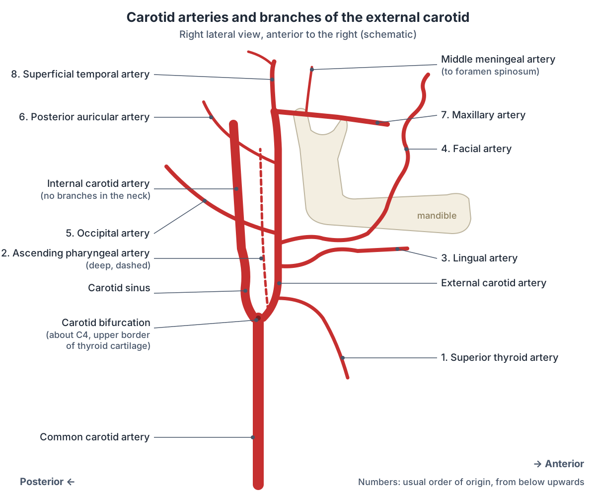
The eight branches of the external carotid artery
| Branch | Where it arises | Main area supplied | Notes |
|---|---|---|---|
| 1. Superior thyroid artery | Front of the ECA at about the level of the hyoid bone | Thyroid gland and larynx | Gives the superior laryngeal artery; arises from the common carotid in about 10% |
| 2. Ascending pharyngeal artery | Back of the ECA near the bifurcation | Pharynx, soft palate, middle ear and the dura of the skull base | Smallest branch; runs up between the ICA and the pharynx |
| 3. Lingual artery | Front of the ECA at about C3 | Tongue and floor of the mouth | Its loop is crossed by the hypoglossal nerve |
| 4. Facial artery | Front of the ECA, just above the lingual artery | Face, lips, palatine tonsil and soft palate | Pulse felt where it crosses the mandible at the front edge of masseter; ends as the angular artery |
| 5. Occipital artery | Back of the ECA, opposite the facial artery | Back of the scalp, upper neck muscles, posterior fossa dura | Runs with the greater occipital nerve to the scalp |
| 6. Posterior auricular artery | Back of the ECA above the digastric and stylohyoid | Auricle, scalp behind the ear, facial nerve in the stylomastoid foramen | May share a trunk with the occipital artery |
| 7. Maxillary artery | Terminal branch, behind the neck of the mandible in the parotid gland | Deep structures of the face, teeth, nasal cavity, palate and the dura | Three parts; gives the middle meningeal and inferior alveolar arteries |
| 8. Superficial temporal artery | Terminal branch, in the parotid gland | Side of the face and scalp | Crosses the zygomatic arch; frontal and parietal branches |
Compiled from the Radiopaedia articles on each branch and TeachMeAnatomy (see references). The order and pattern of branching vary between people.
Facial artery (arteria facialis)
The facial artery (Latin arteria facialis) arises from the external carotid artery in the carotid triangle, just above the lingual artery. It passes deep to the posterior belly of the digastric, then follows a winding course over the lower border of the mandible at the front edge of the masseter muscle and up towards the nasolabial fold. (Radiopaedia: Facial artery.)
In the neck it gives the ascending palatine artery, a tonsillar branch, the submental artery and glandular branches. On the face it gives the inferior and superior labial arteries to the lips and a lateral nasal branch, and it ends as the angular artery at the inner corner of the eye. Its pulse can be felt where it crosses the lower border of the mandible, which is why it is sometimes called the anaesthetist's artery. (Radiopaedia: Facial artery.)
Middle meningeal artery (arteria meningea media)
The middle meningeal artery (arteria meningea media, sometimes searched as the "medial meningeal artery") is a branch of the first part of the maxillary artery, so it belongs to the external carotid system. It runs up between the roots of the auriculotemporal nerve and enters the middle cranial fossa through the foramen spinosum. It is the main artery of the cranial dura mater. (Radiopaedia: Middle meningeal artery.)
Inside the skull it divides into an anterior and a posterior division that groove the inner surface of the skull. The anterior division runs forwards on the greater wing of the sphenoid and passes under the pterion on the side of the skull. (Radiopaedia: Middle meningeal artery.)
A fracture at the pterion, the weakest point of the skull, can tear the middle meningeal artery and cause an extradural (epidural) haematoma: arterial blood collects between the skull and the dura and raises intracranial pressure. (TeachMeAnatomy: Arterial Supply to the Head and Neck.)
Variants are clinically important. The middle meningeal artery sometimes arises from the internal carotid system, most often through the ophthalmic artery, and rarely the ophthalmic artery arises from the middle meningeal artery. (Radiopaedia: Middle meningeal artery.)
Internal carotid artery: course and segments
The internal carotid artery (ICA) runs up through the neck in the carotid sheath without giving any branches. It enters the skull base through the carotid canal of the temporal bone, passes through the cavernous sinus, and ends by dividing into the anterior and middle cerebral arteries. (Radiopaedia: Internal carotid artery.)
Radiologists and surgeons divide the ICA into seven segments (the Bouthillier classification): C1 cervical, C2 petrous, C3 lacerum, C4 cavernous, C5 clinoid, C6 ophthalmic and C7 communicating. The ophthalmic artery arises from C6, and the posterior communicating and anterior choroidal arteries arise from C7 before the terminal division. (Radiopaedia: Internal carotid artery.)
Inside the skull its main branches are the ophthalmic artery to the orbit, forehead and nose, the anterior cerebral artery to the medial surfaces of the cerebral hemispheres, and the middle cerebral artery to their lateral surfaces. The brain arteries page covers these in detail. (TeachMeAnatomy: Arterial Supply to the Head and Neck.)
Subclavian artery branches: thyrocervical trunk and transverse cervical artery
The subclavian arteries also send important branches into the neck. The right subclavian artery is a terminal branch of the brachiocephalic trunk; the left is the third branch of the aortic arch. Each is divided into three parts by scalenus anterior. The first part, medial to the muscle, gives the vertebral artery, the internal thoracic artery and the thyrocervical trunk. The second part, behind the muscle, gives the costocervical trunk. The third part may give the dorsal scapular artery. The artery becomes the axillary artery at the lateral border of the first rib. (Radiopaedia: Subclavian artery.)
The thyrocervical trunk is a short, stout vessel that arises from the front of the first part of the subclavian artery, lateral to the vertebral artery and next to scalenus anterior. Radiopaedia describes three branches: the inferior thyroid artery, the largest, which supplies the thyroid and parathyroid glands, larynx, trachea and oesophagus; the transverse cervical artery; and the suprascapular artery, which joins the scapular anastomosis. (Radiopaedia: Thyrocervical trunk.)
Sources count the branches differently. TeachMeAnatomy lists four branches of the thyrocervical trunk: the inferior thyroid, ascending cervical, transverse cervical and suprascapular arteries. Radiopaedia notes that the ascending cervical artery usually comes off the inferior thyroid artery and arises from the trunk itself in only about 11% of people. (TeachMeAnatomy: Arterial Supply to the Head and Neck.)
The transverse cervical artery (cervicodorsal trunk) is short. It runs laterally across the front of the phrenic nerve and scalenus anterior, then crosses or passes through the trunks of the brachial plexus. It divides into a superficial cervical artery, which runs in front of trapezius next to the accessory nerve, and the dorsal scapular artery. It can also arise directly from the second or third part of the subclavian artery. (Radiopaedia: Transverse cervical artery.)
Vertebral artery segments V1 to V4
The vertebral artery arises from the first part of the subclavian artery. It is divided into four segments: V1 runs from its origin to the transverse foramen of C6; V2 climbs through the transverse foramina from C6 to C2; V3 loops out through the transverse foramen of C1 and around the back of the atlas, then pierces the posterior atlanto-occipital membrane and dura; V4 runs inside the skull in front of the medulla. The two V4 segments unite at the lower border of the pons to form the basilar artery. (Radiopaedia: Vertebral artery.)
Its branches include muscular and spinal branches in the neck, meningeal branches, the anterior and posterior spinal arteries, and the posterior inferior cerebellar artery (PICA), its largest branch. The two vertebral arteries are often unequal: Radiopaedia gives left dominance in about 45% of people, right dominance in about 30% and equal size in about 25%. The left vertebral artery arises directly from the aortic arch in about 5 to 6%. (Radiopaedia: Vertebral artery.)
The vertebral arteries enter the skull through the foramen magnum. Branches from each join to form the anterior spinal artery, which supplies the front of the spinal cord, and the basilar artery supplies the brainstem and cerebellum. (OpenStax Anatomy and Physiology 2e, 13.3: Circulation and the Central Nervous System.)
Vertebral artery segments
| Segment | Name | Course |
|---|---|---|
| V1 | Pre-foraminal | From the subclavian artery to the transverse foramen of C6, between longus colli and scalenus anterior |
| V2 | Foraminal | Up through the transverse foramina of C6 to C2 |
| V3 | Atlantic (extradural) | From C2, loops laterally through the transverse foramen of C1, curves back around the atlas and pierces the posterior atlanto-occipital membrane and dura |
| V4 | Intradural (intracranial) | Up in front of the medulla to the lower border of the pons, where it joins the other vertebral artery to form the basilar artery |
Source: Radiopaedia, Vertebral artery.
Internal and external jugular veins
There is no "jugular artery": the jugular vessels are veins. The large artery in the neck is the carotid artery, and the internal jugular vein runs beside it in the carotid sheath. The internal jugular vein starts at the jugular foramen as the continuation of the sigmoid sinus, runs down deep to sternocleidomastoid and lateral to the common carotid artery, and ends behind the sternal end of the clavicle by joining the subclavian vein to form the brachiocephalic vein. (TeachMeAnatomy: Venous Drainage of the Head and Neck.)
The internal jugular vein receives the facial, lingual, pharyngeal and superior and middle thyroid veins. Higher up, the vagus nerve always lies between the internal carotid artery and the internal jugular vein. (Radiopaedia: Internal jugular vein.)
The external jugular vein is formed by the posterior division of the retromandibular vein and the posterior auricular vein, which unite within the parotid gland near the angle of the mandible. It runs down the side of the neck under the skin, deep to platysma but superficial to sternocleidomastoid, then pierces the deep cervical fascia and drains into the subclavian vein. Its tributaries include the anterior jugular, posterior external jugular, transverse cervical and suprascapular veins. (Radiopaedia: External jugular vein.)
Because the external jugular vein lies just under the skin it is easily injured. Its wall is held open by the surrounding fascia, so if it is cut, air can be sucked in, which is a medical emergency. (TeachMeAnatomy: Venous Drainage of the Head and Neck.)
In summary, blood from the brain and the deep face drains into the internal jugular vein, and blood from the more superficial parts of the head and scalp drains into the external jugular vein. The two jugular veins and the subclavian vein meet to form the brachiocephalic vein on each side. (OpenStax Anatomy and Physiology 2e, 20.5: Circulatory Pathways.)
Clinical notes: why the carotid region matters
The carotid sinus helps to control blood pressure: its baroreceptors detect changes in pressure, and the carotid body senses blood oxygen. Both lie at the carotid bifurcation in the carotid triangle. (TeachMeAnatomy: Arterial Supply to the Head and Neck.)
TeachMeAnatomy lists embolism, often from atherosclerotic plaque in the arteries of the neck, as the most common cause of stroke. (TeachMeAnatomy: Arterial Supply to the Brain.)
An extradural haematoma from a torn middle meningeal artery is the classic emergency linked with this region. TeachMeAnatomy describes blood collecting between the skull and the periosteal layer of the dura, usually as a result of head trauma. (TeachMeAnatomy: The Meninges.)
Carotid arteries in the 3D model
- Brachiocephalic artery
- The brachiocephalic artery (trunk) is the first branch of the aortic arch and supplies the right head, neck and arm.
- Brachiocephalic vein
- The brachiocephalic veins are two large veins that unite to form the superior vena cava.
- Common carotid artery
- The common carotid artery is the main artery of the neck and splits into the external and internal carotid arteries.
- Costocervical trunk
- The costocervical trunk is a branch of the subclavian artery that supplies the neck and upper intercostal spaces.
- Inferior thyroid artery
- The inferior thyroid artery supplies the thyroid and parathyroid glands.
- Internal carotid artery
- The internal carotid artery supplies the brain and the eye.
- Internal jugular vein
- The internal jugular vein is the main vein draining the head and neck.
- Subclavian artery
- The major artery to the upper limb, neck and part of the brain.
- Subclavian vein
- The large vein draining the upper limb into the brachiocephalic vein.
- Suprascapular artery
- A branch of the thyrocervical trunk to the scapular region.
- Thyrocervical trunk
- A short branch of the subclavian artery to the neck.
- Transverse cervical artery
- A branch of the thyrocervical trunk to the neck.
- Vertebral artery
- A branch of the subclavian artery supplying the brain and spinal cord.
Frequently asked questions
What are the 8 branches of the external carotid artery?
From below upwards: superior thyroid, ascending pharyngeal, lingual, facial, occipital and posterior auricular arteries, then the two terminal branches, the maxillary and superficial temporal arteries. A common mnemonic is "Some Anatomists Like Freaking Out Poor Medical Students". The order varies; for example, the lingual and facial arteries share a trunk in about one person in five.
Where does the common carotid artery divide?
Usually at about the level of C4, the upper border of the thyroid cartilage, in the carotid triangle of the neck. The level varies between C3 and C5 in most people and can be as high as the hyoid bone. At the bifurcation the artery splits into the internal carotid artery, which has a dilated carotid sinus at its start, and the external carotid artery.
What is the difference between the internal and external carotid artery?
The internal carotid artery gives no branches in the neck, enters the skull through the carotid canal and supplies the brain and eye. The external carotid artery gives eight branches soon after it starts and supplies the neck, face, scalp, jaw and the dura through the middle meningeal artery. Just above the bifurcation the internal carotid usually lies posterolateral to the external carotid.
Is there a jugular artery?
No. The jugular vessels are veins that drain blood from the head. The pulsing artery felt in the side of the neck is the common carotid artery. It runs in the carotid sheath with the internal jugular vein lateral to it and the vagus nerve between and behind them. The external jugular vein lies more superficially, on top of sternocleidomastoid.
What does the thyrocervical trunk supply?
The thyrocervical trunk is a short branch of the first part of the subclavian artery. Through the inferior thyroid artery it supplies the thyroid and parathyroid glands, larynx, trachea and oesophagus. Through the transverse cervical and suprascapular arteries it supplies muscles of the neck, shoulder and back of the scapula. Some sources also count the ascending cervical artery as a direct branch.
What are the segments of the vertebral artery?
There are four. V1 runs from the subclavian artery to the transverse foramen of C6. V2 climbs through the transverse foramina of C6 to C2. V3 loops through C1 and around the atlas before piercing the dura. V4 lies inside the skull and joins the other vertebral artery at the lower border of the pons to form the basilar artery.
Which artery is damaged in an extradural haematoma?
Usually the middle meningeal artery, a branch of the maxillary artery that enters the skull through the foramen spinosum. Its anterior division runs under the pterion, the thinnest part of the side of the skull, so a fracture there can tear it. Arterial blood then collects between the skull and the dura and raises the pressure inside the skull.
References
- OpenStax. Anatomy and Physiology 2e, §20.5 Circulatory Pathways. Rice University, 2022.
- OpenStax. Anatomy and Physiology 2e, §13.3 Circulation and the Central Nervous System. Rice University, 2022.
- TeachMeAnatomy. Arterial Supply to the Head and Neck.
- TeachMeAnatomy. Arterial Supply to the Brain.
- TeachMeAnatomy. Venous Drainage of the Head and Neck.
- TeachMeAnatomy. The Meninges.
- Radiopaedia. Common carotid artery.
- Radiopaedia. External carotid artery.
- Radiopaedia. Internal carotid artery.
- Radiopaedia. Superior thyroid artery.
- Radiopaedia. Ascending pharyngeal artery.
- Radiopaedia. Lingual artery.
- Radiopaedia. Facial artery.
- Radiopaedia. Occipital artery.
- Radiopaedia. Posterior auricular artery.
- Radiopaedia. Maxillary artery.
- Radiopaedia. Superficial temporal artery.
- Radiopaedia. Middle meningeal artery.
- Radiopaedia. Subclavian artery.
- Radiopaedia. Thyrocervical trunk.
- Radiopaedia. Transverse cervical artery.
- Radiopaedia. Vertebral artery.
- Radiopaedia. Internal jugular vein.
- Radiopaedia. External jugular vein.
- Radiopaedia. Carotid space (carotid sheath).
- Mitsuhashi N. et al. (2009). BodyParts3D: 3D structure database for anatomical concepts. Nucleic Acids Research 37, D782–D785. Data © The Database Center for Life Science, CC BY 4.0.
Related anatomy
- Brain arteriesCircle of Willis and the arteries of the brain: anterior, middle and posterior cerebral, communicating, basilar and…
- Internal jugular veinInternal jugular vein (IJ vein) anatomy: jugular foramen to brachiocephalic vein, carotid sheath relations…
- Dural venous sinusesDural venous sinuses explained: superior sagittal, straight, transverse, sigmoid, occipital and cavernous sinus…
- AortaAorta anatomy explained: ascending aorta, aortic arch and its three branches, thoracic aorta, and every abdominal…
- Neck musclesNeck muscles explained: sternocleidomastoid, suprahyoid, infrahyoid, scalene, prevertebral and suboccipital groups…
- Vertebral arteryVertebral artery: A branch of the subclavian artery supplying the brain and spinal cord. See where it is in an…
Explore the whole body in the interactive 3D human anatomy model, or browse the A–Z list of human structures.