

How many bones are in the foot?
There are 26 bones in each foot: 7 tarsal bones at the back of the foot, 5 metatarsal bones in the middle and 14 phalanges in the toes. The big toe (hallux) has two phalanges, proximal and distal, while each of the other four toes has three: proximal, middle and distal. Both feet together therefore contain 52 bones, before counting sesamoid bones. (OpenStax Anatomy and Physiology 2e, 8.4: Bones of the Lower Limb.)
Clinicians divide the foot into three regions. The hindfoot is the talus and calcaneus; the midfoot is the navicular, cuboid and three cuneiforms; and the forefoot is the metatarsals and phalanges. (TeachMeAnatomy: Bones of the Foot: Tarsals, Metatarsals and Phalanges.)
In addition, two sesamoid bones are always present on the plantar (sole) side of the head of the first metatarsal, where they glide in two grooved facets. They are not usually counted among the 26. (Kenhub: Metatarsal bones.)
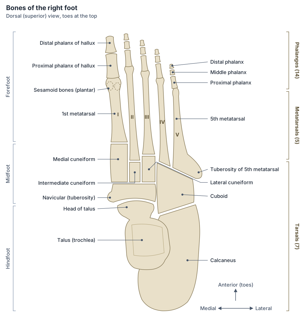
Tarsal bones (the tarsus)
The tarsus is the group of seven tarsal bones that forms the back half of the foot: the talus, calcaneus, navicular, cuboid and the medial, intermediate and lateral cuneiforms. Besides bearing weight, the tarsals give attachment to muscles and help maintain the arches of the foot. (Kenhub: Tarsal bones.)
The tarsals are often described in three rows. The proximal row is the talus above and the calcaneus below; the intermediate row is the navicular on the medial side; and the distal row is the three cuneiforms medially and the cuboid laterally. (TeachMeAnatomy: Bones of the Foot: Tarsals, Metatarsals and Phalanges.)
Bones of the foot at a glance
| Group | Bones | Number per foot | Key features |
|---|---|---|---|
| Tarsals: hindfoot | Talus, calcaneus | 2 | Talus forms the ankle joint and has no muscle attachments; calcaneus is the largest tarsal and forms the heel |
| Tarsals: midfoot | Navicular, cuboid, medial, intermediate and lateral cuneiforms | 5 | Navicular tuberosity for tibialis posterior; cuboid groove for fibularis longus; cuneiforms are wedge-shaped |
| Metatarsals | First to fifth metatarsals (I to V) | 5 | 1st thickest and shortest, 2nd longest, 5th has a palpable tuberosity |
| Phalanges | Proximal, middle and distal phalanges | 14 | Hallux has 2; toes 2 to 5 have 3 each |
| Sesamoids | Two hallucal sesamoids | 2 (not in the 26) | Under the head of the first metatarsal |
Counts from OpenStax; features from TeachMeAnatomy and Kenhub.
Talus and calcaneus: the hindfoot
The talus (ankle bone) is the most superior tarsal bone and transmits body weight from the leg to the foot. Above, it articulates with the tibia and fibula at the ankle joint; below, with the calcaneus at the subtalar joint; and in front, with the navicular at the talonavicular joint. Its upper articular surface is wider in front, which adds stability to the ankle. Many ligaments attach to the talus but no muscles do, so its blood supply is easily disrupted and avascular necrosis is a risk after fractures. (TeachMeAnatomy: Bones of the Foot: Tarsals, Metatarsals and Phalanges.)
The calcaneus (heel bone, sometimes misspelled calcaneous) is the largest tarsal bone. It receives weight from the talus, and a shelf on its medial side, the sustentaculum tali, supports the talus from below. In front it articulates with the cuboid. (OpenStax Anatomy and Physiology 2e, 8.4: Bones of the Lower Limb.)
The back of the calcaneus projects as the calcaneal tuberosity, which forms the heel and receives the calcaneal (Achilles) tendon. (TeachMeAnatomy: Bones of the Foot: Tarsals, Metatarsals and Phalanges.)
Metatarsal bones
The five metatarsals are numbered 1 to 5 (often written I to V) from the medial, big-toe side. Each has a base that articulates with the tarsus, a shaft and a head. The heads articulate with the proximal phalanges at the metatarsophalangeal joints and form the ball of the foot. (OpenStax Anatomy and Physiology 2e, 8.4: Bones of the Lower Limb.)
The first metatarsal is the thickest and shortest; the second is the longest, and its base has four articular facets that meet the three cuneiforms and the third metatarsal. The fifth metatarsal has a tuberosity lateral to its base, which can be seen and felt on the outer border of the foot. Fibularis brevis inserts on this tuberosity and fibularis tertius on the dorsal surface of the base. (Kenhub: Metatarsal bones.)
The metatarsals articulate with the tarsals at the tarsometatarsal joints, with each other at the intermetatarsal joints, and with the proximal phalanges at the metatarsophalangeal joints. (TeachMeAnatomy: Bones of the Foot: Tarsals, Metatarsals and Phalanges.)
Phalanges of the foot (toe bones)
The 14 phalanges are the toe bones. The hallux has a proximal and a distal phalanx, and toes two to five each have a proximal, a middle (intermediate) and a distal phalanx. The joints between adjacent phalanges are the interphalangeal joints. (OpenStax Anatomy and Physiology 2e, 8.4: Bones of the Lower Limb.)
Like the metatarsals, each phalanx has a base, a shaft and a head. The bases of the proximal phalanges articulate with the metatarsal heads. (TeachMeAnatomy: Bones of the Foot: Tarsals, Metatarsals and Phalanges.)
Joints of the foot: subtalar, talonavicular and tarsometatarsal
The subtalar (talocalcaneal) joint lies between the underside of the talar body and the posterior facet on the top of the calcaneus. It is a plane synovial joint whose main stabiliser is the interosseous talocalcaneal ligament in the sinus tarsi. Because of its oblique axis, it is the main site of inversion and eversion of the foot and plays no part in dorsiflexion or plantarflexion. Some texts include the talocalcaneal part of the talocalcaneonavicular joint in a wider "functional" subtalar joint. (TeachMeAnatomy: The Subtalar Joint.)
The transverse tarsal joint, also called the midtarsal or Chopart's joint (and sometimes "mediotarsal"), runs across the foot between the hindfoot and the midfoot. It combines the talonavicular joint, a ball-and-socket part of the talocalcaneonavicular joint, and the calcaneocuboid joint, a saddle joint with its own capsule. Together they allow pronation and supination of the foot. (Kenhub: Transverse tarsal joint.)
The tarsometatarsal joints, together called the Lisfranc joint complex, join the tarsus to the metatarsal bases. The cuboid is the only tarsal bone that takes part in both the Lisfranc and Chopart joints, and its fourth and fifth tarsometatarsal joints are about three times as mobile as the first to third. (Angoules et al. Update on diagnosis and management of cuboid fractures. World J Orthop 2019.)
Main joints of the foot
| Joint | Bones | Type | Main movement |
|---|---|---|---|
| Ankle (talocrural) | Tibia, fibula and talus | Synovial | Dorsiflexion and plantarflexion |
| Subtalar (talocalcaneal) | Talus and calcaneus (posterior facets) | Plane synovial | Inversion and eversion |
| Talonavicular | Head of talus and navicular | Ball-and-socket part of the talocalcaneonavicular joint | Pronation and supination (with calcaneocuboid) |
| Calcaneocuboid | Calcaneus and cuboid | Saddle synovial | Pronation and supination (with talonavicular) |
| Tarsometatarsal (Lisfranc) | Cuneiforms and cuboid with metatarsal bases | Synovial | Limited; 4th and 5th are the most mobile |
| Metatarsophalangeal | Metatarsal heads and proximal phalanges | Synovial | Toe flexion and extension |
| Interphalangeal | Adjacent phalanges | Synovial | Toe flexion and extension |
The talonavicular and calcaneocuboid joints together form the transverse tarsal (Chopart) joint.
Arches of the foot
The foot bones form three arches. The medial longitudinal arch, the highest, is made of the calcaneus, talus, navicular, three cuneiforms and first three metatarsals. The lateral longitudinal arch, made of the calcaneus, cuboid and fourth and fifth metatarsals, is flatter and lies on the ground when standing. The transverse arch runs across the foot through the metatarsal bases, the cuboid and the three cuneiforms. The arches work like springs, bearing weight and absorbing shock. (TeachMeAnatomy: The Arches of the Foot.)
Tendons hold the arches up as well as ligaments. Tibialis posterior is considered the most important muscle for maintaining the medial arch, and damage to its tendon lets the arch collapse; the fibularis longus tendon is the main stabiliser of the lateral arch and, with tibialis posterior, maintains the transverse arch. (Kenhub: Arches of the foot.)
Pes planus (flat foot) is loss of the longitudinal arches. Arches normally form by 2 to 3 years of age, so flat feet in infants are normal. Pes cavus is the opposite, an unusually high medial longitudinal arch that reduces shock absorption. (TeachMeAnatomy: The Arches of the Foot.)
How the foot bones develop
The foot bones ossify in a fixed order. In the classic table of Keibel and Mall, the calcaneus and talus begin to ossify in the sixth fetal month, the cuboid about birth, the lateral cuneiform in the first year, the medial cuneiform in the second to third year, the intermediate cuneiform in the third to fourth year and the navicular in the fourth to fifth year. The metatarsal shafts begin in the 8th to 10th week of pregnancy, and their epiphyses appear between the 3rd and 8th years. (UNSW Embryology: Lower limb ossification table.)
Common foot bone fractures and problems
About half of talar fractures occur at the talar neck, usually from forced dorsiflexion, and they can disrupt the blood supply of the talus, causing avascular necrosis. Calcaneal fractures usually follow axial loading such as a fall from a height onto the heel; they are often comminuted, look shorter and wider on X-ray, often disrupt the subtalar joint and should prompt a check for lumbar spine fractures. (TeachMeAnatomy: Bones of the Foot: Tarsals, Metatarsals and Phalanges.)
Metatarsal fractures are most often caused by a direct blow such as a heavy object falling on the foot. Stress fractures are common in athletes, usually at the necks of the second and third metatarsals and the proximal fifth metatarsal. (TeachMeAnatomy: Bones of the Foot: Tarsals, Metatarsals and Phalanges.)
Fractures of the proximal fifth metatarsal are sorted into three zones. Zone 1 tuberosity avulsions, pulled off by fibularis brevis and the lateral band of the plantar fascia in an inversion injury, make up more than 90 percent and usually heal without surgery. A true Jones fracture is a zone 2 fracture at the junction of the metaphysis and diaphysis, a vascular watershed area where healing is slow and non-union is more common; zone 3 fractures are stress fractures of the proximal shaft. (Chloros et al. Fifth metatarsal fractures: an update on management, complications, and outcomes. EFORT Open Rev 2022.)
A Lisfranc injury tears the Lisfranc ligament between the medial cuneiform and second metatarsal, sometimes with a medial cuneiform fracture, and can let the second metatarsal become unstable. If missed, it can lead to collapse of the arch. (Kenhub: Cuneiform bones.)
Hallux valgus is lateral deviation of the great toe with medial deviation of the first metatarsal; when painful it is called a bunion, and it is more common in females. The first metatarsophalangeal joint is also the classic first site of gout (podagra). (Kenhub: Metatarsal bones.)
How to remember the bones of the foot
Work from back to front along the two columns. Medially, the talus sits on the calcaneus, then come the navicular, the three cuneiforms and the first three metatarsals; laterally, the calcaneus leads to the cuboid and the fourth and fifth metatarsals. Then count the toes: 2 phalanges in the hallux and 3 in each other toe give 14. Seven tarsals, five metatarsals and fourteen phalanges add up to 26. (TeachMeAnatomy: The Arches of the Foot.)
Useful landmarks you can feel on your own foot are the heel (calcaneus), the bump of the fifth metatarsal base on the outer border of the foot and the heads of the metatarsals in the ball of the foot. (OpenStax Anatomy and Physiology 2e, 8.4: Bones of the Lower Limb.)
Foot bones in the 3D model
- Calcaneus
- The largest tarsal bone, forming the heel.
- Cuboid bone
- A cube-shaped tarsal bone on the lateral side of the foot.
- Distal phalanx of big toe
- The end bone of the big toe (hallux), broad and stout.
- Distal phalanx of fourth toe
- The small end bone of the fourth toe (fourth digit of the foot).
- Distal phalanx of little toe
- The small end bone of the little toe (fifth digit of the foot).
- Distal phalanx of second toe
- The small end bone of the second toe (second digit of the foot).
- Distal phalanx of third toe
- The small end bone of the third toe (third digit of the foot).
- Fifth metatarsal bone
- The metatarsal of the little toe. One of the five long bones of the forefoot.
- First metatarsal bone
- The shortest, thickest metatarsal, supporting the big toe. One of the five long bones of the forefoot.
- Fourth metatarsal bone
- A lesser metatarsal on the lateral side of the foot. One of the five long bones of the forefoot.
- Intermediate cuneiform bone
- The smallest of the three cuneiform bones.
- Lateral cuneiform bone
- A wedge-shaped bone of the midfoot.
- Medial cuneiform bone
- The largest of the three cuneiform bones.
- Middle phalanx of fourth toe
- The middle phalanx of the fourth toe (fourth digit of the foot).
- Middle phalanx of little toe
- The middle phalanx of the little toe (fifth digit of the foot).
- Middle phalanx of second toe
- The middle phalanx of the second toe (second digit of the foot).
- Middle phalanx of third toe
- The middle phalanx of the third toe (third digit of the foot).
- Navicular bone of foot
- A boat-shaped tarsal bone on the medial side of the foot.
- Proximal phalanx of big toe
- The proximal of the two phalanges of the big toe (hallux).
- Proximal phalanx of fourth toe
- The first and longest phalanx of the fourth toe (fourth digit of the foot).
- Proximal phalanx of little toe
- The first and longest phalanx of the little toe (fifth digit of the foot).
- Proximal phalanx of second toe
- The first and longest phalanx of the second toe (second digit of the foot).
- Proximal phalanx of third toe
- The first and longest phalanx of the third toe (third digit of the foot).
- Second metatarsal bone
- The longest metatarsal in most people. One of the five long bones of the forefoot.
- Sesamoid bone of foot
- Small bone embedded in a tendon under the head of the first metatarsal.
- Talus
- A tarsal bone that sits between the tibia and fibula above and the calcaneus below.
- Third metatarsal bone
- The middle of the lesser metatarsals. One of the five long bones of the forefoot.
Frequently asked questions
How many bones are in the foot?
Each human foot has 26 bones: 7 tarsals, 5 metatarsals and 14 phalanges, making 52 in both feet. Two sesamoid bones under the head of the first metatarsal are usually counted separately. The ankle joint itself is formed by the tibia and fibula with the talus.
What are the 7 tarsal bones?
The seven tarsal bones are the talus, calcaneus, navicular, cuboid, and the medial, intermediate and lateral cuneiforms. The talus and calcaneus form the hindfoot; the navicular, cuboid and cuneiforms form the midfoot. The calcaneus is the largest tarsal bone and forms the heel, and the talus links the foot to the leg at the ankle.
What is the difference between tarsals and metatarsals?
Tarsals are the seven short, irregular bones of the back half of the foot, including the heel and ankle bones. Metatarsals are the five long bones in front of them that form the middle of the foot and end in the ball of the foot. The tarsometatarsal (Lisfranc) joints connect the two groups.
Which is the largest bone in the foot?
The calcaneus (heel bone) is the largest bone of the foot and the largest tarsal bone. It receives body weight from the talus, forms the heel and gives attachment to the calcaneal (Achilles) tendon at its back. Among the metatarsals, the second is the longest and the first is the thickest.
What is the subtalar joint?
The subtalar joint is the joint between the talus and the calcaneus, below the ankle joint. Anatomically it is the articulation of their posterior facets, a plane synovial joint held mainly by the interosseous talocalcaneal ligament. It is the main joint for inversion and eversion of the foot, not for pointing the foot up or down.
What is the medial cuneiform?
The medial cuneiform is the largest of the three wedge-shaped cuneiform bones, on the inner side of the midfoot. It articulates with the navicular behind and the first and second metatarsals in front. Tibialis anterior and fibularis longus attach to it, and the Lisfranc ligament runs from it to the base of the second metatarsal.
What is a Jones fracture?
A Jones fracture is a fracture of the fifth metatarsal at the junction of its metaphysis and shaft (zone 2), just beyond the tuberosity. This area has a poor, watershed blood supply, so these fractures heal more slowly and fail to unite more often than the common tuberosity avulsion fractures of zone 1.
References
- OpenStax. Anatomy and Physiology 2e, §8.4 Bones of the Lower Limb. Rice University, 2022.
- TeachMeAnatomy. Bones of the Foot: Tarsals, Metatarsals and Phalanges.
- Kenhub. Tarsal bones.
- Kenhub. Cuneiform bones.
- Kenhub. Metatarsal bones.
- TeachMeAnatomy. The Subtalar Joint.
- Kenhub. Transverse tarsal joint: bones, ligaments, movements.
- Angoules AG, Angoules NA, Georgoudis M, Kapetanakis S. Update on diagnosis and management of cuboid fractures. World J Orthop. 2019;10(2):71-80.
- TeachMeAnatomy. The Arches of the Foot.
- Kenhub. Arches of the foot.
- UNSW Embryology. Lower limb ossification table (after Keibel F, Mall FP. Manual of Human Embryology I, 1910).
- Chloros GD, Kakos CD, Tastsidis IK, Giannoudis VP, Panteli M, Giannoudis PV. Fifth metatarsal fractures: an update on management, complications, and outcomes. EFORT Open Rev. 2022;7(1):13-25.
- TeachMeAnatomy. Muscles in the Lateral Compartment of the Leg.
- Mitsuhashi N. et al. (2009). BodyParts3D: 3D structure database for anatomical concepts. Nucleic Acids Research 37, D782–D785. Data © The Database Center for Life Science, CC BY 4.0.
Related anatomy
- Cuboid boneCuboid bone anatomy: where the cuboid sits in the foot, its joints with the calcaneus, cuneiform and metatarsals, the…
- TibiaTibia anatomy explained: where the shinbone is, the proximal tibia, tibial plateau, shaft and medial malleolus, its…
- FibulaFibula anatomy: the head of fibula, shaft and lateral malleolus, the proximal and distal tibiofibular joints and…
- Foot musclesFoot muscle anatomy: extensor digitorum brevis on the dorsum and the four plantar layers, from abductor hallucis to…
- Hand bonesHow many bones are in the human hand? Learn the 8 carpal bones, 5 metacarpals and 14 phalanges by name, plus the thumb…
- Skeletal systemExplore a realistic 3D human skeleton: the 206 bones, axial vs appendicular skeleton, bone types, bony landmarks and…
Explore the whole body in the interactive 3D human anatomy model, or browse the A–Z list of human structures.