

What are the compartments of the leg?
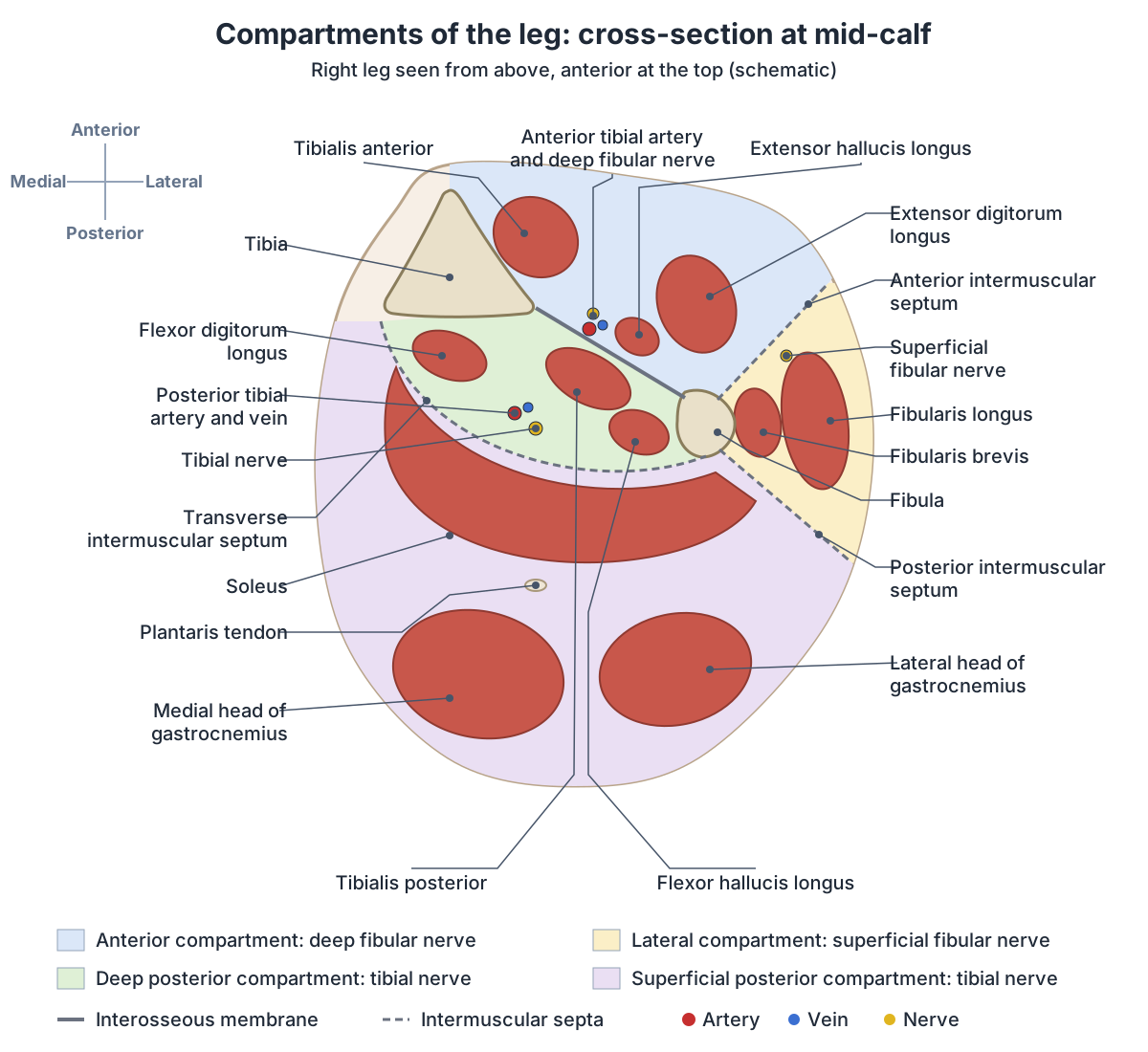
In anatomy, the leg means the segment between the knee and the ankle. Its muscles are divided by deep fascia into compartments, and the leg has three: anterior, lateral and posterior. The posterior compartment is further split into a superficial group (gastrocnemius, soleus and plantaris) and a deep group (popliteus, flexor digitorum longus, flexor hallucis longus and tibialis posterior). (OpenStax Anatomy and Physiology 2e, 11.6: Appendicular Muscles of the Pelvic Girdle and Lower Limbs.)
Each compartment has its own nerve. The anterior compartment is supplied by the deep fibular (peroneal) nerve and the anterior tibial artery. The lateral compartment is supplied by the superficial fibular nerve. Both posterior groups are supplied by the tibial nerve, a branch of the sciatic nerve. (TeachMeAnatomy: Muscles in the Anterior Compartment of the Leg.)
The two fibular nerves come from the common fibular nerve. It passes through a small gap between the parts of fibularis longus that arise from the fibular head and neck, and then divides into its deep and superficial branches. (TeachMeAnatomy: Muscles in the Lateral Compartment of the Leg.)
Grouping by compartment makes the actions easy to remember. The muscles of the anterior compartment are generally responsible for dorsiflexion, lifting the foot, and the muscles of the posterior compartment are generally responsible for plantar flexion, pointing the foot down. (OpenStax Anatomy and Physiology 2e, 11.6: Appendicular Muscles of the Pelvic Girdle and Lower Limbs.)
Muscles of the anterior compartment of the leg
The anterior compartment holds four muscles: tibialis anterior, extensor hallucis longus, extensor digitorum longus and fibularis (peroneus) tertius. All are supplied by the deep fibular nerve and the anterior tibial artery. (TeachMeAnatomy: Muscles in the Anterior Compartment of the Leg.)
Tibialis anterior (often shortened to tib anterior) is the long, thick muscle on the lateral surface of the tibia. It arises from the lateral condyle and the upper half to two-thirds of the lateral surface of the tibia, the interosseous membrane and the deep surface of the crural fascia. Its tendon inserts on the medial and plantar surfaces of the medial cuneiform and the base of the first metatarsal. It is a dorsiflexor of the ankle and an invertor of the foot. (University of Washington Musculoskeletal Atlas: Tibialis anterior.)
Extensor digitorum longus lies lateral to tibialis anterior. It arises from the lateral condyle of the tibia, the upper two-thirds to three-quarters of the medial surface of the fibula and the interosseous membrane. Below the inferior extensor retinaculum its tendon splits into four slips that join the extensor expansions of toes 2 to 5. It extends those toes and dorsiflexes the ankle. (University of Washington Musculoskeletal Atlas: Extensor digitorum longus.)
Extensor hallucis longus lies deep, between the other two. It arises from the anterior surface of the fibula and the interosseous membrane and inserts on the base of the distal phalanx of the great toe, which it extends; it also dorsiflexes the ankle. The University of Washington atlas gives the deep fibular nerve roots for tibialis anterior and both long extensors as L4, L5 and S1. (University of Washington Musculoskeletal Atlas: Extensor hallucis longus.)
Fibularis tertius arises with the lower part of extensor digitorum longus and inserts on the dorsal surface of the base of the fifth metatarsal. It works with extensor digitorum longus to dorsiflex and evert the foot. (University of Washington Musculoskeletal Atlas: Peroneus tertius.)
TeachMeAnatomy notes that fibularis tertius is not present in all individuals, so it is the one anterior compartment muscle that may be missing on dissection. (TeachMeAnatomy: Muscles in the Anterior Compartment of the Leg.)
At the ankle, thick bands of connective tissue, the superior extensor retinaculum (transverse ligament of the ankle) and the inferior extensor retinaculum, hold the tendons of the anterior muscles in place during dorsiflexion. (OpenStax Anatomy and Physiology 2e, 11.6: Appendicular Muscles of the Pelvic Girdle and Lower Limbs.)
The main dorsiflexor of the ankle, in 3D. Tibialis anterior.
Lateral compartment: fibularis (peroneus) longus and brevis
The lateral compartment contains two muscles, fibularis longus and fibularis brevis, still widely called peroneus longus and peroneus brevis. Their common action is eversion, turning the sole outward, and both are supplied by the superficial fibular nerve. (TeachMeAnatomy: Muscles in the Lateral Compartment of the Leg.)
Fibularis brevis (peroneus brevis) arises from the lower two-thirds of the lateral surface of the fibula and the anterior and posterior intermuscular septa. It inserts on the lateral side of the styloid process (tuberosity) at the base of the fifth metatarsal. It everts the foot and plantarflexes the ankle, and the atlas lists its nerve roots as L5, S1 and S2 and its blood supply as the fibular (peroneal) artery. (University of Washington Musculoskeletal Atlas: Peroneus brevis.)
Fibularis longus arises higher, from the head and the upper half to two-thirds of the lateral surface of the fibula. Its tendon runs under the foot to the medial cuneiform and the base of the first metatarsal, so it everts the foot, plantarflexes the ankle and helps support the transverse arch. (University of Washington Musculoskeletal Atlas: Peroneus longus.)
Both tendons pass in a groove behind the lateral malleolus inside one shared synovial sheath. Here the longus tendon lies slightly lateral to and behind the brevis tendon. Lower down, at the peroneal tubercle on the lateral surface of the calcaneus, they separate: brevis passes above the tubercle and longus below it. (Bokwa-Dąbrowska K et al. Peroneus brevis split tear: a pictorial review, Part 1. Eur J Radiol Open. 2025.)
From the anatomical position only a few degrees of eversion are possible. In practice the lateral muscles fix the medial edge of the foot during running and stop the foot rolling into too much inversion, which matters on uneven ground. (TeachMeAnatomy: Muscles in the Lateral Compartment of the Leg.)
Full page: tendon course under the foot, arches, os peroneum and tendon problems. Fibularis (peroneus) longus.
Superficial posterior compartment: gastrocnemius, soleus and plantaris
The superficial muscles of the posterior compartment all insert through the calcaneal (Achilles) tendon on the calcaneus. Gastrocnemius is the most superficial and visible muscle of the calf. Its two heads arise from the femur: the medial head from the back of the medial femoral condyle and the lateral head from the lateral surface of the lateral condyle. (University of Washington Musculoskeletal Atlas: Gastrocnemius.)
The two heads of gastrocnemius unite in a broad aponeurosis that merges with the deep tendon of soleus to form the Achilles tendon, which inserts on the middle third of the posterior surface of the calcaneus. Gastrocnemius is a powerful plantar flexor of the ankle and is supplied by the tibial nerve (S1, S2); each head receives a sural branch of the popliteal artery. (University of Washington Musculoskeletal Atlas: Gastrocnemius.)
Soleus lies deep to gastrocnemius. It arises entirely below the knee, from the back of the fibular head and upper fibula, the middle third of the medial border of the tibia and a tendinous arch between the two bones, and it joins the Achilles tendon. Like gastrocnemius it is a powerful plantar flexor supplied by the tibial nerve (S1, S2). (University of Washington Musculoskeletal Atlas: Soleus.)
Gastrocnemius arises from the femur, so it crosses the knee as well as the ankle: it plantarflexes the ankle and also flexes the knee. Soleus arises from the tibia and fibula and acts only at the ankle. The two converge distally to form the calcaneal tendon, which has a subcutaneous bursa between it and the skin and a deep bursa between it and the calcaneus. (TeachMeAnatomy: Muscles in the Posterior Compartment of the Leg.)
OpenStax describes the action of the calf in everyday terms: gastrocnemius lowers the sole of the foot to the ground, as in foot-tapping or jumping, the opposite of tibialis anterior, which raises it. (OpenStax Anatomy and Physiology 2e, 11.6: Appendicular Muscles of the Pelvic Girdle and Lower Limbs.)
The deep calf muscle that joins the Achilles tendon, in 3D. Soleus.
Plantaris muscle and plantaris tendon
Plantaris is a small muscle with a very long tendon. It arises from the lower part of the lateral supracondylar line of the femur and inserts on the middle third of the posterior calcaneus, just medial to the Achilles tendon. It plantarflexes the ankle and also flexes the knee, and it is supplied by the tibial nerve. (University of Washington Musculoskeletal Atlas: Plantaris.)
Its belly is only 7 to 13 cm long, and its thin tendon runs between gastrocnemius and soleus along the medial side of the Achilles tendon. The tendon is easily mistaken for a nerve during dissection, which earned it the nickname "freshman's nerve". Plantaris is absent in about 7 to 20% of limbs. Spina groups it with gastrocnemius and soleus as the triceps surae and notes that it is rich in muscle spindles, so it may matter more for sensing position than for force. (Spina AA. The plantaris muscle: anatomy, injury, imaging, and treatment. J Can Chiropr Assoc. 2007.)
Because people manage well without it, surgeons often harvest the plantaris tendon as a graft for tendon and ligament reconstruction. A sudden calf pain while running or jumping, sometimes called tennis leg, can come from a torn plantaris, although tears of the medial head of gastrocnemius are far more common: in one series of 141 patients, isolated plantaris rupture accounted for 1.4% of cases. (Spina AA. The plantaris muscle: anatomy, injury, imaging, and treatment. J Can Chiropr Assoc. 2007.)
The slender calf muscle with the long tendon, in 3D. Plantaris.
Deep posterior compartment: popliteus, tibialis posterior and the long toe flexors
Popliteus is a short, flat muscle behind the knee. It arises from the lateral femoral condyle (and the lateral meniscus) and inserts on the back of the tibia above the soleal line. Acting on the knee only, it laterally rotates the femur on the tibia, which unlocks the fully straightened knee so that flexion can begin. (TeachMeAnatomy: Muscles in the Posterior Compartment of the Leg.)
The University of Washington atlas describes the same movement from the other end, as medial rotation of the knee (tibia on femur) with flexion of the leg on the thigh, and gives popliteus a tibial nerve supply from L4, L5 and S1. (University of Washington Musculoskeletal Atlas: Popliteus.)
Tibialis posterior is the deepest muscle of the compartment. It arises from the interosseous membrane and the adjacent posterior surfaces of the tibia and fibula. Its tendon passes behind the medial malleolus and splits: the main slip inserts on the tuberosity of the navicular, and deeper slips reach the cuneiforms and the bases of metatarsals 2 to 4. It is the principal invertor of the foot and also plantarflexes the ankle. (University of Washington Musculoskeletal Atlas: Tibialis posterior.)
Flexor digitorum longus arises from the posterior surface of the tibia below the popliteal line. In the sole its tendon splits into four slips that insert on the bases of the distal phalanges of toes 2 to 5; it flexes those toes and helps plantarflex the ankle. (University of Washington Musculoskeletal Atlas: Flexor digitorum longus.)
Flexor hallucis longus arises from the lower two-thirds of the posterior surface of the fibula and inserts on the base of the distal phalanx of the great toe. It flexes the big toe, helps to supinate the foot and is only a very weak plantar flexor of the ankle. (University of Washington Musculoskeletal Atlas: Flexor hallucis longus.)
The tendons of the deep muscles reach the sole through the tarsal tunnel behind the medial malleolus. From front to back the contents are the tibialis posterior tendon, the flexor digitorum longus tendon, the posterior tibial artery and vein, the tibial nerve and the flexor hallucis longus tendon, remembered as "Tom, Dick and a Very Nervous Harry". (TeachMeAnatomy: The Tarsal Tunnel.)
Muscles of the lower leg: origin, insertion, nerve and action
| Muscle | Compartment | Origin | Insertion | Nerve (roots) | Main action |
|---|---|---|---|---|---|
| Tibialis anterior | Anterior | Lateral condyle and upper lateral surface of tibia, interosseous membrane | Medial cuneiform and base of 1st metatarsal | Deep fibular (L4–S1) | Dorsiflexion, inversion |
| Extensor digitorum longus | Anterior | Lateral tibial condyle, medial surface of fibula, interosseous membrane | Extensor expansions of toes 2–5 | Deep fibular (L4–S1) | Extends toes 2–5, dorsiflexion |
| Extensor hallucis longus | Anterior | Anterior surface of fibula, interosseous membrane | Base of distal phalanx of great toe | Deep fibular (L4–S1) | Extends great toe, dorsiflexion |
| Fibularis tertius (inconstant) | Anterior | Lower medial fibula, with extensor digitorum longus | Dorsal base of 5th metatarsal | Deep fibular | Dorsiflexion, eversion |
| Fibularis (peroneus) longus | Lateral | Head and upper lateral surface of fibula | Medial cuneiform and base of 1st metatarsal | Superficial fibular (L5–S2) | Eversion, weak plantar flexion, supports arches |
| Fibularis (peroneus) brevis | Lateral | Lower two-thirds of lateral fibula | Tuberosity of 5th metatarsal | Superficial fibular (L5–S2) | Eversion, plantar flexion |
| Gastrocnemius | Superficial posterior | Medial and lateral femoral condyles | Calcaneus via calcaneal tendon | Tibial (S1–S2) | Plantar flexion, knee flexion |
| Soleus | Superficial posterior | Posterior fibular head and upper fibula, medial border of tibia | Calcaneus via calcaneal tendon | Tibial (S1–S2) | Plantar flexion |
| Plantaris (absent in some people) | Superficial posterior | Lateral supracondylar line of femur | Calcaneus, medial to calcaneal tendon | Tibial (L5–S2) | Weak plantar flexion and knee flexion |
| Popliteus | Deep posterior | Lateral femoral condyle | Posterior tibia above soleal line | Tibial (L4–S1) | Unlocks the knee, knee flexion |
| Tibialis posterior | Deep posterior | Interosseous membrane, posterior tibia and fibula | Navicular tuberosity, cuneiforms, metatarsals 2–4 | Tibial (L4–L5) | Inversion, plantar flexion |
| Flexor digitorum longus | Deep posterior | Posterior tibia below popliteal line | Distal phalanges of toes 2–5 | Tibial | Flexes toes 2–5, assists plantar flexion |
| Flexor hallucis longus | Deep posterior | Lower two-thirds of posterior fibula | Distal phalanx of great toe | Tibial | Flexes great toe |
Nerve roots as given by the University of Washington Musculoskeletal Atlas; other texts differ slightly. Sources: UW atlas, TeachMeAnatomy.
Dorsiflexion vs plantar flexion: which muscles do each?
Both movements happen at the ankle (talocrural) joint, a hinge joint. Dorsiflexion lifts the front of the foot so that the top of the foot moves towards the front of the leg. Plantar flexion lifts the heel from the ground or points the toes downward. (OpenStax Anatomy and Physiology 2e, 9.5: Types of Body Movements.)
The dorsiflexors are the anterior compartment muscles: tibialis anterior, extensor digitorum longus, extensor hallucis longus and fibularis tertius. Each one dorsiflexes the foot, and the long extensors also extend the toes, so the toes tend to lift when you pull the foot up. (TeachMeAnatomy: Muscles in the Anterior Compartment of the Leg.)
The main plantar flexors are gastrocnemius and soleus, which the University of Washington atlas describes as powerful plantar flexors of the ankle. (University of Washington Musculoskeletal Atlas: Gastrocnemius.)
Several smaller muscles help. Plantaris contributes to plantar flexion and tibialis posterior plantarflexes the foot as well as inverting it. The long toe flexors act mainly on the toes. (TeachMeAnatomy: Muscles in the Posterior Compartment of the Leg.)
Inversion vs eversion of the foot
Inversion turns the sole of the foot towards the midline; eversion turns it away from the midline. Neither movement happens at the ankle joint. They take place at the intertarsal joints between the tarsal bones of the back of the foot, mainly the subtalar joint. (OpenStax Anatomy and Physiology 2e, 9.5: Types of Body Movements.)
Tibialis posterior is the principal invertor of the foot. It also adducts the foot, plantarflexes the ankle and helps to supinate the foot. (University of Washington Musculoskeletal Atlas: Tibialis posterior.)
Tibialis anterior is the other main invertor. It is described as a dorsiflexor of the ankle and an invertor of the foot, so it inverts the foot while lifting it, the opposite of tibialis posterior, which inverts while pointing the foot down. (University of Washington Musculoskeletal Atlas: Tibialis anterior.)
The evertors are the lateral compartment muscles, fibularis longus and fibularis brevis, whose common function is to turn the sole outward. Fibularis tertius from the anterior compartment helps when it is present. (TeachMeAnatomy: Muscles in the Lateral Compartment of the Leg.)
Notice that fibularis longus inserts on the medial cuneiform and the base of the first metatarsal, the same bones as tibialis anterior. Its tendon reaches them by crossing the sole from the lateral side, so the two muscles pull on the same spot from opposite directions: one everts and the other inverts. (University of Washington Musculoskeletal Atlas: Peroneus longus.)
Movements of the ankle and foot and the muscles that produce them
| Movement | Where it happens | Main muscles |
|---|---|---|
| Dorsiflexion | Ankle (talocrural) joint | Tibialis anterior, extensor digitorum longus, extensor hallucis longus, fibularis tertius |
| Plantar flexion | Ankle (talocrural) joint | Gastrocnemius and soleus; helped by plantaris, tibialis posterior, fibularis longus and brevis, flexor digitorum longus |
| Inversion | Subtalar and other intertarsal joints | Tibialis posterior, tibialis anterior |
| Eversion | Subtalar and other intertarsal joints | Fibularis longus, fibularis brevis, fibularis tertius |
Foot drop, Achilles tendon rupture and fibular tendon problems
Foot drop is the inability to dorsiflex the foot at the ankle, so the foot hangs down under gravity. It follows damage to the common fibular nerve, which gives rise to the deep fibular nerve, or damage to the deep fibular nerve itself. Patients may flick the foot outward (an eversion flick) while walking to clear the toes from the ground. (TeachMeAnatomy: Muscles in the Anterior Compartment of the Leg.)
A ruptured calcaneal (Achilles) tendon is a partial or complete tear, usually caused by forceful plantar flexion and more likely after calcaneal tendinitis. The person cannot plantarflex against resistance, and the contracted soleus and gastrocnemius may form a lump in the calf. (TeachMeAnatomy: Muscles in the Posterior Compartment of the Leg.)
The fibular (peroneal) tendons can become inflamed (tenosynovitis), and if the superior peroneal retinaculum that holds them is stripped from the fibula they can dislocate over the lateral malleolus. These problems are covered on the fibularis longus page. (Kenhub: Fibularis longus muscle.)
Leg muscles in the 3D model
- Calcaneal tendon
- The thick tendon joining gastrocnemius and soleus to the heel bone.
- Extensor digitorum longus
- A long extensor in the anterior leg compartment.
- Extensor hallucis longus
- A long extensor of the great toe, lying between tibialis anterior and extensor digitorum longus.
- Fibularis brevis
- A lateral leg muscle lying beneath fibularis longus.
- Fibularis longus
- A long lateral leg muscle whose tendon crosses the sole of the foot.
- Fibularis tertius
- A small muscle of the anterior leg, sometimes absent.
- Flexor digitorum longus
- A deep posterior leg muscle whose tendon passes behind the medial malleolus.
- Flexor hallucis longus
- A deep posterior leg muscle that bends the great toe, running beneath the sustentaculum tali.
- Lateral head of gastrocnemius
- The lateral head of the superficial calf muscle.
- Medial head of gastrocnemius
- The larger medial head of the superficial calf muscle.
- Plantaris
- A small, inconstant calf muscle with a very long thin tendon.
- Popliteus
- A small triangular muscle forming part of the floor of the popliteal fossa.
- Soleus
- A broad flat calf muscle lying deep to gastrocnemius.
- Tibialis anterior
- The main dorsiflexor on the front of the leg.
- Tibialis posterior
- The deepest muscle of the posterior leg compartment.
Frequently asked questions
What muscles are in the anterior compartment of the leg?
Four muscles: tibialis anterior, extensor hallucis longus, extensor digitorum longus and fibularis (peroneus) tertius, which is missing in some people. All are supplied by the deep fibular nerve and the anterior tibial artery. They dorsiflex the ankle, and the long extensors also extend the toes. Tibialis anterior also inverts the foot, while fibularis tertius helps evert it.
What muscles are in the posterior compartment of the leg?
The superficial group is gastrocnemius, soleus and plantaris, which all insert on the calcaneus through or beside the Achilles tendon. The deep group is popliteus, tibialis posterior, flexor digitorum longus and flexor hallucis longus. All are supplied by the tibial nerve. Together they plantarflex the ankle, flex the toes, invert the foot and, for popliteus, unlock the knee.
What is the difference between dorsiflexion and plantar flexion?
Both happen at the ankle joint. Dorsiflexion lifts the front of the foot towards the shin, as when you walk on your heels; the anterior compartment muscles, led by tibialis anterior, produce it. Plantar flexion points the foot down or lifts the heel, as when you stand on tiptoe; gastrocnemius and soleus are the main muscles.
Which muscles invert and evert the foot?
Tibialis posterior is the main invertor, helped by tibialis anterior. The evertors are fibularis (peroneus) longus and brevis in the lateral compartment, helped by fibularis tertius. Inversion and eversion take place at the subtalar and other intertarsal joints, not at the ankle joint itself.
Is peroneus brevis the same as fibularis brevis?
Yes. Peroneus is the older name and fibularis is the current Terminologia Anatomica name; both refer to the same muscle. Fibularis brevis arises from the lower two-thirds of the lateral fibula, runs behind the lateral malleolus with fibularis longus and inserts on the tuberosity at the base of the fifth metatarsal. It everts the foot.
What is the plantaris tendon?
It is the long, thin tendon of the small plantaris muscle, which arises from the lower femur above the lateral condyle. The tendon runs down between gastrocnemius and soleus and along the medial side of the Achilles tendon to the calcaneus. The muscle is absent in about 7 to 20% of limbs, and surgeons often use its tendon as a graft.
Why does damage to the common fibular nerve cause foot drop?
The common fibular nerve divides into the deep fibular nerve, which supplies all the dorsiflexors in the anterior compartment, and the superficial fibular nerve. If it is damaged, the dorsiflexors stop working and the foot hangs down and drags while walking.
References
- OpenStax. Anatomy and Physiology 2e, §11.6 Appendicular Muscles of the Pelvic Girdle and Lower Limbs. Rice University, 2022.
- OpenStax. Anatomy and Physiology 2e, §9.5 Types of Body Movements. Rice University, 2022.
- TeachMeAnatomy. Muscles in the Anterior Compartment of the Leg.
- TeachMeAnatomy. Muscles in the Lateral Compartment of the Leg.
- TeachMeAnatomy. Muscles in the Posterior Compartment of the Leg.
- TeachMeAnatomy. The Tarsal Tunnel: Borders and Contents.
- University of Washington Department of Radiology. Musculoskeletal Atlas: Tibialis anterior.
- University of Washington Department of Radiology. Musculoskeletal Atlas: Extensor digitorum longus.
- University of Washington Department of Radiology. Musculoskeletal Atlas: Extensor hallucis longus.
- University of Washington Department of Radiology. Musculoskeletal Atlas: Peroneus tertius.
- University of Washington Department of Radiology. Musculoskeletal Atlas: Peroneus longus.
- University of Washington Department of Radiology. Musculoskeletal Atlas: Peroneus brevis.
- University of Washington Department of Radiology. Musculoskeletal Atlas: Gastrocnemius.
- University of Washington Department of Radiology. Musculoskeletal Atlas: Soleus.
- University of Washington Department of Radiology. Musculoskeletal Atlas: Plantaris.
- University of Washington Department of Radiology. Musculoskeletal Atlas: Popliteus.
- University of Washington Department of Radiology. Musculoskeletal Atlas: Tibialis posterior.
- University of Washington Department of Radiology. Musculoskeletal Atlas: Flexor digitorum longus.
- University of Washington Department of Radiology. Musculoskeletal Atlas: Flexor hallucis longus.
- Spina AA. The plantaris muscle: anatomy, injury, imaging, and treatment. J Can Chiropr Assoc. 2007;51(3):158–165.
- Kenhub. Fibularis longus muscle.
- Bokwa-Dąbrowska K, Zych R, Mocanu D, Huuskonen M, Dziedzic D, Szaro P. Peroneus brevis split tear: a challenging diagnosis. A pictorial review of magnetic resonance and ultrasound imaging. Part 1. Anatomical basis and clinical insights. Eur J Radiol Open. 2025;14:100633.
- Mitsuhashi N. et al. (2009). BodyParts3D: 3D structure database for anatomical concepts. Nucleic Acids Research 37, D782–D785. Data © The Database Center for Life Science, CC BY 4.0.
Related anatomy
- Fibularis longusFibularis longus (peroneus longus) explained: origin, the tendon's path behind the ankle and under the foot…
- Foot musclesFoot muscle anatomy: extensor digitorum brevis on the dorsum and the four plantar layers, from abductor hallucis to…
- Thigh musclesThigh muscles explained: quadriceps and sartorius at the front, adductor muscles of the inner thigh, hamstrings at the…
- TibiaTibia anatomy explained: where the shinbone is, the proximal tibia, tibial plateau, shaft and medial malleolus, its…
- FibulaFibula anatomy: the head of fibula, shaft and lateral malleolus, the proximal and distal tibiofibular joints and…
- Lower limb nervesLower limb nerves explained: lumbar and sacral plexus diagram, femoral, saphenous, sciatic, tibial, common peroneal…
Explore the whole body in the interactive 3D human anatomy model, or browse the A–Z list of human structures.