

Which nerves supply the lower limb?
Of the four nerve plexuses, one is at the lumbar level and one at the sacral level, and these two supply the lower limb. The lumbar plexus arises from all the lumbar spinal nerves and gives rise to nerves for the pelvic region and the front of the leg. The sacral plexus comes from the lower lumbar nerves L4 and L5 and the sacral nerves S1 to S4. (OpenStax Anatomy and Physiology 2e, 13.4: The Peripheral Nervous System.)
The femoral nerve is one of the major nerves of the lumbar plexus and gives rise to the saphenous nerve, which extends down the leg. The most significant nerve of the sacral plexus is the sciatic nerve, which is really a combination of two nerves, the tibial nerve and the fibular nerve. Sciatica is pain caused by compression or irritation of the sciatic nerve or of any of the spinal nerves that form it. (OpenStax Anatomy and Physiology 2e, 13.4: The Peripheral Nervous System.)
Lumbar plexus: nerves and roots
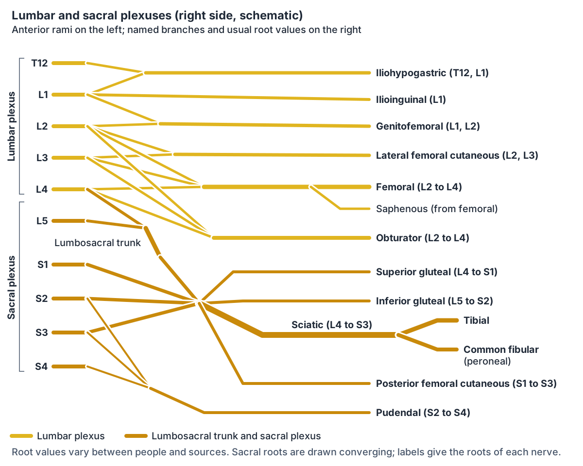
The lumbar plexus is formed by the anterior rami of lumbar spinal nerves L1, L2, L3 and L4, with a contribution from T12. It forms within the substance of the psoas major muscle, in front of the transverse processes of the lumbar vertebrae. (TeachMeAnatomy: The Lumbar Plexus.)
The six main nerves in the lumbar plexus are the iliohypogastric, ilioinguinal, genitofemoral, lateral cutaneous nerve of the thigh, obturator and femoral nerves. TeachMeAnatomy offers the mnemonic "I, I Get Leftovers On Fridays" to recall them. (TeachMeAnatomy: The Lumbar Plexus.)
Iliohypogastric and ilioinguinal nerves
The iliohypogastric nerve (L1, with fibres from T12) crosses quadratus lumborum toward the iliac crest and pierces transversus abdominis. It supplies internal oblique and transversus abdominis and the skin of the posterolateral gluteal region and the pubic region. (TeachMeAnatomy: The Lumbar Plexus.)
The ilioinguinal nerve (L1), sometimes searched as the inguinal nerve, follows the same path but continues through the superficial inguinal ring. It supplies internal oblique and transversus abdominis and the skin of the upper anteromedial thigh and the external genitalia: the root of the penis and front of the scrotum in males, and the mons pubis and labia majora in females. (TeachMeAnatomy: The Lumbar Plexus.)
Genitofemoral nerve (n. genitofemoralis)
The genitofemoral nerve (nervus genitofemoralis, L1 and L2) leaves the psoas major and divides into a genital branch and a femoral branch. The genital branch supplies the cremaster muscle and the skin of the genital region. The femoral branch supplies the skin of the upper anterior thigh. (TeachMeAnatomy: The Lumbar Plexus.)
Lateral femoral cutaneous nerve (lateral cutaneous nerve of thigh)
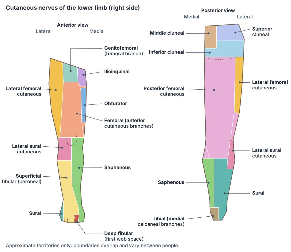
The lateral femoral cutaneous nerve, also called the lateral cutaneous nerve of the thigh, comes from L2 and L3. It is purely sensory. It enters the thigh near the lateral end of the inguinal ligament and supplies the skin of the anterior and lateral thigh down to the knee. (TeachMeAnatomy: The Lumbar Plexus.)
Its anatomy varies. A 2025 review in BJA Education reports that it arises from L2 and L3 in 58.5% of cases, and from L1 and L2 or L2 alone in others. After leaving the lateral border of psoas, it runs over iliacus toward the anterior superior iliac spine (ASIS) and may pass under, through or above the inguinal ligament, on average about 1.73 cm from the ASIS. Below the ligament it splits into anterior and posterior branches to the anterolateral thigh. (Bromley K, Kanakarajan S. Meralgia paraesthetica. BJA Educ. 2025.)
Compression of this nerve, usually near the inguinal ligament, causes meralgia paraesthetica, a common but under-recognised mononeuropathy that produces dysaesthesia (abnormal, often unpleasant sensation) over the anterolateral thigh. Reported causes include direct entrapment, raised abdominal pressure (pregnancy, obesity), tight belts or trousers, metabolic causes such as diabetes, and procedures such as inguinal hernia repair or Caesarean section. (Bromley K, Kanakarajan S. Meralgia paraesthetica. BJA Educ. 2025.)
Femoral nerve and saphenous nerve
The femoral nerve comes from the anterior rami of L2, L3 and L4. It runs down through psoas major on the posterior abdominal wall, passes underneath the inguinal ligament and enters the femoral triangle, where it lies lateral to the femoral vessels. (TeachMeAnatomy: The Femoral Nerve.)
It supplies the muscles of the anterior thigh: iliacus, pectineus, sartorius and the quadriceps femoris group, which extends the knee. Its anterior cutaneous branches supply the skin of the anteromedial thigh. (TeachMeAnatomy: The Femoral Nerve.)
The saphenous nerve (saphenous n.) is a sensory branch of the femoral nerve. It travels through the adductor canal, leaves it before the adductor hiatus and then supplies the skin of the medial side of the leg and foot. (TeachMeAnatomy: The Femoral Nerve.)
Obturator nerve
The obturator nerve receives fibres from the anterior divisions of L2, L3 and L4. It emerges from the medial border of psoas major and enters the medial thigh through the obturator canal, where it divides into anterior and posterior branches. (TeachMeAnatomy: The Obturator Nerve.)
It supplies all the muscles of the medial compartment of the thigh: adductor longus, adductor brevis, gracilis, obturator externus and the adductor part of adductor magnus (the hamstring part is supplied by the sciatic nerve). Its cutaneous branch supplies the skin of the middle part of the medial thigh. It can be damaged in pelvic or abdominal surgery, causing medial thigh numbness and weak adduction. (TeachMeAnatomy: The Obturator Nerve.)
Sacral plexus nerves (plexus sacralis)
The sacral plexus (plexus sacralis) is formed by the anterior rami of S1, S2, S3 and S4, joined by the lumbosacral trunk, which carries fibres from L4 and L5. It lies on the posterior wall of the pelvis, in front of the piriformis muscle. (TeachMeAnatomy: The Sacral Plexus.)
Its main branches are the superior gluteal nerve (L4 to S1), which supplies gluteus medius, gluteus minimus and tensor fasciae latae; the inferior gluteal nerve (L5 to S2), which supplies gluteus maximus; the sciatic nerve (L4 to S3); the posterior femoral cutaneous nerve (S1 to S3); and the pudendal nerve (S2 to S4), which supplies the muscles of the perineum, including the external urethral and anal sphincters, and most of the skin of the perineum. Smaller branches go to piriformis, obturator internus and quadratus femoris. (TeachMeAnatomy: The Sacral Plexus.)
Nerves of the lower limb: roots, motor and sensory supply
| Nerve | Plexus and roots | Main motor supply | Main skin supply |
|---|---|---|---|
| Iliohypogastric | Lumbar; L1 (with T12) | Internal oblique, transversus abdominis | Posterolateral gluteal and pubic regions |
| Ilioinguinal | Lumbar; L1 | Internal oblique, transversus abdominis | Upper anteromedial thigh, external genitalia |
| Genitofemoral | Lumbar; L1, L2 | Cremaster (genital branch) | Genital skin; upper anterior thigh (femoral branch) |
| Lateral femoral cutaneous | Lumbar; L2, L3 | None (sensory) | Anterior and lateral thigh to the knee |
| Femoral | Lumbar; L2 to L4 | Iliacus, pectineus, sartorius, quadriceps femoris | Anteromedial thigh; medial leg and foot via saphenous nerve |
| Obturator | Lumbar; L2 to L4 | Adductors, gracilis, obturator externus | Middle of medial thigh |
| Superior gluteal | Sacral; L4 to S1 | Gluteus medius and minimus, tensor fasciae latae | None |
| Inferior gluteal | Sacral; L5 to S2 | Gluteus maximus | None |
| Sciatic | Sacral; L4 to S3 | Hamstrings, hamstring part of adductor magnus; all leg and foot muscles via branches | None directly (via branches) |
| Tibial | Sciatic; L4 to S3 | Posterior leg muscles, most intrinsic foot muscles | Heel (medial calcaneal), sole (plantar nerves) |
| Common fibular (peroneal) | Sciatic; L4 to S2 | Via superficial and deep fibular nerves | Upper lateral leg (lateral sural cutaneous) |
| Superficial fibular (peroneal) | Common fibular | Fibularis longus and brevis | Anterolateral leg, dorsum of foot except first web space |
| Deep fibular (peroneal) | Common fibular | Tibialis anterior, extensor digitorum longus, extensor hallucis longus | First web space |
| Sural | Tibial and common fibular contributions | None (sensory) | Posterolateral distal leg, lateral foot, heel and fifth toe |
| Posterior femoral cutaneous | Sacral; S1 to S3 | None (sensory) | Lower buttock, posterior thigh, upper back of leg, perineum |
| Pudendal | Sacral; S2 to S4 | Perineal muscles, external urethral and anal sphincters | Most of the perineum |
Based mainly on TeachMeAnatomy, with Kenhub (sural) and Radiopaedia (posterior femoral cutaneous). Root values vary between people.
Sciatic nerve
The sciatic nerve (roots L4 to S3) is the most significant nerve of the sacral plexus. It leaves the pelvis through the greater sciatic foramen, emerges below piriformis into the gluteal region, and descends into the posterior thigh deep to the long head of biceps femoris. At the apex of the popliteal fossa, behind the knee, it ends by dividing into the tibial and common fibular nerves. (TeachMeAnatomy: The Sciatic Nerve.)
It directly supplies the hamstring muscles of the posterior thigh (biceps femoris, semitendinosus and semimembranosus) and the hamstring part of adductor magnus. Through its terminal branches it supplies all the muscles of the leg and foot. It has no direct sensory branches; its branches supply the skin of the lateral leg, the heel and both surfaces of the foot. (TeachMeAnatomy: The Sciatic Nerve.)
Because the nerve passes through the lower medial part of the buttock, intramuscular injections are given only in the upper lateral quadrant of the gluteal region. Compression of the sciatic nerve by piriformis is called piriformis syndrome. (TeachMeAnatomy: The Sciatic Nerve.)
Tibial nerve
The tibial nerve (L4 to S3) arises at the apex of the popliteal fossa and continues down the back of the leg, posterior to the tibia. It passes behind and below the medial malleolus through the tarsal tunnel and divides into the medial and lateral plantar nerves. (TeachMeAnatomy: The Tibial Nerve.)
It supplies the muscles of the posterior leg and, through the plantar nerves, most of the intrinsic muscles of the foot. Its medial calcaneal branches arise in the tarsal tunnel and supply the skin of the heel. The medial plantar nerve supplies the sole of the medial three and a half digits and the lateral plantar nerve the lateral one and a half digits. In the popliteal fossa it gives a cutaneous branch that joins a branch of the common fibular nerve to form the sural nerve. (TeachMeAnatomy: The Tibial Nerve.)
Common fibular (common peroneal) nerve and its superficial and deep branches
The common fibular nerve, still widely called the common peroneal nerve (roots L4 to S2), arises at the apex of the popliteal fossa and wraps around the neck of the fibula, where it divides into the superficial and deep fibular nerves. In the popliteal fossa it gives the lateral sural cutaneous nerve, which supplies the skin of the upper lateral leg, and the sural communicating branch. (TeachMeAnatomy: The Common Fibular Nerve.)
The superficial fibular (superficial peroneal) nerve supplies the muscles of the lateral compartment, fibularis (peroneus) longus and brevis, and the skin of the anterolateral leg and the dorsum of the foot, except the first web space. The deep fibular (deep peroneal) nerve supplies the anterior compartment muscles, tibialis anterior, extensor digitorum longus and extensor hallucis longus, some intrinsic foot muscles, and the skin of the first web space between the big and second toes. (TeachMeAnatomy: The Common Fibular Nerve.)
Because it lies so close to the bone at the neck of the fibula, the common fibular nerve is easily injured by fractures there. Paralysis of the dorsiflexors leaves the foot plantarflexed, a sign called foot drop. (TeachMeAnatomy: The Common Fibular Nerve.)
Sural nerve (nervus suralis)
The sural nerve (nervus suralis) is a sensory nerve of the back of the leg. Kenhub describes it as the merger of the medial sural cutaneous nerve, a branch of the tibial nerve, and the lateral sural cutaneous nerve, a branch of the common fibular nerve; TeachMeAnatomy describes the fibular contribution as the sural communicating branch. The level at which they unite varies widely, from the popliteal fossa down to the ankle. (Kenhub: Sural nerve.)
The sural nerve descends between the two heads of gastrocnemius alongside the small saphenous vein, then passes between the lateral malleolus and the calcaneus into the foot. It supplies the skin of the posterolateral part of the distal third of the leg, the lateral side of the foot, heel and ankle, and the lateral side of the little (fifth) toe. (Kenhub: Sural nerve.)
Posterior femoral cutaneous nerve (posterior cutaneous nerve of thigh)
The posterior femoral cutaneous nerve, also called the posterior cutaneous nerve of the thigh, is a sensory branch of the sacral plexus from S1, S2 and S3. It leaves the pelvis through the greater sciatic foramen below piriformis together with the sciatic nerve, lies deep to gluteus maximus, then runs down the thigh deep to the fascia lata before piercing the fascia in the popliteal fossa. (Radiopaedia: Posterior femoral cutaneous nerve.)
It supplies the skin of the buttock, the posterior thigh, the upper part of the back of the leg and the posterior scrotum or labia. It may receive fibres from S4, and in one study it was duplicated in 18% of cadavers. (Radiopaedia: Posterior femoral cutaneous nerve.)
A 2023 dissection study classified its branches as thigh, gluteal and perineal branches. The gluteal branches, the medial and lateral inferior cluneal nerves, supply the lower half of the gluteal region; the perineal branches supply the lateral perineum; and the thigh branches supply the posterior thigh and upper leg. (Jiamjunyasiri A et al. Origin, course, and distribution of the posterior femoral cutaneous nerve. Anat Sci Int. 2023.)
Cluneal nerves: superior, middle and inferior
The skin of the buttock (clunis) is supplied by three groups of cluneal nerves. The superior cluneal nerves are cutaneous branches of the lumbar dorsal rami. They pass through the thoracolumbar fascia, cross the posterior iliac crest and supply the skin over the upper half of the gluteal muscles. Classic descriptions give their origin as L1 to L3, but a 2017 cadaver study found branches from T12 to L5. (Konno T et al. Anatomical etiology of pseudo-sciatica from superior cluneal nerve entrapment. J Pain Res. 2017.)
In that study, 13 of 81 superior cluneal branches passed through an osteofibrous tunnel at the iliac crest, and 10 of them were visibly entrapped. Superior cluneal nerve entrapment can cause low back pain with pain referred into the leg, sometimes called pseudo-sciatica. (Konno T et al. Anatomical etiology of pseudo-sciatica from superior cluneal nerve entrapment. J Pain Res. 2017.)
The middle cluneal nerves are sensory branches of the dorsal rami of S1 to S3. They run roughly horizontally below the posterior superior iliac spine to supply the skin of the posteromedial buttock, and they can be entrapped where they pass under or through the long posterior sacroiliac ligament. (Aota Y. Entrapment of middle cluneal nerves as an unknown cause of low back pain. World J Orthop. 2016.)
The inferior cluneal nerves are the gluteal branches of the posterior femoral cutaneous nerve, from the sacral plexus. They supply the skin of the lower half of the gluteal region, and their number and course vary from person to person. (Jiamjunyasiri A et al. Origin, course, and distribution of the posterior femoral cutaneous nerve. Anat Sci Int. 2023.)
Frequently asked questions
What are the main nerves of the leg?
The main nerves of the lower limb are the femoral nerve (front of the thigh), the obturator nerve (inner thigh), the superior and inferior gluteal nerves (buttock muscles) and the sciatic nerve (back of the thigh). Below the knee, the sciatic nerve's tibial branch supplies the calf and sole and its common fibular (peroneal) branch supplies the front and outer leg.
Where is the saphenous nerve?
The saphenous nerve is a sensory branch of the femoral nerve. It runs down the thigh through the adductor canal and leaves it before the adductor hiatus. Below the knee it supplies the skin of the medial side of the leg and the medial side of the foot.
What does the sural nerve supply?
The sural nerve is a purely sensory nerve formed from branches of the tibial and common fibular nerves. It runs down the back of the calf with the small saphenous vein and passes between the lateral malleolus and the calcaneus into the foot. It supplies the skin of the lower posterolateral leg, the lateral foot, heel and ankle, and the outer side of the little toe.
What is the difference between the common peroneal and common fibular nerve?
There is no difference: they are two names for the same nerve. Fibular is the current Terminologia Anatomica term, and peroneal is the older name still used in clinics. The common fibular (peroneal) nerve comes from the sciatic nerve, winds around the neck of the fibula and divides into the superficial and deep fibular nerves.
Which nerves come from the lumbar plexus?
The lumbar plexus (L1 to L4, with T12) gives six main nerves: the iliohypogastric, ilioinguinal, genitofemoral, lateral femoral cutaneous, obturator and femoral nerves. The mnemonic "I, I Get Leftovers On Fridays" helps recall them. They supply the lower abdominal wall, the groin, the front and inner thigh and, through the saphenous nerve, the medial leg.
What causes meralgia paraesthetica?
Meralgia paraesthetica is compression of the lateral femoral cutaneous nerve, usually where it passes near the anterior superior iliac spine and the inguinal ligament. It causes dysaesthesia, an abnormal and often unpleasant sensation, over the outer front of the thigh. Causes include tight belts or clothing, pregnancy, obesity, diabetes and some operations, such as inguinal hernia repair.
What do the superior cluneal nerves supply?
The superior cluneal nerves are skin branches of the lumbar dorsal rami, classically L1 to L3. They cross the posterior iliac crest and supply the skin over the upper part of the buttock. Where they pass through a tight tunnel of fascia at the iliac crest they can become entrapped, which is one recognised cause of low back and leg pain.
References
- OpenStax. Anatomy and Physiology 2e, §13.4 The Peripheral Nervous System. Rice University, 2022.
- TeachMeAnatomy. The Lumbar Plexus.
- TeachMeAnatomy. The Sacral Plexus.
- TeachMeAnatomy. The Femoral Nerve.
- TeachMeAnatomy. The Obturator Nerve.
- TeachMeAnatomy. The Sciatic Nerve.
- TeachMeAnatomy. The Tibial Nerve.
- TeachMeAnatomy. The Common Fibular Nerve.
- Kenhub. Sural nerve.
- Radiopaedia.org. Posterior femoral cutaneous nerve.
- Jiamjunyasiri A, Tsutsumi M, Muro S, Akita K. Origin, course, and distribution of the posterior femoral cutaneous nerve and the spatial relationship among its branches. Anat Sci Int. 2023;98(4):540–547.
- Konno T, Aota Y, Kuniya H, et al. Anatomical etiology of "pseudo-sciatica" from superior cluneal nerve entrapment: a laboratory investigation. J Pain Res. 2017;10:2539–2545.
- Aota Y. Entrapment of middle cluneal nerves as an unknown cause of low back pain. World J Orthop. 2016;7(3):167–170.
- Bromley K, Kanakarajan S. Meralgia paraesthetica. BJA Educ. 2025;25(8):302–308.
Related anatomy
- Spinal cordSpinal cord anatomy with labelled diagrams: cross-section, grey matter horns, spinothalamic and dorsal column tracts…
- Upper limb nervesBrachial plexus diagram with roots, trunks, cords and branches; axillary, musculocutaneous, median, ulnar and radial…
- Thigh musclesThigh muscles explained: quadriceps and sartorius at the front, adductor muscles of the inner thigh, hamstrings at the…
- Leg musclesLower leg muscles by compartment: tibialis anterior, fibularis (peroneus) longus and brevis, gastrocnemius, soleus…
- Hip musclesMuscles of the hip explained: gluteus maximus, medius and minimus, TFL, piriformis, obturator internus and the other…
- Foot musclesFoot muscle anatomy: extensor digitorum brevis on the dorsum and the four plantar layers, from abductor hallucis to…
Explore the whole body in the interactive 3D human anatomy model, or browse the A–Z list of human structures.