

What are the paranasal sinuses?
The paranasal sinuses are hollow, air-filled spaces inside some of the bones of the skull. There are four pairs, each named after the bone that contains it: the maxillary, frontal, ethmoid and sphenoid sinuses. They are lined by respiratory mucosa and each communicates with the nasal cavity. (Radiopaedia: Paranasal sinuses.)
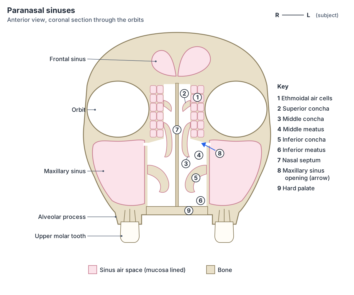
The frontal sinus lies just above the eyebrows in the frontal bone. The maxillary sinuses sit in the maxillae just below the orbits. The ethmoid air cells lie between the upper nasal cavity and the medial wall of each orbit. The sphenoid sinus lies in the body of the sphenoid bone, just in front of and below the sella turcica. (OpenStax Anatomy and Physiology 2e, 7.2: The Skull.)
Because the lining of the sinuses is continuous with the lining of the nose, an infection of the nasal cavity, such as a cold, can spread into them. Inflammation of the sinus lining is called sinusitis; when all the sinuses are involved it is called pansinusitis. (TeachMeAnatomy: The Paranasal Sinuses.)
What do the sinuses do?
The function of the sinuses is still debated. Suggested roles are reducing the weight of the head, helping the immune defence of the nasal cavity, humidifying the air we breathe in and increasing the resonance of the voice. (TeachMeAnatomy: The Paranasal Sinuses.)
OpenStax summarises the two most widely taught roles: the sinuses reduce bone mass and so lighten the skull, and they add resonance to the voice. (OpenStax Anatomy and Physiology 2e, 7.2: The Skull.)
Maxillary sinus
The maxillary sinus (antrum of Highmore) is the largest paranasal sinus. It is a pyramid-shaped space inside the body of the maxilla. Its base faces the lateral wall of the nose and its apex points out towards the zygomatic process. Its roof is the floor of the orbit and its floor is the alveolar process that carries the upper teeth. Its back wall forms the front of the pterygopalatine fossa. (Radiopaedia: Maxillary sinus.)
In a large sinus the floor dips down into the alveolar process, so the roots of the upper molars can project into it. The sinus has extensions called recesses: infraorbital (upwards), zygomatic (laterally), alveolar (downwards) and sometimes palatine. (Radiopaedia: Maxillary sinus.)
Unlike the other sinuses, the opening (ostium) of the maxillary sinus is at the upper end of the sinus, high on its medial wall. It drains into the ethmoidal infundibulum and then through the hiatus semilunaris into the middle meatus. Its lymph drains to the submandibular lymph nodes. (Radiopaedia: Maxillary sinus.)
OpenStax points out that the maxillary sinuses are the ones most commonly involved in sinus infections. Because their connection to the nasal cavity is high on the medial wall, they are difficult to drain. (OpenStax Anatomy and Physiology 2e, 7.2: The Skull.)
The maxillary sinus is supplied by the superior alveolar nerves, the greater palatine nerve and the infraorbital nerve, and by small branches of the facial, maxillary, infraorbital and greater palatine arteries. It is present at birth and keeps developing until about age 14. (Radiopaedia: Maxillary sinus.)
Frontal sinus
The frontal sinuses lie in the frontal bone on either side of the midline, behind the brow ridges. They usually form two chambers separated by a septum and are almost always asymmetrical. About 10% of people have three or more chambers. Their neighbours are the orbit below and the anterior cranial fossa behind. (Radiopaedia: Frontal sinus.)
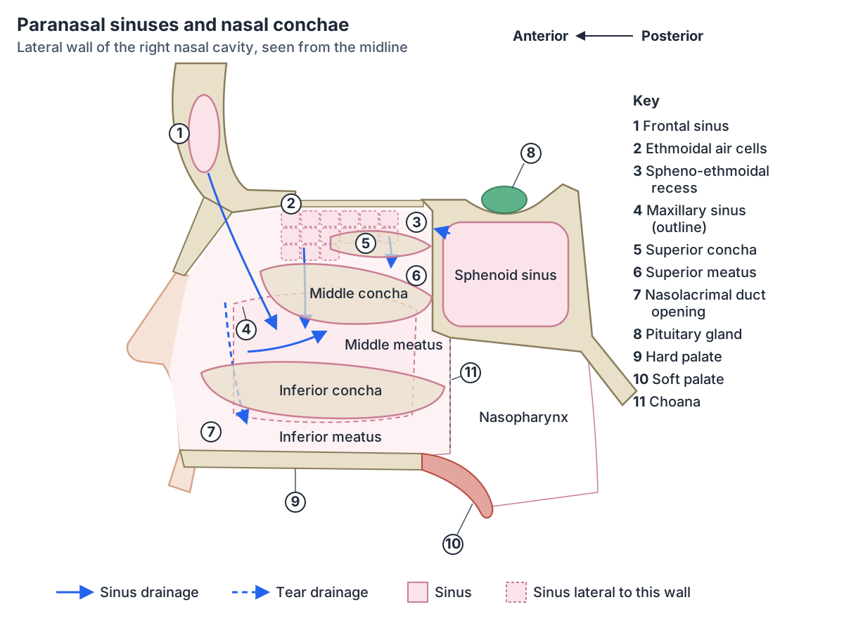
Each frontal sinus drains from its lower medial corner through the frontal recess into the middle meatus, either via the ethmoidal infundibulum and hiatus semilunaris or directly. The route varies between people. Older texts call this channel the frontonasal duct. (Radiopaedia: Frontal sinus.)
TeachMeAnatomy describes the frontal sinuses as the most superior sinuses, roughly triangular, draining via the frontonasal duct into the hiatus semilunaris of the middle meatus, and supplied by the supraorbital nerve. (TeachMeAnatomy: The Paranasal Sinuses.)
The frontal sinuses are not present at birth. They start to pneumatise (fill with air as the bone hollows out) at about 1 to 2 years. One or both may fail to develop: aplasia or hypoplasia affects one side in about 4% and both sides in about 5% of people. Their nerve supply is the supraorbital and supratrochlear nerves, and their arteries are the supratrochlear, supraorbital and anterior ethmoidal arteries. (Radiopaedia: Frontal sinus.)
Ethmoid sinus (ethmoidal air cells)
The ethmoid sinus is not one cavity but a collection of 3 to 18 small air cells on each side, separated by thin bony walls, inside the ethmoid labyrinth, which sits between the orbit and the nasal cavity. (Radiopaedia: Ethmoidal air cells.)
The cells are grouped into anterior and posterior groups, divided by the basal lamella, the lateral attachment of the middle concha. Anterior cells drain into the middle meatus through the hiatus semilunaris and ethmoidal bulla. Posterior cells drain into the superior meatus. Older texts describe a separate middle group; it is now counted with the anterior cells. (Radiopaedia: Ethmoidal air cells.)
TeachMeAnatomy still uses the three-group scheme: anterior cells drain to the hiatus semilunaris, middle cells to the lateral wall of the middle meatus (onto the ethmoidal bulla) and posterior cells to the superior meatus. The ethmoid cells are supplied by the anterior and posterior ethmoidal nerves and arteries. (TeachMeAnatomy: The Paranasal Sinuses.)
The ethmoid cells form in fetal life by folding of the cartilaginous nasal capsule rather than by bone hollowing out, so they are present at birth. They grow quickly up to age 4 and mature between about 8 and 12 years. (Radiopaedia: Ethmoidal air cells.)
Sphenoid sinus
The sphenoid sinus is the most posterior paranasal sinus. It lies in the body of the sphenoid bone, in front of and below the sella turcica. A septum, which is often off the midline, divides it into two halves. (Radiopaedia: Sphenoid sinus.)
It has important neighbours. Above are the sella turcica with the pituitary gland and the cavernous sinus; to each side are the cavernous sinus and cranial cavity; below and in front is the nasal cavity. The optic nerve, internal carotid artery and pituitary gland can make ridges in its walls, which matters to surgeons. It drains into the roof of the nasal cavity through the spheno-ethmoidal recess. (Radiopaedia: Sphenoid sinus.)
Surgeons use the sphenoid sinus as a corridor to the pituitary gland. In endoscopic transsphenoidal surgery, usually done for pituitary adenomas, instruments pass through the nose and the sphenoid sinus to reach the gland. (TeachMeAnatomy: The Paranasal Sinuses.)
The sphenoid sinus begins to pneumatise at about age 2 and develops more slowly than the others. Its extent varies: radiologists classify it as conchal, presellar or sellar depending on how far the air reaches under the sella. It is supplied by the posterior ethmoidal and sphenopalatine arteries and the posterior ethmoidal nerve. (Radiopaedia: Sphenoid sinus.)
Nasal conchae and meatuses
The nasal conchae (turbinates) are curved bony plates that project downward from the lateral wall of each nasal cavity. They swirl incoming air, which helps to warm and moisten it. The inferior concha is the largest. The middle and superior conchae, the superior being the smallest, are both parts of the ethmoid bone. (OpenStax Anatomy and Physiology 2e, 7.2: The Skull.)
The inferior nasal concha is a separate facial bone that articulates with the maxilla and the palatine bone. A small fourth concha, the supreme concha, is sometimes present above the superior concha. (Radiopaedia: Nasal concha.)
The space under each concha is a meatus. The inferior meatus lies between the inferior concha and the floor of the nose and receives the nasolacrimal duct, which drains tears. The middle meatus lies between the inferior and middle conchae. The superior meatus lies between the middle and superior conchae. Above and behind the superior concha is the spheno-ethmoidal recess. (TeachMeAnatomy: The Nasal Cavity.)
The nasal septum divides the nasal cavity into right and left halves from the nostrils to the choanae. Its front part is the septal (quadrangular) cartilage; behind, the perpendicular plate of the ethmoid forms the upper part and the vomer the lower part. Some septal deviation is extremely common. (Radiopaedia: Nasal septum.)
Where do the sinuses drain?
The frontal, anterior ethmoid and maxillary sinuses all drain into the middle meatus through a shared drainage area called the ostiomeatal complex. The posterior ethmoid cells drain into the superior meatus. The sphenoid sinuses drain into the spheno-ethmoidal recess. (Radiopaedia: Paranasal sinuses.)
The middle concha projects over the openings of the maxillary and ethmoid sinuses and shields them from the direct force of airflow. (Radiopaedia: Nasal concha.)
TeachMeAnatomy lists the same pattern: the frontal, maxillary and anterior ethmoidal sinuses open into the middle meatus, the posterior ethmoidal sinuses open at the superior meatus, and the nasolacrimal duct opens into the inferior meatus. (TeachMeAnatomy: The Nasal Cavity.)
The four paranasal sinuses at a glance
| Sinus | Bone | Opens into | Present at birth? | Sensory nerves |
|---|---|---|---|---|
| Maxillary | Body of the maxilla | Middle meatus, via the ethmoidal infundibulum and hiatus semilunaris | Yes; keeps growing until about age 14 | Anterior, middle and posterior superior alveolar nerves, infraorbital nerve, greater palatine nerve (all from V2) |
| Frontal | Frontal bone | Middle meatus, via the frontal recess (route varies) | No; pneumatisation begins at about 1 to 2 years | Supraorbital and supratrochlear nerves (V1) |
| Ethmoidal air cells | Ethmoid labyrinth | Anterior cells: middle meatus. Posterior cells: superior meatus | Yes; grow fast to age 4, mature by about 8 to 12 years | Anterior and posterior ethmoidal nerves (V1), maxillary nerve branches |
| Sphenoid | Body of the sphenoid | Spheno-ethmoidal recess, above and behind the superior concha | No; pneumatisation starts at about age 2 | Posterior ethmoidal nerve, orbital branches of the pterygopalatine ganglion |
From Radiopaedia and TeachMeAnatomy (see references). Size, number of chambers and drainage routes vary between people.
How the sinuses develop and vary
At birth only the maxillary sinuses and ethmoid air cells are present. The frontal and sphenoid sinuses pneumatise later in childhood. (Radiopaedia: Paranasal sinuses.)
Size and septation vary between people and between the two sides. Named variants include agger nasi and other frontal recess cells, Haller cells, concha bullosa (an air-filled middle concha) and maxillary sinus hypoplasia. Absence of the frontal sinus is common, but total absence of all sinuses is very rare. Very large sphenoid sinuses can leave the internal carotid artery or optic nerve with little or no bony covering. (Radiopaedia: Paranasal sinuses.)
Sinus nerves, sinusitis and toothache
The sinuses get their sensation from the trigeminal nerve: the frontal and ethmoid sinuses mainly from ophthalmic (V1) branches, the maxillary sinus from maxillary (V2) branches, and the sphenoid sinus from both. The maxillary nerve also supplies the upper teeth, so maxillary sinusitis can feel like toothache. (TeachMeAnatomy: The Paranasal Sinuses.)
CT is the imaging method of choice for the sinuses because it shows the thin bone walls and drainage channels before surgery. Plain X-rays have largely been replaced. (Radiopaedia: Paranasal sinuses.)
Frequently asked questions
What are the four paranasal sinuses?
The maxillary, frontal, ethmoid and sphenoid sinuses. Each is paired and named after the bone that contains it. The maxillary sinus is the largest. The ethmoid sinus is really a group of small air cells. All four open into the nasal cavity and are lined by the same respiratory mucosa as the nose.
Which sinus is the largest?
The maxillary sinus, inside the body of the maxilla below each orbit. Its roof is the orbital floor and its floor is the alveolar process above the upper teeth. Its opening is high on its medial wall, which makes it difficult to drain, and it is the sinus most often involved in infections.
Which sinuses drain into the middle meatus?
The frontal sinus, the maxillary sinus and the anterior ethmoid air cells. They drain through the ostiomeatal complex, including the hiatus semilunaris. The posterior ethmoid cells drain into the superior meatus, the sphenoid sinus into the spheno-ethmoidal recess, and the nasolacrimal duct (not a sinus) into the inferior meatus.
Which sinuses are present at birth?
The maxillary sinuses and the ethmoid air cells. The frontal sinuses begin to pneumatise at about 1 to 2 years and the sphenoid sinuses at about 2 years. The maxillary sinus keeps growing until about age 14 and the ethmoid cells mature at about 8 to 12 years.
What are the nasal conchae?
Curved bony shelves on the lateral wall of each nasal cavity that swirl, warm and moisten inhaled air. The superior and middle conchae belong to the ethmoid bone; the inferior concha, the largest, is a separate bone. The passage under each concha is a meatus, where the sinuses and nasolacrimal duct open.
Why can sinusitis cause toothache?
The maxillary nerve supplies both the lining of the maxillary sinus and the upper teeth, and the roots of the upper molars can sit close to or project into the sinus floor. Inflammation of the maxillary sinus can therefore be felt as pain in the upper back teeth.
References
- Betts JG, et al. Anatomy and Physiology 2e, §7.2 The Skull. OpenStax, Rice University, 2022.
- TeachMeAnatomy. The Paranasal Sinuses: Structure and Function.
- TeachMeAnatomy. The Nasal Cavity: Structure, Vasculature, Innervation.
- Radiopaedia. Paranasal sinuses (reference article).
- Radiopaedia. Maxillary sinus (reference article).
- Radiopaedia. Frontal sinus (reference article).
- Radiopaedia. Sphenoid sinus (reference article).
- Radiopaedia. Ethmoidal air cells (reference article).
- Radiopaedia. Nasal concha (reference article).
- Radiopaedia. Nasal septum (reference article).
Related anatomy
- Skull bonesLearn the 22 bones of the skull, the 8 cranial bones of the neurocranium and the coronal, sagittal, lambdoid and…
- MaxillaMaxilla bone anatomy: the body and four processes, palatine and alveolar processes, maxillary sinus, articulations, Le…
- Ethmoid boneThe ethmoid bone (os ethmoidale) explained: cribriform plate, crista galli, perpendicular plate, ethmoid sinus air…
- Sphenoid boneThe sphenoid bone (os sphenoidale): sella turcica and hypophyseal fossa, greater and lesser wings, pterygoid process…
- Frontal boneThe frontal bone (os frontale) explained: squamous, orbital and nasal parts, supraorbital margin and foramen, frontal…
- Skull foraminaSkull foramina explained: the three cranial fossae, foramen magnum, jugular foramen and every other opening, with a…
Explore the whole body in the interactive 3D human anatomy model, or browse the A–Z list of human structures.