

The cranial cavity and its three fossae
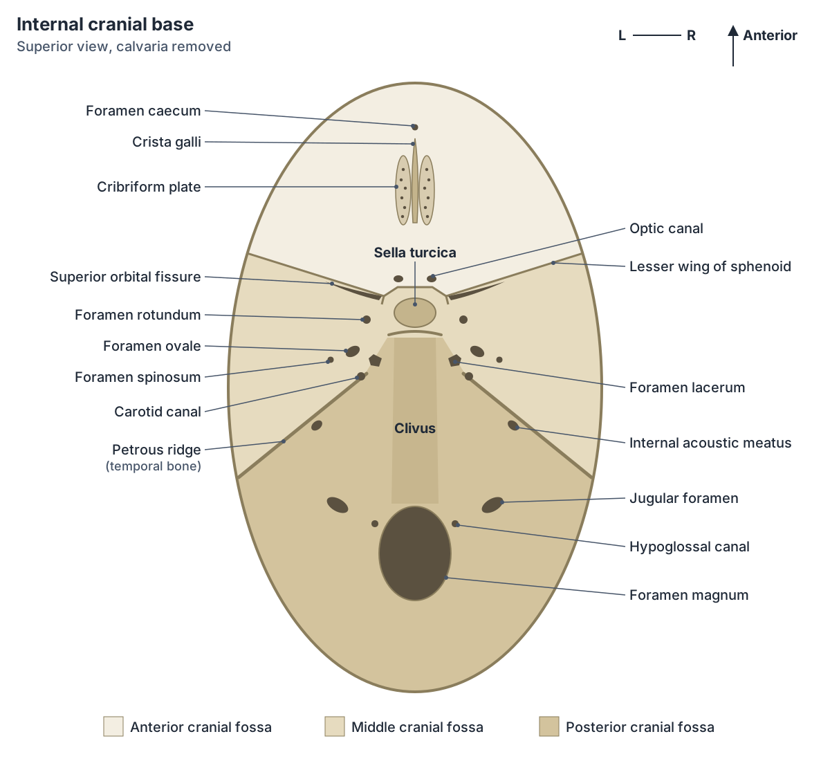
The cranial cavity is the space inside the skull that holds the brain. Its floor is not flat. It steps down from front to back as three large hollows: the anterior cranial fossa, the middle cranial fossa and the posterior cranial fossa. From anterior to posterior the fossae become deeper. (OpenStax Anatomy and Physiology 2e, 7.2: The Skull.)
The anterior cranial fossa lies over the orbits and holds the frontal lobes. The middle cranial fossa is butterfly-shaped, narrow in the midline and broad at the sides, and holds the temporal lobes. The posterior cranial fossa is the deepest and most posterior part of the cranial cavity and holds the cerebellum. (OpenStax Anatomy and Physiology 2e, 7.2: The Skull.)
The borders between the fossae are bony ridges. The lesser wings of the sphenoid and the limbus of the sphenoid separate the anterior from the middle fossa. The upper border of the petrous part of the temporal bone (the petrous ridge) and the dorsum sellae separate the middle from the posterior fossa. (TeachMeAnatomy: Middle Cranial Fossa.)
Anterior cranial fossa: cribriform plate and olfactory foramina
The anterior cranial fossa is formed by the frontal, ethmoid and sphenoid bones. Its floor is made by the frontal bone, the ethmoid bone, the front of the body of the sphenoid and the lesser wings of the sphenoid. It is the shallowest of the three fossae. (TeachMeAnatomy: Anterior Cranial Fossa.)
In the midline stands the crista galli (Latin for cock's comb), an upward spike of the ethmoid that anchors the falx cerebri. On each side of it is the cribriform plate, a thin sheet of bone that supports the olfactory bulb. Its many small holes (the olfactory or cribriform foramina) let olfactory nerve fibres (cranial nerve I) pass up from the roof of the nasal cavity. (TeachMeAnatomy: Anterior Cranial Fossa.)
Just in front of the ethmoid, the frontal bone has a small foramen caecum, which transmits an emissary vein from the nasal cavity to the superior sagittal sinus. (Kenhub: Skull foramina, fissures and contents.)
The cribriform plate is the thinnest part of the anterior cranial fossa, so it is the part most likely to break in a head injury. A fracture here can tear the olfactory fibres and cause loss of smell (anosmia), or let cerebrospinal fluid leak into the nose (CSF rhinorrhoea). (TeachMeAnatomy: Anterior Cranial Fossa.)
Middle cranial fossa: optic canal to foramen spinosum
The floor of the middle cranial fossa is formed by the body and greater wings of the sphenoid and the squamous and petrous parts of the temporal bones. Its central part, on the body of the sphenoid, is the saddle-shaped sella turcica, whose hollow (the hypophysial fossa) holds the pituitary gland. Its two lateral parts hold the temporal lobes. (TeachMeAnatomy: Middle Cranial Fossa.)
The optic canal lies at the front corner of the sella turcica. It carries the optic nerve (cranial nerve II) and the ophthalmic artery into the orbit. Lateral to it, the superior orbital fissure is a slit between the lesser and greater wings of the sphenoid. It carries the oculomotor (III), trochlear (IV) and abducens (VI) nerves, the branches of the ophthalmic nerve (V1), the ophthalmic veins and sympathetic fibres. (TeachMeAnatomy: Middle Cranial Fossa.)
Three foramina in the greater wing of the sphenoid form a curved line on each side. Foramen rotundum carries the maxillary nerve (V2) forward into the pterygopalatine fossa. Foramen ovale, posterolateral to it, carries the mandibular nerve (V3) and the accessory meningeal artery. Foramen spinosum, posterolateral to the ovale, carries the middle meningeal artery and vein and a meningeal branch of V3. (TeachMeAnatomy: Middle Cranial Fossa.)
These three branches of the trigeminal nerve have distinct jobs. The nerve through foramen rotundum supplies sensation to the cheek, nose and upper teeth. The nerve through foramen ovale supplies the lateral head, cheek, chin and lower teeth. The artery entering through foramen spinosum supplies the coverings of the brain. (OpenStax Anatomy and Physiology 2e, 7.2: The Skull.)
Two small openings on the front of the petrous temporal bone, the hiatus for the greater petrosal nerve and the hiatus for the lesser petrosal nerve, carry these small branches of the facial and glossopharyngeal nerves. (TeachMeAnatomy: Middle Cranial Fossa.)
Foramen lacerum and the carotid canal
The foramen lacerum is an irregular gap at the junction of the sphenoid, the apex of the petrous temporal bone and the basilar part of the occipital bone. It is not a true foramen within one bone. In life it is filled with cartilage, so the large opening seen on a dry skull is partly an artefact. (Radiopaedia: Foramen lacerum.)
Only small structures pass through the cartilage: meningeal branches of the ascending pharyngeal artery and emissary veins. The greater petrosal nerve reaches the foramen, joins the deep petrosal nerve and leaves as the nerve of the pterygoid canal. The internal carotid artery runs across the top of the foramen lacerum but does not pass through it. (Radiopaedia: Foramen lacerum.)
The carotid canal is a passage about 2 cm long inside the petrous temporal bone. It starts on the underside of the skull just in front of the jugular fossa, runs upward, turns about 90 degrees forward and medially, and opens into the middle cranial fossa just above the foramen lacerum. It carries the internal carotid artery with its venous and sympathetic plexuses. (Radiopaedia: Carotid canal.)
Posterior fossa of the skull
The posterior cranial fossa is formed by the occipital bone and the two temporal bones. It holds the brainstem (midbrain, pons and medulla) and the cerebellum. Its floor is made by the mastoid part of the temporal bone and the squamous, condylar and basilar parts of the occipital bone. (TeachMeAnatomy: Posterior Cranial Fossa.)
The internal acoustic meatus opens on the posterior surface of the petrous temporal bone. It carries the facial nerve (VII), the vestibulocochlear nerve (VIII) and the labyrinthine artery to the inner ear. (TeachMeAnatomy: Posterior Cranial Fossa.)
The jugular foramen lies between the petrous temporal bone in front and the occipital bone behind. It carries the glossopharyngeal (IX), vagus (X) and accessory (XI) nerves. The sigmoid sinus drains through it to become the internal jugular vein, and the inferior petrosal sinus and meningeal branches of the ascending pharyngeal and occipital arteries also pass through. (TeachMeAnatomy: Posterior Cranial Fossa.)
The hypoglossal canal lies just above the anterolateral margin of the foramen magnum and carries the hypoglossal nerve (XII). (TeachMeAnatomy: Posterior Cranial Fossa.)
What passes through the foramen magnum?
The foramen magnum is the large opening in the occipital bone where the brainstem becomes the spinal cord. It transmits the medulla oblongata and its meninges, the two vertebral arteries, the anterior and posterior spinal arteries, the spinal roots of the accessory nerve (which ascend into the skull here and leave again through the jugular foramen) and dural veins. (TeachMeAnatomy: Posterior Cranial Fossa.)
Raised pressure inside the skull can push the cerebellar tonsils down through the foramen magnum. This is called tonsillar herniation or coning. It compresses the medulla, which contains the cardiac and respiratory centres, and can be fatal. (TeachMeAnatomy: Posterior Cranial Fossa.)
Foramina of the cranial base and what passes through them
| Opening | Bone and fossa | Main contents |
|---|---|---|
| Foramen caecum | Frontal bone, anterior fossa (in front of the crista galli) | Emissary vein from the nasal cavity to the superior sagittal sinus |
| Cribriform foramina (olfactory foramina) | Cribriform plate of the ethmoid, anterior fossa | Olfactory nerve fibres (CN I); anterior ethmoidal nerve |
| Optic canal | Lesser wing and body of the sphenoid, middle fossa | Optic nerve (CN II), ophthalmic artery |
| Superior orbital fissure | Between the lesser and greater wings of the sphenoid, middle fossa | Oculomotor (CN III), trochlear (CN IV) and abducens (CN VI) nerves; lacrimal, frontal and nasociliary branches of the ophthalmic nerve (V1); ophthalmic veins; sympathetic fibres |
| Foramen rotundum | Greater wing of the sphenoid, middle fossa | Maxillary nerve (V2), to the pterygopalatine fossa |
| Foramen ovale | Greater wing of the sphenoid, middle fossa | Mandibular nerve (V3), accessory meningeal artery; sometimes the lesser petrosal nerve and an emissary vein |
| Foramen spinosum | Greater wing of the sphenoid, middle fossa | Middle meningeal artery and vein, meningeal branch of V3 |
| Foramen lacerum | Gap between sphenoid, petrous temporal and occipital bones, middle fossa | Filled with cartilage in life; small meningeal arteries and emissary veins. The greater petrosal nerve crosses it; the internal carotid artery runs over its upper surface |
| Carotid canal | Petrous part of the temporal bone; opens into the middle fossa | Internal carotid artery with its venous and sympathetic plexuses |
| Internal acoustic meatus | Petrous part of the temporal bone, posterior fossa | Facial nerve (CN VII), vestibulocochlear nerve (CN VIII), labyrinthine artery |
| Jugular foramen | Between the petrous temporal and occipital bones, posterior fossa | Glossopharyngeal (CN IX), vagus (CN X) and accessory (CN XI) nerves; internal jugular vein (from the sigmoid sinus); inferior petrosal sinus; meningeal arteries |
| Hypoglossal canal | Occipital bone, at the anterolateral margin of the foramen magnum | Hypoglossal nerve (CN XII) |
| Foramen magnum | Occipital bone, posterior fossa | Medulla oblongata and meninges, vertebral arteries, anterior and posterior spinal arteries, spinal roots of the accessory nerve, dural veins |
Compiled from TeachMeAnatomy, Kenhub and Radiopaedia (see references). Small variations exist between individuals.
Stylomastoid foramen, external acoustic meatus and palate foramina
The stylomastoid foramen is on the underside of the petrous temporal bone, between the base of the styloid process and the mastoid process. It is the lower end of the facial canal. The facial nerve leaves the skull here to supply the muscles of facial expression, and the stylomastoid artery enters. (Radiopaedia: Stylomastoid foramen.)
The external acoustic meatus (external auditory canal) runs from the opening on the side of the head to the eardrum. It is about 2.5 cm long and S-shaped. Its outer third is cartilage and its inner two-thirds is bone, formed by the tympanic and squamous parts of the temporal bone. Do not confuse it with the internal acoustic meatus, which opens inside the cranial cavity and carries nerves. (Radiopaedia: External auditory canal.)
On the hard palate, the incisive canal opens in the midline behind the central incisors at the incisive foramen (inside the incisive fossa). It carries the nasopalatine nerve and a terminal branch of the descending (greater) palatine artery. Further back, the greater palatine foramen lies medial to the third molar and carries the greater palatine nerve and vessels; the lesser palatine foramina, in the pyramidal process of the palatine bone, carry the lesser palatine nerves. (TeachMeAnatomy: The Palate.)
The palatine process of the maxilla carries the incisive fossa and incisive canals. The anterior surface of the body of the maxilla also has a depression called the incisive fossa (alongside a canine fossa), so check which one a question means. (Radiopaedia: Maxilla.)
The choanae are the two posterior openings of the nasal cavity into the nasopharynx. The posterior border of the vomer, which does not articulate with any bone, separates them in the midline. (Radiopaedia: Vomer.)
In choanal atresia the choanae fail to form because the bucconasal membrane does not break down. About two-thirds of cases affect one side only and may be noticed late. When both sides are blocked, a newborn can be in respiratory distress, because infants are obligate nose breathers. (Radiopaedia: Choanal atresia.)
Supraorbital, infraorbital, mental and mandibular foramina
Three foramina on the front of the face carry sensory nerves to the skin. The supraorbital foramen sits near the middle of the upper rim of the orbit and passes a sensory nerve to the skin of the forehead. The infraorbital foramen, below the orbit on the maxilla, passes a sensory nerve to the face below the orbit. The mental foramen, on the mandible, passes a sensory nerve to the chin. (OpenStax Anatomy and Physiology 2e, 7.2: The Skull.)
The mandibular foramen is on the inner surface of the mandibular ramus. The inferior alveolar nerve and artery enter it, run forward in the mandibular canal to supply the lower teeth, and leave through the mental foramen below the second premolar, where the nerve becomes the mental nerve to the lower lip and chin. (TeachMeAnatomy: The Mandible.)
Orbit anatomy: walls and fissures
Each orbit is built from seven bones. The frontal bone forms the roof. The zygomatic bone forms the lateral wall and lateral floor. The maxilla forms most of the medial floor, with a small part from the palatine bone. The ethmoid and lacrimal bones form much of the medial wall, and the sphenoid forms the back of the orbit. (OpenStax Anatomy and Physiology 2e, 7.2: The Skull.)
The inferior orbital fissure lies in the floor of the orbit, below the superior orbital fissure. It is bordered by the greater wing of the sphenoid above, the maxilla and the orbital process of the palatine bone below, and the zygomatic bone laterally. It carries the infraorbital and zygomatic nerves (from V2), the infraorbital artery and vein and a branch of the inferior ophthalmic vein. At its medial end it meets the pterygomaxillary fissure at a right angle. (Radiopaedia: Inferior orbital fissure.)
Temporal fossa and infratemporal fossa
On the side of the skull above the zygomatic arch is a shallow space called the temporal fossa. Below the arch and deep to the ramus of the mandible is the infratemporal fossa. Both contain muscles that move the mandible during chewing. (OpenStax Anatomy and Physiology 2e, 7.2: The Skull.)
The infratemporal fossa is bounded laterally by the ramus of the mandible, medially by the lateral pterygoid plate, in front by the back wall of the maxilla and above by the greater wing of the sphenoid, which contains foramen ovale and foramen spinosum. It holds the medial and lateral pterygoid muscles, branches of the mandibular nerve (including the inferior alveolar and lingual nerves), the chorda tympani, the otic ganglion, the maxillary artery and the pterygoid venous plexus. (TeachMeAnatomy: The Infratemporal Fossa.)
Pterygopalatine fossa, pterygomaxillary fissure and sphenopalatine foramen
The pterygopalatine fossa is a small space between the maxilla in front, the pterygoid process of the sphenoid behind and the perpendicular plate of the palatine bone medially. It is a junction box: it contains the maxillary nerve, the pterygopalatine ganglion and the last part of the maxillary artery. (TeachMeAnatomy: The Pterygopalatine Fossa.)
It connects to many neighbouring regions. Foramen rotundum and the pterygoid canal lead back to the middle cranial fossa. The inferior orbital fissure leads up to the orbit. The greater palatine canal leads down to the mouth. The sphenopalatine foramen, its only medial opening, leads into the nasal cavity at the superior meatus and carries the sphenopalatine artery and vein and the nasopalatine nerve. Laterally, the pterygomaxillary fissure opens into the infratemporal fossa. (TeachMeAnatomy: The Pterygopalatine Fossa.)
The pterygomaxillary fissure lies between the back of the maxilla and the lateral pterygoid plate. The maxillary artery enters the pterygopalatine fossa through it, and the posterior superior alveolar nerve (a branch of the maxillary nerve) passes out through it from the pterygopalatine fossa to the infratemporal fossa. (Radiopaedia: Pterygomaxillary fissure.)
Openings outside the cranial cavity
| Opening | Where it is | Main contents |
|---|---|---|
| Stylomastoid foramen | Under the petrous temporal bone, between the styloid and mastoid processes | Facial nerve (CN VII), stylomastoid artery |
| External acoustic meatus | Temporal bone, lateral skull | Air canal to the tympanic membrane (eardrum) |
| Supraorbital foramen | Frontal bone, upper margin of the orbit | Sensory nerve to the skin of the forehead |
| Infraorbital foramen | Maxilla, below the orbit | Infraorbital nerve and vessels |
| Mental foramen | Mandible, below the second premolar | Mental nerve |
| Mandibular foramen | Inner surface of the mandibular ramus | Inferior alveolar nerve and artery |
| Incisive canal and foramen | Midline of the hard palate behind the central incisors | Nasopalatine nerve, terminal branch of the descending (greater) palatine artery |
| Greater palatine foramen | Hard palate, medial to the third molar | Greater palatine nerve and vessels |
| Lesser palatine foramina | Pyramidal process of the palatine bone | Lesser palatine nerves |
| Inferior orbital fissure | Floor of the orbit, between the greater wing of the sphenoid and the maxilla | Infraorbital and zygomatic nerves (V2), infraorbital artery and vein, branch of the inferior ophthalmic vein |
| Pterygomaxillary fissure | Between the maxilla and the lateral pterygoid plate | Posterior superior alveolar nerve, end of the maxillary artery |
| Sphenopalatine foramen | Medial wall of the pterygopalatine fossa | Sphenopalatine artery and vein, nasopalatine nerve |
Skull foramina in the 3D model
- Ethmoid
- A delicate cube-shaped bone between the orbits.
- Frontal bone
- The bone of the forehead and the roof of the orbits.
- Mandible
- The lower jaw bone.
- Maxilla
- The upper jaw bone, paired.
- Occipital bone
- The bone forming the back and base of the skull.
- Palatine bone
- An L-shaped bone behind the maxilla.
- Sphenoid bone
- A butterfly-shaped bone at the base of the skull.
- Temporal bone
- A paired bone forming part of the side and base of the skull.
Frequently asked questions
What passes through the foramen magnum?
The medulla oblongata and its meninges, the two vertebral arteries, the anterior and posterior spinal arteries, the spinal roots of the accessory nerve and dural veins. The spinal cord continues from the medulla just below the opening. The accessory nerve roots enter the skull through the foramen magnum and leave through the jugular foramen.
What passes through the jugular foramen?
Three cranial nerves, the glossopharyngeal (IX), vagus (X) and accessory (XI), plus the internal jugular vein, which begins here as the continuation of the sigmoid sinus. The inferior petrosal sinus and small meningeal arteries also pass. The foramen lies between the petrous temporal bone and the occipital bone in the posterior cranial fossa.
What passes through foramen rotundum, ovale and spinosum?
All three are in the greater wing of the sphenoid. Foramen rotundum carries the maxillary nerve (V2). Foramen ovale carries the mandibular nerve (V3) and the accessory meningeal artery. Foramen spinosum carries the middle meningeal artery and vein and a meningeal branch of V3. From front to back they run rotundum, ovale, spinosum, each posterolateral to the one before.
What is the difference between the internal and external acoustic meatus?
The internal acoustic meatus opens inside the skull on the petrous temporal bone and carries the facial and vestibulocochlear nerves and the labyrinthine artery. The external acoustic meatus is the ear canal on the outside of the head, about 2.5 cm long, leading to the eardrum. Both are in the temporal bone.
Why is the foramen lacerum called a false foramen?
It is a gap between the sphenoid, temporal and occipital bones rather than a hole within one bone, and in life it is filled with cartilage. Only small meningeal arteries and emissary veins pass through. The internal carotid artery crosses over its upper surface after leaving the carotid canal but does not run through it.
Where is the pterygopalatine fossa?
It is a small space deep in the side of the face, between the back of the maxilla and the pterygoid process of the sphenoid, with the palatine bone on its medial side. It holds the maxillary nerve, the pterygopalatine ganglion and the end of the maxillary artery, and opens into the orbit, nose, mouth, cranial cavity and infratemporal fossa.
Which bones form the three cranial fossae?
The anterior fossa is formed by the frontal, ethmoid and sphenoid bones. The middle fossa is formed mainly by the sphenoid and the two temporal bones. The posterior fossa is formed by the occipital bone and the two temporal bones. Each fossa is deeper than the one in front of it.
References
- Betts JG, et al. Anatomy and Physiology 2e, §7.2 The Skull. OpenStax, Rice University, 2022.
- TeachMeAnatomy. The Cranial Foramina.
- TeachMeAnatomy. The Anterior Cranial Fossa: Boundaries and Contents.
- TeachMeAnatomy. The Middle Cranial Fossa: Boundaries and Contents.
- TeachMeAnatomy. The Posterior Cranial Fossa: Boundaries and Contents.
- TeachMeAnatomy. The Pterygopalatine Fossa.
- TeachMeAnatomy. The Infratemporal Fossa: Borders and Contents.
- TeachMeAnatomy. The Palate: Hard Palate, Soft Palate, Uvula.
- TeachMeAnatomy. The Mandible: Structure, Attachments, Fractures.
- Kenhub. Skull: Foramina, fissures and contents.
- Radiopaedia. Stylomastoid foramen (reference article).
- Radiopaedia. Carotid canal (reference article).
- Radiopaedia. Foramen lacerum (reference article).
- Radiopaedia. Inferior orbital fissure (reference article).
- Radiopaedia. Pterygomaxillary fissure (reference article).
- Radiopaedia. Incisive canal (reference article).
- Radiopaedia. External auditory canal (reference article).
- Radiopaedia. Vomer (reference article).
- Radiopaedia. Choanal atresia (reference article).
- Radiopaedia. Maxilla (reference article).
- Mitsuhashi N. et al. (2009). BodyParts3D: 3D structure database for anatomical concepts. Nucleic Acids Research 37, D782–D785. Data © The Database Center for Life Science, CC BY 4.0.
Related anatomy
- Skull bonesLearn the 22 bones of the skull, the 8 cranial bones of the neurocranium and the coronal, sagittal, lambdoid and…
- Sphenoid boneThe sphenoid bone (os sphenoidale): sella turcica and hypophyseal fossa, greater and lesser wings, pterygoid process…
- Temporal boneThe temporal bone (os temporale) explained: petrous part and inner ear, mastoid process, zygomatic and styloid…
- Occipital boneThe occipital bone (os occipitale) explained: what passes through the foramen magnum, the external occipital…
- Cranial nervesThe 12 cranial nerves in order with a labelled diagram: type, skull foramen, functions and test for CN I to CN XII…
- Paranasal sinusesParanasal sinuses anatomy: the maxillary, frontal, ethmoid and sphenoid sinuses, where each drains, the nasal conchae…
Explore the whole body in the interactive 3D human anatomy model, or browse the A–Z list of human structures.