

What are the abdominal muscles?
The anterior and lateral abdominal wall is made of three flat muscle layers plus a vertical pair of muscles at the front. From outside in, the flat layers are the external oblique, the internal oblique and the transversus abdominis. Their fibres run in different directions. The rectus abdominis muscles are a pair of long muscles running vertically on either side of the midline. (OpenStax Anatomy and Physiology 2e, 11.4: Axial Muscles of the Abdominal Wall and Thorax.)
Each flat muscle ends at the front in an aponeurosis (a broad, flat tendon). These aponeuroses enclose rectus abdominis as the rectus sheath and interlace in the midline as the linea alba, a fibrous band running from the xiphoid process of the sternum to the pubic symphysis. (TeachMeAnatomy: The Anterolateral Abdominal Wall.)
Together the abdominal muscles flex and twist the trunk and bend it sideways, and they compress the abdomen. Compression is used in forceful breathing out, defecation, urination and childbirth. The back of the abdominal wall has its own muscles, including quadratus lumborum, which bends the trunk to the side. (OpenStax Anatomy and Physiology 2e, 11.4: Axial Muscles of the Abdominal Wall and Thorax.)
Abdominal wall muscles: origin, insertion, nerve and action
| Muscle | Origin | Insertion | Innervation | Main action |
|---|---|---|---|---|
| External oblique | Outer surfaces of ribs 5–12 | Iliac crest, pubic tubercle, linea alba (via aponeurosis) | Thoracoabdominal nerves T7–T11, subcostal nerve T12 | Compresses abdomen; rotates trunk to opposite side; lateral flexion |
| Internal oblique | Inguinal ligament, iliac crest, thoracolumbar fascia | Ribs 10–12, linea alba (via aponeurosis) | T7–T11, T12, branches of lumbar plexus | Compresses abdomen; rotates trunk to same side |
| Transversus abdominis | Inguinal ligament, costal cartilages 7–12, iliac crest, thoracolumbar fascia | Conjoint tendon, xiphoid process, linea alba, pubic crest | T7–T11, T12, branches of lumbar plexus | Compresses abdominal contents |
| Rectus abdominis | Pubic crest | Xiphoid process, costal cartilages 5–7 | Thoracoabdominal nerves T7–T11, subcostal nerve T12 | Flexes vertebral column; compresses abdomen; depresses ribs |
| Pyramidalis | Pubic crest and symphysis | Linea alba | Subcostal nerve T12 | Tenses linea alba |
| Quadratus lumborum (posterior wall) | Iliac crest, iliolumbar ligament | Rib 12, transverse processes L1–L4 | Subcostal nerve T12, anterior rami L1–L4 | Fixes rib 12; lateral flexion of trunk |
Sources: TeachMeAnatomy (anterolateral abdominal wall); OpenStax 11.4; Kenhub (quadratus lumborum).
External oblique (obliquus externus abdominis)
The external oblique (obliquus externus abdominis) is the largest and most superficial of the three flat muscles. It arises from ribs 5 to 12, and its fibres run inferomedially (down and towards the midline). It inserts on the iliac crest and pubic tubercle, and its aponeurosis joins the linea alba. One external oblique rotates the trunk to the opposite side, so the right external oblique turns the trunk to the left. It is supplied by the thoracoabdominal nerves (T7 to T11) and the subcostal nerve (T12). (TeachMeAnatomy: The Anterolateral Abdominal Wall.)
OpenStax compares the external oblique fibre direction to sliding four fingers into the front pockets of your trousers, while the internal oblique fibres follow the direction the thumbs point. Both obliques twist the waist and bend the trunk to the side. (OpenStax Anatomy and Physiology 2e, 11.4: Axial Muscles of the Abdominal Wall and Thorax.)
The lower border of the external oblique aponeurosis is rolled up on itself to form the inguinal ligament, which runs from the anterior superior iliac spine to the pubic tubercle and forms the floor of the inguinal canal. (TeachMeAnatomy: The Inguinal Canal.)
Internal oblique (internal abdominal oblique)
The internal oblique (internal abdominal oblique, obliquus internus abdominis) lies deep to the external oblique. Its fibres run superomedially (up and towards the midline), at right angles to those of the external oblique. It arises from the inguinal ligament, the iliac crest and the thoracolumbar (lumbodorsal) fascia, and inserts on ribs 10 to 12 and, through its aponeurosis, into the linea alba. (TeachMeAnatomy: The Anterolateral Abdominal Wall.)
Acting together, the two internal obliques compress the abdomen. One internal oblique on its own rotates the trunk to the same side, so the right internal oblique works with the left external oblique to turn the trunk to the right. It is supplied by the thoracoabdominal nerves (T7 to T11), the subcostal nerve (T12) and branches of the lumbar plexus. (TeachMeAnatomy: The Anterolateral Abdominal Wall.)
The internal oblique also contributes to the spermatic cord: the cremaster muscle and cremasteric fascia that surround the cord are continuations of the internal oblique. (Kenhub: Inguinal canal.)
Transversus abdominis
Transversus abdominis is the deepest flat muscle. Its fibres run horizontally (transversely) around the abdomen. It arises from the inguinal ligament, the costal cartilages of ribs 7 to 12, the iliac crest and the thoracolumbar fascia, and inserts into the conjoint tendon, the xiphoid process, the linea alba and the pubic crest. Its main action is to compress the abdominal contents. Its nerve supply is the same as that of the internal oblique. (TeachMeAnatomy: The Anterolateral Abdominal Wall.)
At the back, the aponeuroses of transversus abdominis and internal oblique blend with the thoracolumbar fascia at a seam called the lateral raphe. This connects the abdominal wall to the sheath around the deep back muscles, so the abdominal and back muscles work together as a girdle around the trunk. (Willard FH et al. The thoracolumbar fascia: anatomy, function and clinical considerations. J Anat. 2012.)
Rectus abdominis and pyramidalis
Rectus abdominis is the paired 'six-pack' muscle. It arises from the pubic crest and inserts on the xiphoid process and the costal cartilages of ribs 5 to 7. It is supplied by the thoracoabdominal nerves (T7 to T11) and the subcostal nerve (T12). It helps compress the abdominal organs, stabilises the pelvis during walking and depresses the ribs. (TeachMeAnatomy: The Anterolateral Abdominal Wall.)
The main action of rectus abdominis is flexion of the vertebral column, as in a sit-up. Each muscle is crossed by three transverse bands of collagen fibres, the tendinous intersections, which divide it into the segments that give the 'six-pack' look. (OpenStax Anatomy and Physiology 2e, 11.4: Axial Muscles of the Abdominal Wall and Thorax.)
Pyramidalis is a small triangular muscle in front of the lower end of rectus abdominis. It arises from the pubic crest and pubic symphysis, inserts into the linea alba and tenses it. It is supplied by the subcostal nerve (T12). (TeachMeAnatomy: The Anterolateral Abdominal Wall.)
Layers of the abdominal wall
From outside in, the anterior abdominal wall has four main layers: skin, superficial fascia, the muscles with their fascia, and the parietal peritoneum that lines the abdominal cavity. (TeachMeAnatomy: The Anterolateral Abdominal Wall.)
Above the umbilicus the superficial fascia is a single sheet. Below the umbilicus it has two layers: a fatty superficial layer (Camper's fascia) and a membranous deep layer (Scarpa's fascia). Superficial vessels and nerves run between them. Deep to transversus abdominis is a well-formed layer called the transversalis fascia. (TeachMeAnatomy: The Anterolateral Abdominal Wall.)
Radiopaedia lists nine layers through the lateral part of the wall, from superficial to deep: skin, subcutaneous fat (Camper's fascia), superficial (Scarpa's) fascia, external oblique, internal oblique, transversus abdominis, transversalis fascia, extraperitoneal (preperitoneal) fat and parietal peritoneum. Nearer the midline, the flat muscles give way to their aponeuroses, which continue medially around rectus abdominis as the rectus sheath. (Radiopaedia: Anterior abdominal wall.)
The rectus sheath and the arcuate line
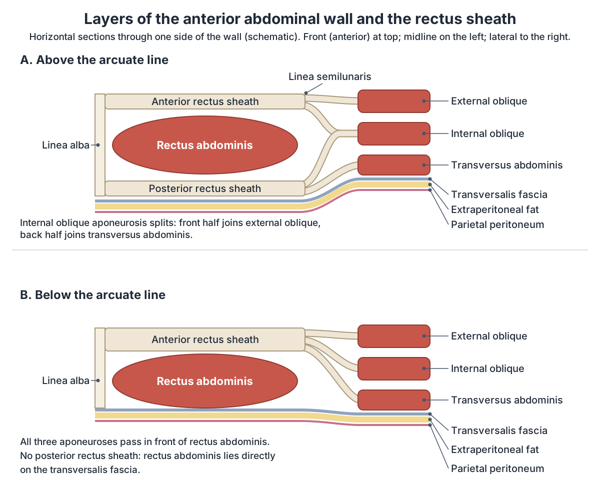
The rectus sheath is formed by the aponeuroses of the three flat muscles. In the upper part, the anterior wall is made of the external oblique aponeurosis and half of the internal oblique aponeurosis, and the posterior wall is made of the other half of the internal oblique aponeurosis and the transversus abdominis aponeurosis. (TeachMeAnatomy: The Anterolateral Abdominal Wall.)
The arcuate line marks the boundary between the upper three-quarters and the lower quarter of rectus abdominis. Below it, all three aponeuroses pass in front of the muscle and there is no posterior wall: the back of rectus abdominis lies directly on the transversalis fascia. (Kenhub: Rectus sheath.)
The sheath contains rectus abdominis and pyramidalis, the superior and inferior epigastric arteries and veins (which join near the umbilicus), lymphatic vessels and the ends of the lower intercostal nerves (T7 to T11) and the subcostal nerve (T12). Because there is no strong posterior wall below the arcuate line, bleeding from the epigastric vessels into the sheath (a rectus sheath haematoma) can spread more easily in the lower abdomen. (Kenhub: Rectus sheath.)
Linea alba, linea semilunaris and abdominal incisions
The linea alba runs from the xiphoid process to the pubic symphysis, where the flat muscle aponeuroses interlace in the midline. The linea semilunaris is the curved lateral border of rectus abdominis, running from the 9th rib to the pubic tubercle. A midline incision through the linea alba is used in surgery because it has a poor blood supply and avoids major nerves, so blood loss is small. (TeachMeAnatomy: The Anterolateral Abdominal Wall.)
Diastasis recti abdominis is an abnormal separation of the two rectus abdominis muscles along the linea alba. It is common after pregnancy, with reported postpartum incidence ranging from about 21% to nearly 30%. One proposed definition treats a separation of 2 cm or more as clinically significant, but there is no global consensus guideline. (Du Y et al. Diastasis recti abdominis: a comprehensive review. Hernia. 2025.)
A Spigelian hernia passes through a weakness in the fascia along the linea semilunaris. The opening is narrow, which raises the risk that the trapped bowel will be strangulated. (Kenhub: Rectus sheath.)
Inguinal canal anatomy
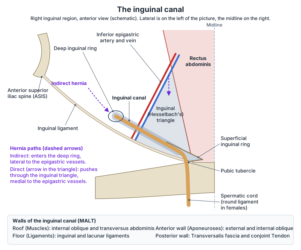
The inguinal canal is a short oblique passage through the lower part of the anterior abdominal wall, just above and parallel to the inguinal ligament. It is about 3 to 5 cm long in an adult. It begins at the deep inguinal ring, midway along the inguinal ligament (halfway between the anterior superior iliac spine and the pubic tubercle), and ends at the superficial inguinal ring, a V-shaped gap in the external oblique aponeurosis about 1 cm above and lateral to the pubic tubercle. (Kenhub: Inguinal canal.)
The deep ring is formed where the transversalis fascia is pushed out to cover the contents of the canal, and it lies lateral to the inferior epigastric vessels. The superficial ring is formed by the external oblique; intercrural fibres stop it from widening. (TeachMeAnatomy: The Inguinal Canal.)
The walls are remembered with the mnemonic MALT. Roof (M for muscles): internal oblique and transversus abdominis. Anterior wall (A for aponeuroses): the aponeuroses of the external and internal oblique. Floor (L for ligaments): the inguinal and lacunar ligaments. Posterior wall (T for transversalis fascia and tendon): the transversalis fascia and the conjoint tendon. (Kenhub: Inguinal canal.)
In males the canal carries the spermatic cord, and in females the round ligament of the uterus. In both sexes it also carries the ilioinguinal nerve, which enters part-way along and leaves through the superficial ring, and the genital branch of the genitofemoral nerve. The ilioinguinal nerve is the nerve most at risk during inguinal hernia repair. (TeachMeAnatomy: The Inguinal Canal.)
The spermatic cord picks up a covering from each layer it passes: the internal spermatic fascia from the transversalis fascia, the cremaster muscle and cremasteric fascia from the internal oblique, and the external spermatic fascia from the external oblique aponeurosis. (Kenhub: Inguinal canal.)
Direct and indirect inguinal hernias
An indirect inguinal hernia enters the canal through the deep ring, lateral to the inferior epigastric vessels. It is caused by failure of the processus vaginalis (an outpouching of peritoneum formed during development) to regress, and it is the more common type. A direct inguinal hernia is acquired, usually in adulthood, from weakness of the abdominal wall; it pushes through the posterior wall medial to the inferior epigastric vessels. (TeachMeAnatomy: The Inguinal Canal.)
Direct hernias come through the inguinal (Hesselbach's) triangle. Its borders are the lateral border of rectus abdominis medially, the inferior epigastric vessels laterally and the medial part of the inguinal ligament inferiorly. An indirect hernia can run the whole length of the canal. (Kenhub: Inguinal canal.)
Muscles of the posterior abdominal wall
Quadratus lumborum is a flat muscle of the posterior abdominal wall beside the lumbar spine. It arises from the iliac crest and the iliolumbar ligament and inserts on the lower border of rib 12 and the transverse processes of L1 to L4. It is supplied by the subcostal nerve (T12) and the anterior rami of L1 to L4. Acting together the two muscles fix the 12th ribs during breathing in and help extend the trunk; one muscle alone bends the trunk to its own side. (Kenhub: Quadratus lumborum muscle.)
Psoas major and iliacus also lie on the posterior abdominal wall but act mainly on the hip; they are shown in the 3D model for orientation. The thoracolumbar fascia separates the deep back muscles from these posterior abdominal wall structures. (Willard FH et al. The thoracolumbar fascia: anatomy, function and clinical considerations. J Anat. 2012.)
The hip flexor that runs from the lumbar spine to the femur. Psoas major.
Abdominal muscles in the 3D model
- External oblique
- The largest and most superficial lateral abdominal wall muscle.
- Iliacus
- A fan-shaped muscle filling the iliac fossa.
- Linea alba
- A fibrous midline band running down the abdominal wall.
- Psoas major
- A long muscle of the posterior abdominal wall that forms the main part of iliopsoas.
Frequently asked questions
What are the 4 main abdominal muscles?
The four main muscles of the anterior abdominal wall are rectus abdominis, external oblique, internal oblique and transversus abdominis. The three flat muscles lie in layers, with external oblique outermost and transversus abdominis deepest, and their aponeuroses form the rectus sheath around rectus abdominis. The small pyramidalis lies in front of the lower rectus abdominis.
Which way do the internal and external oblique fibres run?
External oblique fibres run down and towards the midline, like fingers sliding into front trouser pockets. Internal oblique fibres lie deep to them and run up and towards the midline, at right angles to the external oblique. Transversus abdominis fibres, deepest of all, run horizontally around the abdomen.
What is the arcuate line?
The arcuate line is the lower edge of the posterior layer of the rectus sheath, at the junction of the upper three-quarters and lower quarter of rectus abdominis, below the umbilicus. Above it, the internal oblique aponeurosis splits around rectus abdominis. Below it, all three aponeuroses pass in front, so rectus abdominis rests directly on the transversalis fascia.
What passes through the inguinal canal?
In males the inguinal canal carries the spermatic cord; in females it carries the round ligament of the uterus. In both sexes it also contains the ilioinguinal nerve, which leaves through the superficial ring, and the genital branch of the genitofemoral nerve. The canal is about 3 to 5 cm long in adults.
What are the walls of the inguinal canal?
Use MALT. The roof is formed by muscles (internal oblique and transversus abdominis), the anterior wall by aponeuroses (external and internal oblique), the floor by ligaments (inguinal and lacunar) and the posterior wall by the transversalis fascia and the conjoint tendon.
What is the difference between a direct and an indirect inguinal hernia?
An indirect hernia enters the deep inguinal ring lateral to the inferior epigastric vessels and follows the canal; it results from a processus vaginalis that did not close and is the more common type. A direct hernia pushes through the weakened posterior wall medial to those vessels, within the inguinal (Hesselbach's) triangle, and is usually acquired in adulthood.
What does the pyramidalis muscle do?
Pyramidalis is a small triangular muscle that lies in front of the lower end of rectus abdominis, inside the rectus sheath. It runs from the pubic crest and pubic symphysis up to the linea alba, and its action is to tense the linea alba. It is supplied by the subcostal nerve (T12).
References
- OpenStax. Anatomy and Physiology 2e, §11.4 Axial Muscles of the Abdominal Wall and Thorax. Rice University, 2022.
- TeachMeAnatomy. The Anterolateral Abdominal Wall.
- TeachMeAnatomy. The Inguinal Canal.
- Kenhub. Inguinal canal.
- Kenhub. Rectus sheath.
- Radiopaedia. Anterior abdominal wall.
- Kenhub. Quadratus lumborum muscle.
- Willard FH, Vleeming A, Schuenke MD, Danneels L, Schleip R. The thoracolumbar fascia: anatomy, function and clinical considerations. J Anat. 2012;221(6):507–536.
- Du Y, Huang M, Wang S, et al. Diastasis recti abdominis: a comprehensive review. Hernia. 2025;29:222.
- Mitsuhashi N. et al. (2009). BodyParts3D: 3D structure database for anatomical concepts. Nucleic Acids Research 37, D782–D785. Data © The Database Center for Life Science, CC BY 4.0.
Related anatomy
- Back musclesBack muscles explained layer by layer: trapezius, rhomboids, serratus posterior, erector spinae, multifidus and…
- Psoas majorPsoas major and iliopsoas explained: lumbar attachments, the iliacus, hip and trunk flexion, the lumbar plexus inside…
- PelvisPelvis bones explained: ilium, ischium and pubis, obturator foramen, pelvic ligaments and male vs female pelvis, with…
- Rib cageRib cage anatomy: 12 pairs of ribs, true, false and floating ribs, parts of a rib, sternum and jugular notch, costal…
- Male reproductive systemMale reproductive anatomy with labelled diagrams: scrotum, testes, epididymis, ductus deferens, seminal gland…
- Muscular systemExplore an interactive 3D muscle model: how muscles move bones, how they are named, the upper body and leg muscles…
Explore the whole body in the interactive 3D human anatomy model, or browse the A–Z list of human structures.