

Male reproductive organs: an overview
The testes are the paired male gonads. They produce sperm and androgens, mainly testosterone. Sperm leave each testis through a series of ducts, the epididymis, the ductus (vas) deferens and the ejaculatory duct, and reach the urethra, which runs through the penis. (OpenStax Anatomy and Physiology 2e, 27.1: Anatomy and Physiology of the Testicular Reproductive System.)
Sperm make up only about 5 percent of the volume of semen. The rest is fluid from the accessory glands: the paired seminal glands, the single prostate and the paired bulbourethral glands. (OpenStax Anatomy and Physiology 2e, 27.1: Anatomy and Physiology of the Testicular Reproductive System.)
The 3D model on this page is built from BodyParts3D, an adult male data set. It includes the testes, epididymides, ductus deferentes (labelled deferent duct), seminal glands, prostate, the corpora cavernosa and corpus spongiosum, the glans and the main penile vessels. It does not include the bulbourethral glands, the scrotal layers or the individual perineal muscles, so the diagrams below show these.
Male reproductive organs and their functions
| Organ | Where it is | Main function |
|---|---|---|
| Testis (testicle) | Scrotum, one on each side | Makes sperm (seminiferous tubules) and testosterone (Leydig cells) |
| Epididymis | Back of each testis | Stores sperm and lets them mature |
| Ductus (vas) deferens | Spermatic cord, inguinal canal, pelvis behind the bladder | Carries sperm to the ejaculatory duct |
| Seminal gland (seminal vesicle) | Behind the bladder, above the prostate | Alkaline, fructose-rich fluid, about 60 to 70 percent of semen |
| Prostate | Below the bladder, in front of the rectum | Milky alkaline fluid that helps semen clot and then liquefy |
| Bulbourethral (Cowper) glands | Deep perineal pouch beside the membranous urethra | Alkaline mucus that lubricates and cleans the urethra |
| Penis | Root in the perineum, body hanging in front of the scrotum | Carries the urethra; erectile tissue for intercourse |
| Scrotum | Behind the penis | Holds the testes 2 to 4 °C below core temperature |
Summary from OpenStax 27.1 and TeachMeAnatomy (see references).
Scrotum anatomy
The scrotum is a skin-covered muscular sac that hangs behind the penis and holds the testes. Sperm production is most efficient at 2 to 4 °C below core body temperature, which is why the testes sit outside the abdominal cavity. (OpenStax Anatomy and Physiology 2e, 27.1: Anatomy and Physiology of the Testicular Reproductive System.)
Under the skin is the dartos muscle, which forms the subcutaneous layer and continues inwards as the scrotal septum and divides the scrotum into two compartments, one for each testis. The dartos and the cremaster muscles raise the testes towards the body in the cold and let them hang lower when it is warm. A ridge in the midline of the skin is called the raphe. (OpenStax Anatomy and Physiology 2e, 27.1: Anatomy and Physiology of the Testicular Reproductive System.)
The scrotal raphe marks where the paired genital swellings fused during development. The scrotum is supplied by local vessels: the anterior scrotal artery (from the external pudendal artery) and the posterior scrotal artery (from the internal pudendal artery). Its skin is supplied by the ilioinguinal and genitofemoral nerves in front and by branches of the pudendal nerve and the posterior femoral cutaneous nerve behind. (TeachMeAnatomy: The Scrotum.)
Lymph from the scrotal skin drains to the superficial inguinal nodes. This differs from the testes, whose vessels and lymphatics come down from the abdomen, so a scrotal skin problem and a testicular problem spread to different lymph nodes. (TeachMeAnatomy: The Scrotum.)
Testicles (testes) anatomy
Each testis is about 4 to 5 cm long. It is covered on the outside by the tunica vaginalis, a serous membrane with parietal and visceral layers, and beneath that by the tunica albuginea, a tough capsule of dense connective tissue. (OpenStax Anatomy and Physiology 2e, 27.1: Anatomy and Physiology of the Testicular Reproductive System.)
Septa from the tunica albuginea divide each testis into 300 to 400 lobules. The lobules contain the seminiferous tubules, where sperm develop. Sertoli (sustentacular) cells line the tubules, support the developing germ cells and form the blood-testis barrier. Leydig (interstitial) cells lie between the tubules and make about 6 to 7 mg of testosterone a day. (OpenStax Anatomy and Physiology 2e, 27.1: Anatomy and Physiology of the Testicular Reproductive System.)
The testicular arteries arise directly from the abdominal aorta and descend through the inguinal canal in the spermatic cord. Veins leave the testis as a network around the artery, the pampiniform plexus, which forms the testicular vein. The left testicular vein drains into the left renal vein; the right drains straight into the inferior vena cava. (TeachMeAnatomy: The Testes and Epididymis.)
Because the testes develop on the posterior abdominal wall and descend later, their lymph drains to the lumbar and para-aortic nodes beside the aorta, not to the inguinal nodes. (TeachMeAnatomy: The Testes and Epididymis.)
Epididymis: where sperm mature
Sperm leave the seminiferous tubules through the straight tubules (tubuli recti) into the rete testis, and then through 15 to 20 efferent ductules that cross the tunica albuginea into the epididymis. (OpenStax Anatomy and Physiology 2e, 27.1: Anatomy and Physiology of the Testicular Reproductive System.)
The epididymis is a single, tightly coiled duct on the back of the testis. It has a head, formed by the efferent ductules, a body and a tail. The tail is where the ductus deferens begins. Its job is to store sperm and let them mature. (TeachMeAnatomy: The Testes and Epididymis.)
Uncoiled, the epididymis would be about 6 m long. Sperm take on average 12 days to pass through it, and they are stored in the tail until ejaculation. (OpenStax Anatomy and Physiology 2e, 27.1: Anatomy and Physiology of the Testicular Reproductive System.)
Ductus deferens (vas deferens) and the path of sperm
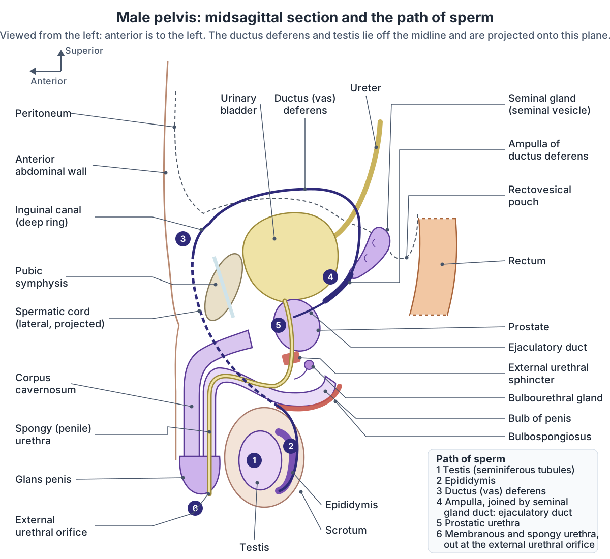
In order, sperm travel through the seminiferous tubules, rete testis and efferent ductules, the epididymis, the ductus deferens, the ejaculatory duct and finally the urethra, which carries them through the prostate and penis to the outside. (OpenStax Anatomy and Physiology 2e, 27.1: Anatomy and Physiology of the Testicular Reproductive System.)
The ductus deferens (vas deferens) is a paired muscular tube about 30 to 45 cm long. It continues from the tail of the epididymis, ascends behind the testis in the posterior part of the spermatic cord, passes through the inguinal canal and enters the abdomen at the deep inguinal ring, where it hooks around the inferior epigastric artery. (Radiopaedia: Ductus deferens.)
Inside the pelvis it crosses the external iliac vessels, runs backwards along the side wall of the pelvis, and crosses above the ureter to reach the back of the bladder. There it widens into the spindle-shaped ampulla, which lies medial to the seminal gland. The ampulla joins the duct of the seminal gland to form the ejaculatory duct, which passes through the prostate and opens into the prostatic urethra. (Radiopaedia: Ductus deferens.)
The wall of the ductus deferens has three layers of smooth muscle (inner longitudinal, middle circular and outer longitudinal), which contract strongly to move sperm during ejaculation. (TeachMeAnatomy: The Spermatic Cord.)
A vasectomy is a contraceptive operation that ties (ligates) both ductus deferentes. Rarely, the ductus deferens is absent from birth (agenesis), which is often associated with absence of the seminal gland and of a kidney. (Radiopaedia: Ductus deferens.)
Spermatic cord
The spermatic cord runs from the deep inguinal ring, lateral to the inferior epigastric vessels, through the inguinal canal and out of the superficial inguinal ring, to end at the back of the testis. (TeachMeAnatomy: The Spermatic Cord.)
It has three coverings, each taken from a layer of the abdominal wall as the testis descended: the external spermatic fascia (from the external oblique aponeurosis), the cremaster muscle and fascia (from the internal oblique) and the internal spermatic fascia (from the transversalis fascia). (TeachMeAnatomy: The Spermatic Cord.)
Its contents include the ductus deferens, three arteries (testicular artery, cremasteric artery and artery to the ductus deferens), the pampiniform plexus of veins, the genital branch of the genitofemoral nerve, autonomic nerves, lymphatic vessels and the closed remnant of the processus vaginalis. The pampiniform plexus acts as a counter-current heat exchanger that cools arterial blood before it reaches the testis. (TeachMeAnatomy: The Spermatic Cord.)
Stroking the upper inner thigh makes the cremaster contract and lift the testis on that side. This cremasteric reflex uses the ilioinguinal nerve as its sensory limb and the genital branch of the genitofemoral nerve as its motor limb. (TeachMeAnatomy: The Spermatic Cord.)
Seminal gland (seminal vesicle)
The official English term in Terminologia Anatomica is seminal gland, with seminal vesicle as a synonym. The older name is discouraged because the organ is not a reservoir for semen but a gland that secretes a large part of the seminal plasma. (IFAA Terminologia Anatomica (TA98): Seminal gland.)
Each seminal gland is a tightly coiled tube about 4 to 5 cm long (10 to 15 cm if stretched out). It lies behind the base of the bladder and above and behind the prostate, with the ampulla of the ductus deferens on its medial side. Its upper tip is covered by the peritoneum of the rectovesical pouch. (Radiopaedia: Seminal vesicle.)
The seminal glands produce about 60 percent of the seminal fluid. Their secretion is rich in fructose, which supports sperm survival. (Radiopaedia: Seminal vesicle.)
TeachMeAnatomy gives a higher share, about 70 percent of semen volume, and lists the components: an alkaline fluid that neutralises acidity in the urethra and vagina, fructose for energy, prostaglandins, and clotting factors that help semen stay in the female tract. (TeachMeAnatomy: The Seminal Vesicles.)
The glands are separated from the rectum by the rectovesical pouch and the rectoprostatic fascia, and their arteries come from branches of the internal iliac artery (the inferior vesical, internal pudendal and middle rectal arteries). (TeachMeAnatomy: The Seminal Vesicles.)
Prostate anatomy and zones
The prostate is a single gland about the size of a walnut. It sits at the base of the bladder, in front of the rectum. It secretes an alkaline, milky fluid that helps semen coagulate and then liquefy after ejaculation. (OpenStax Anatomy and Physiology 2e, 27.1: Anatomy and Physiology of the Testicular Reproductive System.)
The prostate surrounds the proximal urethra. It is shaped like an upside-down pyramid: the base at the top is continuous with the bladder, and the apex at the bottom ends at the urogenital diaphragm. Behind, it is separated from the rectum by Denonvilliers' fascia. Radiopaedia gives an average size of about 3 × 4 × 2 cm. The two ejaculatory ducts pierce its posterior surface and run through it to the prostatic urethra. (Radiopaedia: Prostate.)
Clinicians divide the gland into zones (McNeal's zones) rather than lobes. TeachMeAnatomy gives the peripheral zone at the back as the largest, about 65 percent of the volume; the central zone around the ejaculatory ducts as about 25 percent; and the transition zone around the urethra as about 5 to 10 percent. The anterior fibromuscular stroma in front has no glands and is sometimes counted as a fourth zone. (TeachMeAnatomy: The Prostate Gland.)
Radiopaedia gives slightly different shares (peripheral zone about 70 percent, transition zone about 5 percent in a young adult). About 70 percent of prostate cancers arise in the peripheral zone and about 20 percent in the transition zone. Benign prostatic hyperplasia (BPH) arises in the transition zone, which is why it squeezes the urethra. (Radiopaedia: Prostate.)
Because the peripheral zone faces the rectum, it is the part felt in a digital rectal examination. Prostatic ducts open into the prostatic urethra on each side of the seminal colliculus (verumontanum). The nerves for erection run in neurovascular bundles beside the gland, which surgeons try to spare during radical prostatectomy. (TeachMeAnatomy: The Prostate Gland.)
Zones of the prostate
| Zone | Approximate share | Position | Clinical note |
|---|---|---|---|
| Peripheral zone | 65 to 70% | Back and sides, facing the rectum | Site of about 70% of cancers; felt on rectal examination |
| Central zone | About 25% | Base of the gland, around the ejaculatory ducts | Ducts enter the urethra obliquely |
| Transition zone | 5 to 10% | Around the proximal urethra | Site of benign prostatic hyperplasia and about 20% of cancers |
| Anterior fibromuscular stroma | Non-glandular | Front of the gland | Fibromuscular tissue, no glands |
Percentages differ slightly between TeachMeAnatomy and Radiopaedia; both ranges are shown.
Bulbourethral (Cowper) glands
The bulbourethral glands, also called Cowper's glands, are two pea-shaped glands in the deep perineal pouch. They lie behind and to the side of the membranous urethra, just above the bulb of the penis. Their ducts pierce the perineal membrane and open into the first part of the spongy urethra. (TeachMeAnatomy: The Bulbourethral Glands.)
During sexual arousal they release a thick, alkaline mucus that lubricates the urethra and helps clear urine residue from it before ejaculation. (TeachMeAnatomy: The Bulbourethral Glands.)
This pre-ejaculate fluid can pick up sperm already present in the urethra. (OpenStax Anatomy and Physiology 2e, 27.1: Anatomy and Physiology of the Testicular Reproductive System.)
Penis anatomy: root, body and glans
The penis has three parts. The root is the fixed part in the superficial perineal pouch; it is not visible from outside and contains the two crura, the bulb and two pairs of muscles. The body (shaft) is the free part, hanging from the pubic symphysis. The glans is the cone-shaped end, formed by the expanded end of the corpus spongiosum, and carries the external urethral orifice. (TeachMeAnatomy: The Penis.)
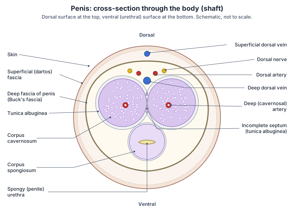
The body contains three columns of erectile tissue. The two corpora cavernosa continue forwards from the crura, which are attached to the ischial (ischiopubic) ramus on each side, and form the dorsal part of the penis. The corpus spongiosum continues forwards from the midline bulb, lies on the ventral (urethral) side and carries the urethra. It fills at lower pressure than the corpora cavernosa, so the urethra is not squeezed shut during erection. (TeachMeAnatomy: The Penis.)
The glans has a raised rim, the corona. The prepuce (foreskin) covers the glans, and the frenulum is a small fold on its urethral (underside) aspect. The paired corpora cavernosa are separated by a median fibrous septum, and the corpus spongiosum lies in a groove on their ventral surface. (Radiopaedia: Penis.)
Two ligaments hold the penis to the front of the pelvis: the suspensory ligament, a condensation of deep fascia attached to the pubic symphysis, and the fundiform ligament, which runs down from the linea alba and slings around the penis. (TeachMeAnatomy: The Penis.)
Cross-section of the penis: layers, vessels and nerves
From the outside in, a section through the shaft shows skin, loosely attached to the fascia below; a superficial fascia (Colles' fascia, continuous with Scarpa's fascia of the abdominal wall); the deep fascia of the penis (Buck's fascia), which holds all three erectile bodies together; and the tunica albuginea, a capsule around each corpus cavernosum that fuses in the midline. (TeachMeAnatomy: The Penis.)
Kenhub calls the superficial layer the dartos fascia and notes that it carries the superficial veins, while the deep dorsal vein, dorsal arteries and dorsal nerves run together in the dorsal groove beneath Buck's fascia. The tunica albuginea has an inner circular layer around each corpus cavernosum and an outer longitudinal layer around both, and forms an incomplete septum between them. A deep (cavernosal) artery runs through the centre of each corpus cavernosum and gives off helicine branches that open into its blood spaces (sinusoids). (Kenhub: Penis.)
In the classic description (Gray's Anatomy), each dorsal artery lies between the deep dorsal vein, medially, and the dorsal nerve, on its lateral side. So, from the midline outwards, the order is vein, artery, nerve. (IMAIOS e-Anatomy: Dorsal artery of penis.)
Recent anatomical studies suggest the venous pattern is more complex than the classic picture of one deep dorsal vein flanked by two arteries, with additional cavernosal and para-arterial veins between the tunica albuginea and Buck's fascia. (Hsieh CH et al. Advances in understanding of mammalian penile evolution, human penile anatomy and human erection physiology. Med Sci Monit. 2012.)
Blood supply and nerves of the penis
All the arteries of the penis come from the internal pudendal artery, a branch of the anterior division of the internal iliac artery. There are three: the dorsal arteries, the deep (cavernosal) arteries and the artery of the bulb (bulbourethral artery). (TeachMeAnatomy: The Penis.)
The deep dorsal vein drains the cavernous spaces of the erectile tissue and empties into the prostatic venous plexus. The superficial dorsal veins drain superficial structures such as the skin. (TeachMeAnatomy: The Penis.)
Sensation comes through the dorsal nerve of the penis, the terminal branch of the pudendal nerve (S2 to S4). Autonomic cavernous nerves from the pelvic (inferior hypogastric) plexus supply the erectile tissue: parasympathetic fibres (S2 to S4) cause erection, and sympathetic fibres (T11 to L2) cause ejaculation and the return to the flaccid state. (TeachMeAnatomy: The Penis.)
During erection, nerve endings and endothelial cells release nitric oxide, which relaxes the smooth muscle of the penile arteries. Blood fills the erectile chambers, which press on the thin-walled veins and reduce outflow. (OpenStax Anatomy and Physiology 2e, 27.1: Anatomy and Physiology of the Testicular Reproductive System.)
Lymph from the penile skin drains to the superficial inguinal nodes, from the glans to the deep inguinal and external iliac nodes, and from the erectile tissue and penile urethra to the internal iliac nodes. (TeachMeAnatomy: The Penis.)
Bulbospongiosus and ischiocavernosus muscles
The bulbospongiosus (musculus bulbospongiosus) lies in the superficial perineal pouch and covers the bulb of the penis. It arises from the perineal body and the median raphe, and its fibres wrap around the bulb and corpus spongiosum to insert on the dorsum of the penis and the perineal membrane. It is supplied by the pudendal nerve and the perineal artery. (Radiopaedia: Bulbospongiosus muscle.)
In men the bulbospongiosus empties the last urine and semen from the spongy urethra, and in the final stages of erection it compresses the veins of the bulb. In women the same muscle covers the bulbs of the vestibule and narrows the vaginal orifice. (Radiopaedia: Bulbospongiosus muscle.)
The ischiocavernosus muscles cover the crura on each side. They push blood from the crura into the corpora cavernosa and help maintain erection. Both pairs of muscles are supplied by the pudendal nerve. (TeachMeAnatomy: The Penis.)
The pudendal nerve leaves the pelvis through the greater sciatic foramen, re-enters through the lesser sciatic foramen and runs forwards in the pudendal (Alcock's) canal before dividing into the inferior rectal nerve, the perineal nerve and the dorsal nerve of the penis. Besides bulbospongiosus and ischiocavernosus, it supplies the external urethral sphincter, the external anal sphincter and part of levator ani. (TeachMeAnatomy: The Pudendal Nerve.)
The male urethra
The male urethra is about 15 to 20 cm long and carries both urine and semen. The prostatic part runs through the prostate; it is the widest part and receives the ejaculatory and prostatic ducts. The membranous part passes through the deep perineal pouch, is surrounded by the external urethral sphincter and is the narrowest part. The spongy (penile) part runs through the bulb and corpus spongiosum, receives the bulbourethral gland ducts and widens into the navicular fossa in the glans before ending at the external urethral orifice. (TeachMeAnatomy: The Urethra.)
The external urethral sphincter is skeletal muscle under voluntary control. In men it forms a ring around the membranous urethra just below the prostate. The internal urethral sphincter at the bladder neck is smooth muscle; during orgasm sympathetic nerves contract it so that semen does not flow backwards into the bladder (retrograde ejaculation). (Kenhub: Urethral sphincters.)
Clinical relevance
Testicular torsion is a twist of the spermatic cord that can cut off the testicular artery. It causes sudden severe testicular pain, with the testis often lying higher than normal, and is a surgical emergency because a delay of a few hours can cause loss of the testis. Ultrasound with colour Doppler is used to assess it. (TeachMeAnatomy: The Spermatic Cord.)
A varicocele is a dilated pampiniform plexus. It is more common on the left, because the left testicular vein is longer and joins the left renal vein at a right angle. A hydrocele is fluid in the tunica vaginalis and, unlike blood, lets light shine through (transillumination). (TeachMeAnatomy: The Testes and Epididymis.)
Benign prostatic hyperplasia becomes more common with age and causes storage symptoms (frequency, urgency, waking at night to pass urine) and voiding symptoms (hesitancy and a weak stream). Prostate cancer often causes few symptoms early on; PSA, a protease made by the prostate, is used as a tumour marker but also rises with inflammation and other causes. (TeachMeAnatomy: The Prostate Gland.)
Phimosis is a foreskin too tight to pull back over the glans. Priapism is a painful erection lasting without stimulation; more than four hours is an emergency. Erectile dysfunction is most often vascular in origin. (TeachMeAnatomy: The Penis.)
Male reproductive system in the 3D model
- Corpus cavernosum of penis
- The corpora cavernosa are the paired columns of erectile tissue forming the dorsal part of the penis.
- Corpus spongiosum of penis
- The corpus spongiosum is the single column of erectile tissue surrounding the penile urethra.
- Deep dorsal vein of penis
- The deep dorsal vein is the main vein of the penis, draining the glans and erectile bodies.
- Deferent duct
- The deferent duct (vas deferens) is a thick-walled muscular tube carrying sperm from the epididymis toward the urethra.
- Dorsal artery of penis
- The dorsal artery of the penis supplies the skin and glans.
- Epididymis
- The epididymis is a coiled tube on the posterior testis where sperm mature and are stored.
- Glans penis
- The glans penis is the cone-shaped distal end of the penis.
- Prostate
- The prostate is a walnut-sized gland surrounding the first part of the male urethra.
- Seminal vesicle
- The seminal vesicles are paired glands that produce much of the fluid of semen.
- Superficial dorsal vein of penis
- A vein draining the skin of the penis.
- Testicular artery
- A paired branch of the abdominal aorta to the testis.
- Testicular vein
- A vein draining the testis.
- Testis
- The testis is the male gonad that produces sperm and testosterone.
- Urethra
- The urethra is the tube carrying urine out of the bladder; in males it also carries semen.
Frequently asked questions
What is the path of sperm in the male reproductive system?
Sperm form in the seminiferous tubules of the testis, pass through the rete testis and efferent ductules into the epididymis, where they mature and are stored. At ejaculation they travel along the ductus (vas) deferens, through the ejaculatory duct inside the prostate, and then through the prostatic, membranous and spongy parts of the urethra to the external urethral orifice.
What is the difference between the testicles and the testes?
There is no difference: testicle is the everyday word and testis (plural testes) is the anatomical term. Each testis is about 4 to 5 cm long, lies in the scrotum, and produces both sperm, in the seminiferous tubules, and testosterone, in the Leydig cells between the tubules.
What is the ductus deferens?
The ductus deferens, also called the vas deferens, is a muscular tube about 30 to 45 cm long on each side. It carries sperm from the tail of the epididymis up the spermatic cord, through the inguinal canal, over the ureter and behind the bladder, where its ampulla joins the seminal gland duct to form the ejaculatory duct.
Where is the prostate located?
The prostate lies in the pelvis just below the bladder, behind the pubic symphysis and in front of the rectum. It surrounds the first part of the urethra. Because its peripheral zone faces the rectum, a doctor can feel it with a gloved finger during a digital rectal examination.
What are the three erectile bodies of the penis?
The penis has two corpora cavernosa, which lie side by side on the dorsal side and make up most of the shaft, and one corpus spongiosum on the ventral side, which surrounds the urethra and expands into the glans at the tip and the bulb at the root. Each corpus cavernosum is wrapped in tunica albuginea and contains a deep artery.
What does the bulbospongiosus muscle do?
The bulbospongiosus covers the bulb of the penis in the superficial perineal pouch. It contracts to empty the last drops of urine and semen from the spongy urethra and, by compressing the veins of the bulb, helps maintain erection. It is supplied by the pudendal nerve. In women it covers the vestibular bulbs and narrows the vaginal opening.
What is the seminal gland?
Seminal gland is the current Terminologia Anatomica name for the seminal vesicle. The two seminal glands are coiled tubes about 5 cm long behind the bladder. They do not store sperm; they secrete an alkaline, fructose-rich fluid that forms about 60 to 70 percent of semen. Each gland's duct joins the ductus deferens to form an ejaculatory duct.
References
- OpenStax. Anatomy and Physiology 2e, §27.1 Anatomy and Physiology of the Testicular Reproductive System. Rice University, 2022.
- Jones O. The Scrotum. TeachMeAnatomy.
- Jones O. The Testes and Epididymis. TeachMeAnatomy.
- Jones O. The Spermatic Cord. TeachMeAnatomy.
- Jones O. The Prostate Gland. TeachMeAnatomy.
- Jones O. The Seminal Vesicles. TeachMeAnatomy.
- Jones O. The Bulbourethral Glands. TeachMeAnatomy.
- Jones O. The Penis. TeachMeAnatomy.
- Jones O. The Urethra. TeachMeAnatomy.
- Jones O. The Pudendal Nerve. TeachMeAnatomy.
- Ductus deferens. Radiopaedia reference article.
- Prostate. Radiopaedia reference article.
- Seminal vesicle. Radiopaedia reference article.
- Penis. Radiopaedia reference article.
- Bulbospongiosus muscle. Radiopaedia reference article.
- Penis. Kenhub anatomy library.
- Urethral sphincters. Kenhub anatomy library.
- Dorsal artery of penis. IMAIOS e-Anatomy (text adapted from Gray's Anatomy, 1918).
- Federative International Programme for Anatomical Terminology. Terminologia Anatomica (TA98): 09.3.06.001 Seminal gland. IFAA.
- Hsieh CH, Liu SP, Hsu GL, et al. Advances in understanding of mammalian penile evolution, human penile anatomy and human erection physiology: clinical implications for physicians and surgeons. Med Sci Monit. 2012;18(7):RA118-RA125.
- Mitsuhashi N. et al. (2009). BodyParts3D: 3D structure database for anatomical concepts. Nucleic Acids Research 37, D782–D785. Data © The Database Center for Life Science, CC BY 4.0.
Related anatomy
- Female reproductive systemLabelled female reproductive system diagrams: uterus anatomy and layers, uterine tube parts, ovaries, ligaments…
- PelvisPelvis bones explained: ilium, ischium and pubis, obturator foramen, pelvic ligaments and male vs female pelvis, with…
- ProstateProstate: The prostate is a walnut-sized gland surrounding the first part of the male urethra. See where it is in an…
- TestisTestis: The testis is the male gonad that produces sperm and testosterone. See where it is in an interactive 3D human…
- UreterWhat do the ureters do? Ureter function, course, relations, the three narrowings where kidney stones lodge, blood…
- KidneyWhere are your kidneys? Kidney location on the back (T12 to L3), coverings, relations, cortex, medulla, calyces, renal…
Explore the whole body in the interactive 3D human anatomy model, or browse the A–Z list of human structures.