

What bones make up the pelvis?
The bony pelvis consists of the right and left hip bones, the sacrum and the coccyx. The two hip bones meet each other at the front, and at the back each one joins the sacrum. Together the two hip bones form the pelvic girdle, which attaches each lower limb to the axial skeleton through the sacrum. (OpenStax Anatomy and Physiology 2e, 8.3: The Pelvic Girdle and Pelvis.)
Each adult hip bone (also called the coxal, innominate or pelvic bone) is made of the ilium, ischium and pubis. In a child these three parts are separate and are joined by a Y-shaped triradiate cartilage. TeachMeAnatomy gives fusion as beginning at about 15 to 17 years, while OpenStax describes it as happening during the late teenage years. (TeachMeAnatomy: The Hip Bone (Ilium, Ischium, Pubis).)
The three parts meet at the acetabulum, the deep cup on the outer surface of the hip bone that receives the head of the femur to form the hip joint. The hip bone takes part in three joints: the sacroiliac joint at the back, the pubic symphysis at the front and the hip joint at the side. (Kenhub: Hip bone.)
In the 3D model the hip bones and sacrum are highlighted. The model is based on BodyParts3D, an adult male data set, and it does not include the coccyx or the pelvic ligaments, so the ligaments are described in the text and table below.
Ilium: the largest part of the hip bone
The ilium is the fan-shaped upper part and the largest part of the hip bone. Its curved upper margin is the iliac crest, which you can feel at the waistline. The crest ends at the front in the anterior superior iliac spine (ASIS) and at the back in the posterior superior iliac spine (PSIS). Below each of these lies a smaller spine: the anterior inferior iliac spine (AIIS) and the posterior inferior iliac spine (PIIS). (OpenStax Anatomy and Physiology 2e, 8.3: The Pelvic Girdle and Pelvis.)
The ilium has a smaller lower body, which forms part of the acetabulum, and a broad upper wing (ala). The iliac tubercle lies on the outer lip of the crest towards its front end. The anterior inferior iliac spine, just above the acetabulum, gives origin to the straight head of rectus femoris and to the iliofemoral ligament of the hip. The outer gluteal surface is marked by the anterior, posterior and inferior gluteal lines, and on the inner surface the iliac tuberosity gives attachment to ligaments. (Kenhub: Hip bone.)
The inner surface of the wing is the shallow iliac fossa, where iliacus arises. The outer gluteal surface is convex and gives attachment to the gluteal muscles. (TeachMeAnatomy: The Hip Bone (Ilium, Ischium, Pubis).)
Behind the iliac fossa is the large, roughened auricular surface, which joins the sacrum at the sacroiliac joint. The arcuate line is a ridge at the lower edge of the iliac fossa, and the greater sciatic notch is the large U-shaped notch on the back edge of the lower ilium. (OpenStax Anatomy and Physiology 2e, 8.3: The Pelvic Girdle and Pelvis.)
Ischium: the bone you sit on
The ischium forms the back and lower part of the hip bone. It has a body, which forms part of the acetabulum, and a ramus. The ischial ramus joins the inferior pubic ramus; the two together are called the ischiopubic ramus, which forms the lower border of the obturator foramen. (Kenhub: Hip bone.)
The ischial ramus is a narrow bar of bone that projects forward and upward from the ischial tuberosity. Above the tuberosity, the slightly curved back edge of the ischium is the lesser sciatic notch. (OpenStax Anatomy and Physiology 2e, 8.3: The Pelvic Girdle and Pelvis.)
The ischial tuberosity is the large, rough lump at the bottom of the ischium. It bears the body's weight when you sit and gives attachment to the hamstrings (semimembranosus, semitendinosus and the long head of biceps femoris), to adductor magnus and to the sacrotuberous ligament. (Kenhub: Hip bone.)
The ischial spine is a pointed projection on the back edge of the ischium. It separates the greater sciatic notch above from the lesser sciatic notch below, and the sacrospinous ligament attaches to it. (OpenStax Anatomy and Physiology 2e, 8.3: The Pelvic Girdle and Pelvis.)
Pubis (os pubis): body, rami and pecten pubis
The pubis is the front part of the hip bone. It has a body and two arms, the superior and inferior pubic rami. The rounded upper border of the body is the pubic crest, which ends laterally at the pubic tubercle, the medial attachment of the inguinal ligament. (Kenhub: Hip bone.)
The superior pubic ramus runs laterally from the body to the acetabulum, where it joins the ilium. A ridge along its upper edge is the pecten pubis (pectineal line). The pecten pubis continues into the arcuate line of the ilium, and together with the pubic crest and the sacral promontory these ridges form the pelvic brim. (Kenhub: Hip bone.)
The inferior pubic ramus (the lower ramus of the pubic bone) joins the ischial ramus below the obturator foramen. The inferior pubic ramus gives attachment to adductor magnus, adductor brevis and gracilis. (Kenhub: Hip bone.)
The two pubic bodies meet in the midline at the pubic symphysis. The pubic arch is formed by the symphysis, the pubic bodies and the inferior pubic rami, and the angle between the two ischiopubic rami below the symphysis is the subpubic angle. (OpenStax Anatomy and Physiology 2e, 8.3: The Pelvic Girdle and Pelvis.)
Parts of the hip bone and their main landmarks
| Part | Position | Main landmarks |
|---|---|---|
| Ilium | Upper, largest part | Iliac crest, ASIS, AIIS, PSIS, PIIS, iliac fossa, gluteal lines, auricular surface, arcuate line, greater sciatic notch |
| Ischium | Back and lower part | Ischial tuberosity, ischial spine, lesser sciatic notch, ischial ramus |
| Pubis | Front part | Body, pubic crest, pubic tubercle, superior ramus with pecten pubis, inferior ramus, symphyseal surface |
| All three | Meet at the side of the hip bone | Acetabulum; ischium and pubis surround the obturator foramen |
Acetabulum and obturator foramen
The acetabulum has a smooth, crescent-shaped lunate surface that articulates with the femoral head, and a central rough acetabular fossa that does not. Its rim is incomplete below at the acetabular notch. In life the socket is deepened by the acetabular labrum and the notch is bridged by the transverse acetabular ligament. (Kenhub: Hip bone.)
The obturator foramen is the large opening in the lower front of the hip bone, between the pubis and ischium. It is largely filled by a sheet of connective tissue (the obturator membrane) that gives attachment to muscles on both its inner and outer surfaces. (OpenStax Anatomy and Physiology 2e, 8.3: The Pelvic Girdle and Pelvis.)
A small gap at the top of the membrane, the obturator canal, carries the obturator nerve, artery and vein from the pelvic cavity to the medial thigh. (Kenhub: Hip bone.)
Sacroiliac joint, pubic symphysis and pelvic ligaments
The sacroiliac joint is a synovial joint between the ala of the sacrum and the auricular surface of the ilium. Its fibrous capsule is thickened in front and behind to form the sacroiliac ligaments, and only limited movement is possible. (Texas Tech University HSC El Paso, Anatomy Tables: Joints and Ligaments of the Pelvis & Perineum.)
The pubic symphysis is a secondary cartilaginous joint. The joint surfaces are covered with hyaline cartilage and joined by fibrocartilage. It is normally almost immobile but loosens during pregnancy, as do the ligaments of the sacroiliac joints. (Kenhub: The pelvis.)
The iliolumbar ligament runs from the transverse process of the fifth lumbar vertebra (L5) to the iliac crest and stabilises the lumbosacral joint, the joint between L5 and the first sacral segment. (Texas Tech University HSC El Paso, Anatomy Tables: Joints and Ligaments of the Pelvis & Perineum.)
The sacrospinous ligament runs from the lateral sacrum and coccyx to the ischial spine. The sacrotuberous ligament runs from the same area to the ischial tuberosity. Together they turn the greater and lesser sciatic notches into the greater and lesser sciatic foramina. (Texas Tech University HSC El Paso, Anatomy Tables: Joints and Ligaments of the Pelvis & Perineum.)
The sacrotuberous ligament is a thick band that also arises from the back of the ilium. Its main job is to stabilise the sacroiliac joint, and its lower edge forms part of the boundary of the pelvic outlet. (Kenhub: Sacrotuberous ligament.)
Piriformis, the sciatic nerve, the superior and inferior gluteal vessels and nerves, the pudendal nerve and the internal pudendal vessels leave the pelvis through the greater sciatic foramen. The tendon of obturator internus, its nerve, the pudendal nerve and the internal pudendal vessels pass through the lesser sciatic foramen. (Kenhub: The pelvis.)
Ligaments of the pelvis
| Ligament | Attachments | Role |
|---|---|---|
| Iliolumbar | Transverse process of L5 to iliac crest | Stabilises the lumbosacral joint |
| Sacroiliac (anterior and posterior) | Thickenings of the sacroiliac joint capsule | Support the sacroiliac joint |
| Sacrospinous | Lateral sacrum and coccyx to ischial spine | Forms the greater and lesser sciatic foramina |
| Sacrotuberous | Posterior ilium, lateral sacrum and coccyx to ischial tuberosity | Stabilises the sacroiliac joint; borders the pelvic outlet |
Attachments from Texas Tech anatomy tables, Kenhub and OpenStax.
Pelvic inlet, outlet, true and false pelvis
The pelvic brim divides the pelvis into two parts. Above it is the greater (false) pelvis, which belongs to the abdominal cavity and holds parts of the small and large intestines. Below it is the lesser (true) pelvis, which contains the bladder and other pelvic organs. (OpenStax Anatomy and Physiology 2e, 8.3: The Pelvic Girdle and Pelvis.)
The pelvic inlet is bounded by the sacral promontory and the sacral alae behind, the arcuate and pectineal lines at the sides and the pubic symphysis in front. The pelvic outlet is bounded by the tip of the coccyx behind, the ischial tuberosities and the lower edge of the sacrotuberous ligaments at the sides, and the pubic arch in front. (TeachMeAnatomy: The Pelvic Girdle.)
In the anatomical position the pelvis is tilted forward, so that the anterior superior iliac spines and the pubic tubercles lie in the same vertical plane. (OpenStax Anatomy and Physiology 2e, 8.3: The Pelvic Girdle and Pelvis.)
Male vs female pelvis: the key differences
The female pelvis is adapted for childbirth. It is wider, its bones are lighter, the distance between the anterior superior iliac spines is greater and the ischial tuberosities are farther apart, which enlarges the pelvic outlet. The male pelvis has thicker, heavier bones. (OpenStax Anatomy and Physiology 2e, 8.3: The Pelvic Girdle and Pelvis.)
The subpubic angle is usually greater than 80 degrees in females and less than 70 degrees in males. The pelvic inlet is round or oval in females and heart-shaped in males, and the greater sciatic notch is broader in females and narrower and deeper in males. (OpenStax Anatomy and Physiology 2e, 8.3: The Pelvic Girdle and Pelvis.)
The female lesser pelvis is shorter, wider and shallower, while the male lesser pelvis is longer, narrower and deeper. The female sacrum is shorter and wider, and OpenStax describes it as less curved, with a promontory that projects less into the pelvic cavity. Because of all these features, the hip bone is the most reliable bone for estimating sex from a skeleton. (OpenStax Anatomy and Physiology 2e, 8.3: The Pelvic Girdle and Pelvis.)
Sources do not agree on the curve of the female sacrum. TeachMeAnatomy describes the typical female (gynaecoid) sacrum as shorter and more curved with a less pronounced promontory, and gives the female subpubic arch as over 80 to 90 degrees. Both sources agree that the female inlet is oval and the male inlet heart-shaped. (TeachMeAnatomy: The Pelvic Girdle.)
Obstetricians also group pelvic shapes into four types. The gynaecoid type, with a round or slightly oval inlet and a wide subpubic angle, is the most common in women. The android type, with a heart-shaped inlet and a narrow subpubic angle, is the most common in men. The anthropoid type has an inlet that is longer from front to back, and the rare platypelloid type has a flat, wide inlet. (Kenhub: The pelvis.)
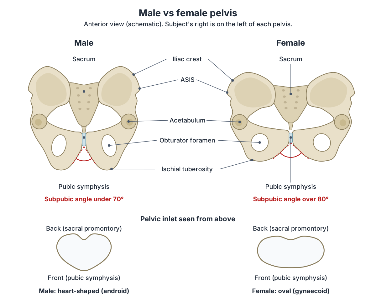
Male vs female pelvis
| Feature | Typical male | Typical female |
|---|---|---|
| Bones | Thicker, heavier | Lighter, wider |
| Subpubic angle | Less than 70° | Greater than 80° |
| Pelvic inlet | Heart-shaped | Round or oval |
| Greater sciatic notch | Narrow and deep | Broad |
| Lesser pelvis | Longer, narrower, deeper | Shorter, wider, shallower |
| Ischial tuberosities | Closer together | Farther apart |
| Sacrum | Longer, narrower | Shorter, wider |
Based on OpenStax Anatomy and Physiology 2e, §8.3. Individuals vary and many pelves fall between these patterns.
Pelvic bone labelling: landmarks you can feel
Several pelvic landmarks can be felt through the skin. The iliac crest lies at the waistline, the ASIS at the front of the hip, and the PSIS often shows as a dimple in the lower back. The ischial tuberosities take your weight when you sit. (OpenStax Anatomy and Physiology 2e, 8.3: The Pelvic Girdle and Pelvis.)
Clinicians use these points for measurements. The mid-inguinal point, where the femoral pulse is felt, lies halfway between the ASIS and the pubic symphysis, while the midpoint of the inguinal ligament lies halfway between the ASIS and the pubic tubercle. True leg length is measured from the ASIS to the medial malleolus. (TeachMeAnatomy: The Hip Bone (Ilium, Ischium, Pubis).)
Because the iliac crest lies just under the skin, it is a common site for bone marrow biopsy, in which a needle is passed through the cortex of the crest to sample marrow. (Kenhub: Hip bone.)
Pelvic fractures
Fractures of the pubic rami are common in older people with osteoporosis after a simple fall from standing. When they are the only injury they are usually treated without surgery and take about 6 to 8 weeks to heal. (TeachMeAnatomy: The Hip Bone (Ilium, Ischium, Pubis).)
High-energy injuries, such as road traffic collisions, can break the ring of the pelvis at the acetabulum and sacroiliac joint. These fractures may be unstable, can injure the bladder and urethra, and can cause life-threatening bleeding. In major trauma a pelvic binder is placed around the hips at the level of the greater trochanters to stabilise the pelvis and limit bleeding. (TeachMeAnatomy: The Hip Bone (Ilium, Ischium, Pubis).)
Pelvis in the 3D model
Frequently asked questions
What are the three bones of the hip bone?
Each hip bone is made of the ilium, ischium and pubis. The ilium is the large upper wing, the ischium is the lower back part you sit on, and the pubis is the front part that meets the other side at the pubic symphysis. They are separate in children, joined by triradiate cartilage, and fuse at the acetabulum during the teenage years.
What is the difference between the pelvis and the hip bone?
A hip bone is one of the paired bones on each side, formed from the fused ilium, ischium and pubis. The pelvis is the whole bony ring: both hip bones, the sacrum and the coccyx. The two hip bones alone make up the pelvic girdle, which attaches the legs to the spine through the sacrum.
What passes through the obturator foramen?
The obturator foramen is almost closed by the obturator membrane. Only a small gap at its top, the obturator canal, lets structures through: the obturator nerve, artery and vein, which travel from the pelvis to the muscles and skin of the medial thigh. The membrane itself gives attachment to the obturator muscles.
How can you tell a male pelvis from a female pelvis?
Look at the subpubic angle and the pelvic inlet. In a typical female pelvis the subpubic angle is wider than 80 degrees and the inlet is round or oval. In a typical male pelvis the angle is under 70 degrees and the inlet is heart-shaped. The female pelvis is also wider and shallower, with broader greater sciatic notches.
What is the subpubic angle?
The subpubic angle is the angle formed below the pubic symphysis where the two ischiopubic rami meet. It is one of the clearest sex differences in the skeleton: usually greater than 80 degrees in females and less than 70 degrees in males, according to OpenStax. A wider angle gives a larger pelvic outlet for childbirth.
What do the sacrospinous and sacrotuberous ligaments do?
Both ligaments run from the side of the sacrum and coccyx to the ischium: the sacrospinous to the ischial spine and the sacrotuberous to the ischial tuberosity. They stabilise the sacroiliac joint and turn the sciatic notches into the greater and lesser sciatic foramina, through which nerves and vessels such as the sciatic and pudendal nerves pass.
References
- OpenStax. Anatomy and Physiology 2e, §8.3 The Pelvic Girdle and Pelvis. Rice University, 2022.
- TeachMeAnatomy. The Hip Bone: Ilium, Ischium, Pubis.
- TeachMeAnatomy. The Pelvic Girdle: Structure, Function, Assessment.
- Kenhub. Hip bone.
- Kenhub. The pelvis.
- Kenhub. Sacrotuberous ligament.
- Texas Tech University Health Sciences Center El Paso. Anatomy Tables: Joints and Ligaments of the Pelvis & Perineum.
- Mitsuhashi N. et al. (2009). BodyParts3D: 3D structure database for anatomical concepts. Nucleic Acids Research 37, D782–D785. Data © The Database Center for Life Science, CC BY 4.0.
Related anatomy
- Hip boneHip bone: The large irregular bone of the pelvis formed by the fusion of the ilium, ischium and pubis. See where it is…
- SacrumSacrum anatomy explained: promontory, ala, sacral foramina, crests and hiatus, joints and ligaments, sacrum vs coccyx…
- FemurFemur bone labelled: head, neck, trochanters, linea aspera, condyles and epicondyles, with muscle attachments, blood…
- Hip musclesMuscles of the hip explained: gluteus maximus, medius and minimus, TFL, piriformis, obturator internus and the other…
- Vertebral columnHuman spine anatomy: cervical, thoracic and lumbar vertebrae, curvatures, discs, facet joints and spinal ligaments…
- Female reproductive systemLabelled female reproductive system diagrams: uterus anatomy and layers, uterine tube parts, ovaries, ligaments…
Explore the whole body in the interactive 3D human anatomy model, or browse the A–Z list of human structures.