

The anatomical position: the starting point
Every plane and directional term is defined from the anatomical position. In this position the body stands upright with the feet at shoulder width and parallel, toes pointing forward, the upper limbs held at the sides and the palms of the hands facing forward. (OpenStax Anatomy and Physiology 2e, 1.6: Anatomical Terminology.)
Anatomists use these terms as if the body were in the anatomical position even when it is not. A patient lying face down (prone) or face up (supine) still has an anterior surface that is the front of the body, and the heart is still superior to the stomach. Right and left always mean the subject's own right and left, not the viewer's. (OpenStax Anatomy and Physiology 2e, 1.6: Anatomical Terminology.)
That last rule matters for every diagram on this page. When you face a person or an anterior view, their right side is on your left. The figures here mark the subject's right with an R so you can check yourself.
What are the anatomical planes?
A plane is an imaginary two-dimensional surface that passes through the body. A section is the flat surface you would see if the body or an organ were actually cut along that plane. There are three basic planes, set at right angles to each other: the sagittal, frontal (coronal) and transverse planes. (OpenStax Anatomy and Physiology 2e, 1.6: Anatomical Terminology.)
The sagittal plane runs vertically and divides the body or an organ into right and left sides. The frontal plane runs vertically at right angles to the sagittal plane and divides the body into an anterior (front) and a posterior (back) portion. The transverse plane runs horizontally and divides the body into upper and lower portions. (OpenStax Anatomy and Physiology 2e, 1.6: Anatomical Terminology.)
Kenhub gives the same three planes with their common synonyms: median or midsagittal for the sagittal plane through the midline, coronal for the frontal plane, and axial or horizontal for the transverse plane, which lies at right angles to both the sagittal and frontal planes. (Kenhub: Anatomical terminology.)
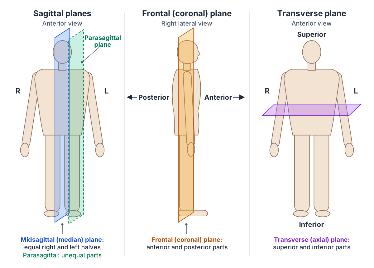
The anatomical planes at a glance
| Plane | Other names | Orientation | Divides the body into |
|---|---|---|---|
| Midsagittal | Median plane | Vertical, through the midline | Equal right and left halves |
| Parasagittal | Sagittal (off midline) | Vertical, parallel to the midsagittal plane | Unequal right and left parts |
| Frontal | Coronal | Vertical, at right angles to the sagittal plane | Anterior and posterior parts |
| Transverse | Axial, horizontal, cross section | Horizontal, at right angles to the other two | Superior and inferior parts |
Sagittal plane: midsagittal vs parasagittal
There are many possible sagittal planes, because a vertical plane can cut the body anywhere from one side to the other. If the plane runs directly down the middle of the body it is the midsagittal or median plane, and it divides the body into equal right and left halves. If it divides the body into unequal right and left sides it is a parasagittal plane. (OpenStax Anatomy and Physiology 2e, 1.6: Anatomical Terminology.)
So the midsagittal plane is one particular sagittal plane, and every other sagittal plane parallel to it is parasagittal. Kenhub describes a sagittal plane as any vertical plane passing through the body parallel to the midline, dividing it into left and right parts; only the one through the centre is the median (midsagittal) plane. (Kenhub: Anatomical terminology.)
The midline defined by the median plane is also the reference for the terms medial and lateral. TeachMeAnatomy describes the midline as a line in the sagittal plane that splits the right and left halves evenly. (TeachMeAnatomy: Anatomical Terms of Location.)
Frontal (coronal) plane
The frontal plane divides the body or an organ into an anterior (front) portion and a posterior (back) portion. It is often called the coronal plane. Like the sagittal plane it is vertical, but it faces the opposite way: it runs from one side of the body to the other. (OpenStax Anatomy and Physiology 2e, 1.6: Anatomical Terminology.)
Kenhub defines the frontal (coronal) plane as a vertical plane at right angles to the sagittal plane. Because it runs from side to side, a single coronal slice can include structures of both the right and left sides of the body. (Kenhub: Anatomical terminology.)
Transverse (axial) plane and the axial view
The transverse plane divides the body or an organ horizontally into upper (superior) and lower (inferior) portions. Images made in transverse planes are called cross sections. (OpenStax Anatomy and Physiology 2e, 1.6: Anatomical Terminology.)
In radiology the transverse plane is usually called the axial or transaxial plane: a horizontal plane perpendicular to the long axis of the body. Radiopaedia notes that some authors keep the two words apart, using axial for the plane perpendicular to the long axis of the CT or MRI scanner and transverse for the plane perpendicular to the long axis of the body. The two are usually the same, but not when a patient cannot lie in the standard position. (Radiopaedia: Radiographic positioning terminology.)
When someone searches for an axial view, they almost always mean this kind of horizontal slice, such as an axial CT image through the abdomen. The same word appears elsewhere in anatomy, so do not confuse an axial view with the axial skeleton, which is made up of all the bones of the head, neck, chest and back. (OpenStax Anatomy and Physiology 2e, 7.1: Divisions of the Skeletal System.)
Planes and axes in anatomy: how joints move
Movements happen in a plane and around an axis, and the axis is always at right angles to the plane. In Kinetic Anatomy the sagittal plane rotates about a frontal horizontal axis (bending the knee is an example), the frontal plane rotates about a sagittal horizontal axis (raising the arm out to the side), and the horizontal (transverse) plane rotates about a vertical or longitudinal axis (turning the head from side to side). (Human Kinetics: Anatomical planes and axes of movement (excerpt from Kinetic Anatomy, 5th ed.).)
OpenStax links the main movements to these planes. Flexion and extension, which decrease and increase the angle between bones, typically take place in the sagittal plane. Abduction, which moves a limb laterally away from the midline, and adduction, which brings it back toward or across the midline, occur in the coronal plane. (OpenStax Anatomy and Physiology 2e, 9.5: Types of Body Movements.)
Rotation, such as medial (internal) and lateral (external) rotation of the limbs, happens in the horizontal plane around the vertical axis. At the ankle, dorsiflexion and plantarflexion are sagittal-plane movements, while inversion and eversion of the foot are frontal-plane movements. (Human Kinetics: Anatomical planes and axes of movement (excerpt from Kinetic Anatomy, 5th ed.).)
Planes, axes and the movements they contain
| Plane of movement | Axis of rotation | Movements | Example |
|---|---|---|---|
| Sagittal | Frontal (side-to-side) horizontal axis | Flexion, extension; dorsiflexion, plantarflexion | Bending the knee |
| Frontal (coronal) | Sagittal (front-to-back) horizontal axis | Abduction, adduction; inversion, eversion | Raising the arm out to the side |
| Transverse (horizontal) | Vertical (longitudinal) axis | Medial and lateral rotation | Turning the head side to side |
Directional terms: superior, medial, proximal and the rest
Directional terms come in opposite pairs. Anterior (ventral) means the front or toward the front of the body, and posterior (dorsal) means the back or toward the back. Superior (cranial) means above or higher than another part, and inferior (caudal) means below or lower. (OpenStax Anatomy and Physiology 2e, 1.6: Anatomical Terminology.)
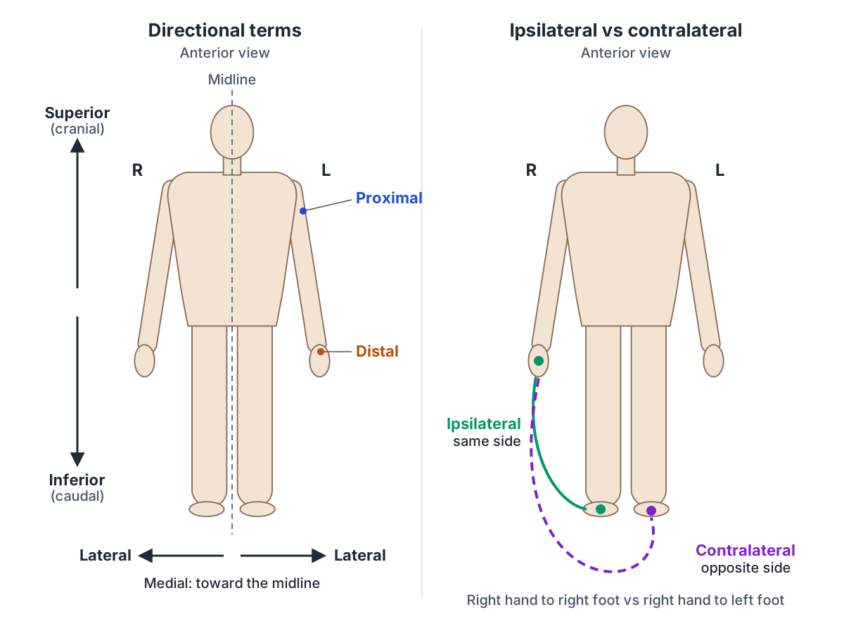
Medial means toward the middle of the body and lateral means toward the side. Proximal describes a position in a limb nearer to its point of attachment to the trunk, and distal a position farther from it. Superficial means closer to the surface of the body and deep means farther from it. (OpenStax Anatomy and Physiology 2e, 1.6: Anatomical Terminology.)
Examples make the pairs easier to remember. The heart lies posterior to the sternum, the nose is superior to the mouth, the eye is lateral to the nose, the wrist is distal to the elbow and the knee is proximal to the ankle. Proximal and distal are used for structures with a beginning and an end, such as limbs and blood vessels. (TeachMeAnatomy: Anatomical Terms of Location.)
Anatomical terms are built on Greek and Latin roots. OpenStax points out that because these languages are no longer used in everyday conversation, the meanings of their words do not drift over time, which keeps the terminology stable. (OpenStax Anatomy and Physiology 2e, 1.6: Anatomical Terminology.)
Directional terms and their opposites
| Term | Meaning | Opposite | Example |
|---|---|---|---|
| Anterior (ventral) | Toward the front | Posterior (dorsal) | The sternum is anterior to the heart |
| Superior (cranial) | Above, toward the head | Inferior (caudal) | The nose is superior to the mouth |
| Medial | Toward the midline | Lateral | The nose is medial to the eye |
| Proximal | Nearer the limb's attachment | Distal | The elbow is proximal to the wrist |
| Superficial | Closer to the body surface | Deep | Skin is superficial to muscle |
| Ipsilateral | On the same side | Contralateral | Right hand and right foot are ipsilateral |
Ipsilateral vs contralateral (and bilateral)
Ipsilateral means on the same side of the body, and contralateral means on the opposite side. Your right hand and right foot are ipsilateral to each other; your right hand and left foot are contralateral. (Radiopaedia: Radiographic positioning terminology.)
The terms are used most in neurology. Axons of the corticobulbar tract are ipsilateral, meaning they project from the cortex to motor nuclei on the same side of the nervous system. The axons of the corticospinal tract are largely contralateral: they cross the midline and synapse on the opposite side of the body. Axons that act on both sides at once are described as bilateral. (OpenStax Anatomy and Physiology 2e, 14.3: Motor Responses.)
Most spinal pathways that connect to the brain are contralateral, so the right side of the body is connected to the left side of the brain, while cranial nerve systems are mostly ipsilateral, with some exceptions. (OpenStax Anatomy and Physiology 2e, 14.1: Sensory Perception.)
Because most corticospinal fibres cross to the opposite side, the right motor cortex of the cerebrum controls muscles on the left side of the body, and the left motor cortex controls the right side. (OpenStax Anatomy and Physiology 2e, 14.3: Motor Responses.)
Clinicians also use the pair for paired organs. A tumour in the left breast with an enlarged lymph node in the left axilla describes ipsilateral spread; a finding in the right axilla would be contralateral. Unilateral means affecting one side only, and bilateral means affecting both.
Anterior, posterior and lateral views of the human body
Anatomical illustrations and 3D models are usually shown from standard directions named with the same terms. An anterior view shows the body from the front, a posterior view from the back, and a lateral (side) view from the right or left. A superior view looks down from above, as when you look at the top of the skull. (OpenStax Anatomy and Physiology 2e, 1.6: Anatomical Terminology.)
In a right lateral view the subject's right side faces you and the anterior surface points to one side of the picture, so the frontal plane appears edge-on as a vertical line, as in the centre panel of the planes diagram. X-ray projections are named by the direction the central ray travels: in an AP (anteroposterior) projection it passes from anterior to posterior, perpendicular to the coronal plane, in a PA projection from posterior to anterior, and in a lateral projection from one side of the body to the other. (Radiopaedia: Radiographic positioning terminology.)
On EasyAnatomyLab's 3D human model you can switch between anterior, posterior, left and right views. Try finding a structure in one view and then predicting where it will appear in another: it is one of the fastest ways to make the planes and directional terms automatic.
Frequently asked questions
What are the three anatomical planes?
The three anatomical planes are the sagittal plane, which divides the body into right and left parts; the frontal or coronal plane, which divides it into anterior and posterior parts; and the transverse or axial plane, which divides it into superior and inferior parts. All three are defined with the body in the anatomical position and are at right angles to one another.
What is the difference between midsagittal and parasagittal?
Both are vertical sagittal planes that separate right from left. The midsagittal, or median, plane runs exactly through the midline and creates equal right and left halves. A parasagittal plane runs parallel to it but off the midline, so it creates unequal right and left parts. There is one midsagittal plane but countless parasagittal planes.
What is the difference between ipsilateral and contralateral?
Ipsilateral means on the same side of the body and contralateral means on the opposite side. The right arm and right leg are ipsilateral; the right arm and left leg are contralateral. In neurology most motor and sensory pathways from the body cross the midline, so the left side of the brain controls and senses the right side of the body.
Is the coronal plane the same as the frontal plane?
Yes. Coronal and frontal are two names for the same vertical plane, which runs from side to side and divides the body into anterior (front) and posterior (back) portions. Radiologists tend to say coronal, as in a coronal MRI image, while many anatomy textbooks say frontal.
What is an axial view?
An axial view is an image in the transverse plane: a horizontal slice through the body at right angles to its long axis, as in most CT scans. It shows structures as a cross section, separating what lies above the slice from what lies below. Some radiologists define axial by the scanner's long axis rather than the body's, but the two usually match.
In which plane does flexion occur?
Flexion and extension typically take place in the sagittal plane, rotating around a side-to-side (frontal) axis. Bending the elbow or knee and nodding the head are examples. Abduction and adduction occur in the frontal plane around a front-to-back axis, and medial and lateral rotation occur in the transverse plane around a vertical axis.
References
- OpenStax. Anatomy and Physiology 2e, §1.6 Anatomical Terminology. Rice University, 2022.
- OpenStax. Anatomy and Physiology 2e, §7.1 Divisions of the Skeletal System. Rice University, 2022.
- OpenStax. Anatomy and Physiology 2e, §9.5 Types of Body Movements. Rice University, 2022.
- OpenStax. Anatomy and Physiology 2e, §14.1 Sensory Perception. Rice University, 2022.
- OpenStax. Anatomy and Physiology 2e, §14.3 Motor Responses. Rice University, 2022.
- Rad A. Anatomical terminology. Kenhub.
- TeachMeAnatomy. Anatomical Terms of Location.
- Shetty A, Bell DJ, et al. Radiographic positioning terminology. Radiopaedia.org (rID 29801).
- Koldenhoven RM, Behnke RS. Kinetic Anatomy, 5th ed. Human Kinetics. Excerpt: Anatomical planes and axes of movement.
Related anatomy
- Skeletal systemExplore a realistic 3D human skeleton: the 206 bones, axial vs appendicular skeleton, bone types, bony landmarks and…
- Types of jointsTypes of joints explained: fibrous, cartilaginous and synovial (diarthrosis) joints, a labelled synovial joint diagram…
- Muscular systemExplore an interactive 3D muscle model: how muscles move bones, how they are named, the upper body and leg muscles…
- Brain anatomyBrain anatomy explained: cerebrum, lobes, gyri, insula, corpus callosum, thalamus, hippocampus, brainstem and…
- Spinal cordSpinal cord anatomy with labelled diagrams: cross-section, grey matter horns, spinothalamic and dorsal column tracts…
Explore the whole body in the interactive 3D human anatomy model, or browse the A–Z list of human structures.