

Where is the spinal cord and how long is it?
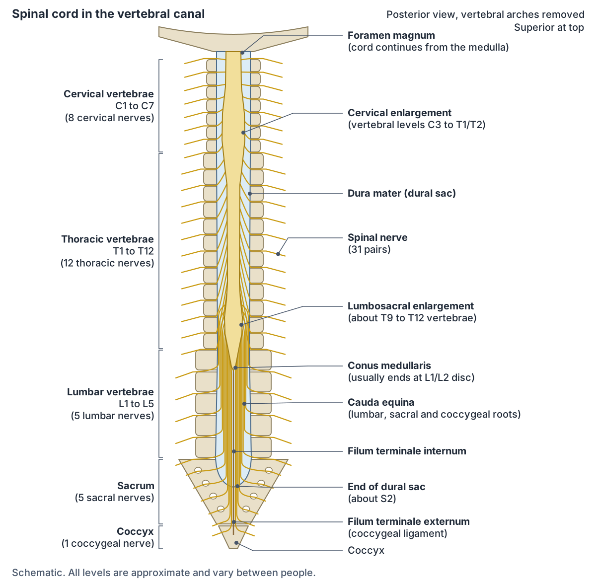
The spinal cord extends from the foramen magnum at the base of the skull to the L1/L2 vertebra, where it ends as the conus medullaris (medullary cone). Because of this, the cord fills only the upper two thirds of the vertebral canal in an adult. (Kenhub: Spinal cord.)
Radiopaedia gives the adult cord a length of about 42 to 45 cm, a diameter of about 1 cm and a mass of about 35 g. Unlike the brain, its grey matter lies on the inside, in an H or butterfly shape around the central canal, and its white matter tracts lie on the outside. (Radiopaedia: Spinal cord.)
The cord stops short because it does not grow significantly longer after the first or second year of life while the vertebral column keeps growing. As a result, the sacral part of the cord sits at the level of the upper lumbar vertebrae, and the nerve roots for the lowest segments have to run down the canal before they reach their exit holes. (OpenStax Anatomy and Physiology 2e, 13.2: The Central Nervous System.)
Regions, segments and the cervical and lumbar enlargements
The cord is divided into cervical, thoracic, lumbar and sacral regions, plus a single coccygeal segment. There are 8 cervical, 12 thoracic, 5 lumbar, 5 sacral and 1 coccygeal segments, and each segment gives rise to one pair of spinal nerves: 31 pairs in total. (Kenhub: Spinal cord.)
The cord is not the same width all the way down. The cervical and lumbar regions have enlargements because they contain larger populations of neurons, which serve the upper and lower limbs. (OpenStax Anatomy and Physiology 2e, 13.2: The Central Nervous System.)
The cervical enlargement occupies cord segments C5 to T1 and gives rise to the spinal nerves that form the brachial plexus. Because the cord is shorter than the vertebral column, it lies at the C3 to T1/T2 vertebral levels, and it is widest, about 38 mm in circumference, at the C6 level. Its lower counterpart is the lumbosacral (lumbar) enlargement, which supplies the lumbar and sacral plexuses. (Radiopaedia: Cervical enlargement.)
Sources give slightly different ranges for the enlargements. Dafny's Neuroscience Online describes the cervical enlargement as extending between C3 and T1 and the lumbar enlargement between L1 and S2, while Radiopaedia's spinal cord article places the lumbar expansion at about T9 to T12. The same textbook notes that in the thoracic and upper lumbar regions the cord segment is about three segments higher than the vertebra of the same number. (Dafny N. Anatomy of the Spinal Cord. Neuroscience Online, McGovern Medical School (UTHealth).)
Conus medullaris, cauda equina and filum terminale
The conus medullaris is the tapered, lowest part of the spinal cord. In children and adults it most often ends at the L1/2 intervertebral disc, and a level at or above the L2/3 disc can be considered normal. An ending at L3 is equivocal: it can be a normal variant or a sign of a tethered cord. (Radiopaedia: Conus medullaris.)
Below the conus, the lumbar, sacral and coccygeal nerve roots descend in the canal as individual rootlets, collectively called the cauda equina because the bundle resembles a horse's tail. (The phrase cauda medullaris is sometimes searched, but the correct terms are conus medullaris for the tip of the cord and cauda equina for the roots below it.) (Radiopaedia: Conus medullaris.)
The filum terminale is a thin filament of connective tissue that runs down from the apex of the conus. Its upper three quarters, the filum terminale internum, lies inside the dural sac with the cauda equina. Its lower quarter, the filum terminale externum, fuses with the dura and continues as the coccygeal ligament to attach to the dorsal surface of the coccyx, anchoring the lower end of the cord. (Radiopaedia: Filum terminale.)
The dural sac usually ends at or below the S1/S2 disc space, although Radiopaedia notes that in 11.3% of people it ends above S1. (Radiopaedia: Filum terminale.)
Spinal meninges and the denticulate ligaments (ligamenta denticulata)
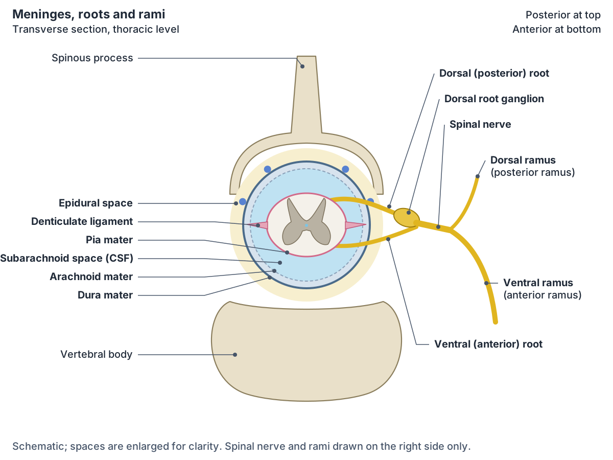
Like the brain, the spinal cord is wrapped in three meninges. From outside in they are the dura mater, a thick fibrous protective sheath; the arachnoid mater, a thin layer that forms a loose sac; and the pia mater, a thin membrane attached directly to the surface of the cord. The subarachnoid space between the arachnoid and pia contains cerebrospinal fluid (CSF), which cushions the brain and spinal cord. (OpenStax Anatomy and Physiology 2e, 13.3: Circulation and the Central Nervous System.)
The spinal dura forms a dural sac (thecal sac) from the foramen magnum to about S2. The subarachnoid space is especially large in the lumbar region, where it forms the lumbar cistern around the cauda equina. Below the conus, the pia continues as the filum terminale. (Radiopaedia: Spinal meninges.)
The spinal epidural space lies outside the dura, between it and the walls of the canal: the vertebral bodies, discs and posterior longitudinal ligament in front, and the laminae and ligamentum flavum behind. It runs from the foramen magnum to the sacral hiatus and contains fat, the internal vertebral venous plexus, small arteries and the spinal nerve roots. Unlike in the skull, it is a real space, separate from the cranial epidural space. (Radiopaedia: Spinal epidural space.)
The denticulate ligaments (ligamenta denticulata) are triangular sideways extensions of the pia mater that anchor the cord to the dura. There are usually 21 pairs, although the number ranges from 18 to 24. Each arises from the side of the cord between the ventral and dorsal roots and attaches by its tip to the dura. The highest pair inserts at the margin of the foramen magnum and the lowest lies at the level of the conus medullaris, between the T12 and L1 roots. They stabilise the cord within the vertebral canal. (Radiopaedia: Denticulate ligaments.)
Because the cord ends in the upper lumbar region, a needle can be passed through the dura and arachnoid lower down to withdraw CSF without hitting the cord. This procedure is a lumbar puncture. (OpenStax Anatomy and Physiology 2e, 13.3: Circulation and the Central Nervous System.)
Cross-section of the spinal cord: grey matter horns and white matter columns
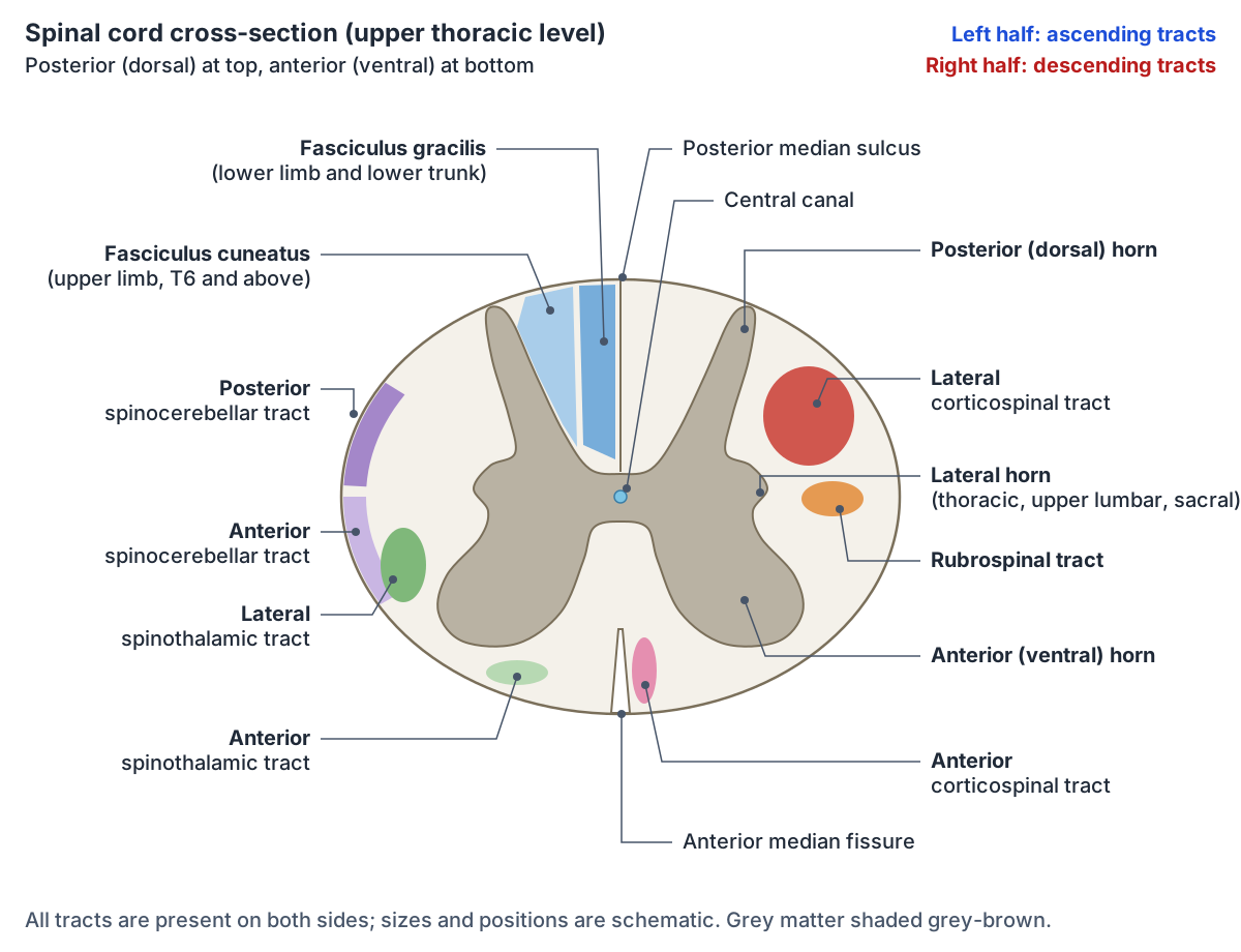
In a cross-section the grey matter (US spelling: gray matter) looks like an ink-blot or butterfly and is divided into horns. The posterior (dorsal) horn processes sensory information. The anterior (ventral) horn contains the motor neurons that send signals to skeletal muscles; some of the largest neurons in the cord are these multipolar motor neurons. The lateral horn is found only in the thoracic, upper lumbar and sacral regions and contains the cell bodies of autonomic motor neurons. (OpenStax Anatomy and Physiology 2e, 13.2: The Central Nervous System.)
The front of the cord is marked in the midline by the deep anterior median fissure and the back by the posterior median sulcus. Sensory axons enter on the posterior side through the dorsal (posterior) nerve root, and motor axons leave on the anterior side through the ventral (anterior) root. In general the posterior parts of the cord are sensory and the anterior parts are motor. (OpenStax Anatomy and Physiology 2e, 13.2: The Central Nervous System.)
The white matter around the grey matter is organised into columns, also called funiculi. On each side there is a posterior column between the posterior horns, an anterior column between the anterior horns and a lateral column at the side. The posterior columns contain only ascending (sensory) tracts, while the anterior and lateral columns contain both ascending and descending tracts. (OpenStax Anatomy and Physiology 2e, 13.2: The Central Nervous System.)
Some grey matter nuclei are confined to particular levels. For example, the dorsal nucleus of Clarke, a column of cells in the base of the dorsal horn, is found only in segments C8 to L3. (Dafny N. Anatomy of the Spinal Cord. Neuroscience Online, McGovern Medical School (UTHealth).)
Dorsal column and medial lemniscus pathway
The dorsal column-medial lemniscus (DCML) pathway carries fine touch, vibration and proprioception. Its first-order axons ascend on the same side in the dorsal column. Signals from the upper limb, T6 and above, travel in the fasciculus cuneatus, the lateral part of the dorsal column, and synapse in the nucleus cuneatus of the medulla. Signals from the lower limb travel in the fasciculus gracilis, the medial part, and synapse in the nucleus gracilis. (TeachMeAnatomy: The Ascending Tracts.)
The second-order neurons decussate (cross the midline) within the medulla oblongata and ascend on the opposite side as a bundle called the medial lemniscus. These axons end in the thalamus, and the third neuron projects to the postcentral gyrus of the cerebral cortex. So within the spinal cord, the dorsal columns carry information from the same side of the body. (OpenStax Anatomy and Physiology 2e, 14.2: Central Processing.)
TeachMeAnatomy names the thalamic relay as the ventral posterolateral nucleus, from which third-order fibres ascend to the primary sensory cortex. (TeachMeAnatomy: The Ascending Tracts.)
Kenhub describes the crossing fibres in more detail: second-order axons leave the gracile and cuneate nuclei, sweep ventromedially as the internal arcuate fibres, cross the midline and then turn upward as the medial lemniscus. In the cord, the fasciculi gracilis and cuneatus run on each side in the dorsal funiculi. (Kenhub: Ascending tracts of the spinal cord.)
Spinothalamic tract (anterolateral system)
The spinothalamic pathway, also called the anterolateral system, has two tracts. The lateral spinothalamic tract carries pain and temperature, and the anterior spinothalamic tract carries crude touch and pressure. The first-order neurons ascend one to two vertebral levels and synapse at the tip of the dorsal horn, in the substantia gelatinosa. (TeachMeAnatomy: The Ascending Tracts.)
The key difference from the dorsal columns is where the pathway crosses. In the spinothalamic pathway the second neuron, whose cell body lies in the spinal cord grey matter, sends its axon across the midline within the spinal cord at the same level, and the axon then ascends to the thalamus on the opposite side. The thalamic neurons project to the somatosensory cortex. (OpenStax Anatomy and Physiology 2e, 14.2: Central Processing.)
In both pathways the second neuron is contralateral, so each side of the brain receives touch, pain and temperature from the opposite side of the body. What differs is the level of crossing: the brain stem for the dorsal columns, the spinal cord itself for the spinothalamic tract. This is why the two pathways are worth learning side by side. (OpenStax Anatomy and Physiology 2e, 14.2: Central Processing.)
Kenhub places the lateral spinothalamic tract in the lateral funiculus, while the anterior spinothalamic tract ascends in the anterior and anterolateral funiculi. (Kenhub: Ascending tracts of the spinal cord.)
Spinocerebellar tracts
The spinocerebellar tracts carry unconscious proprioception to the cerebellum, which uses it to coordinate movement and posture. The posterior spinocerebellar tract carries proprioceptive information from the lower limbs to the cerebellum on the same side, and the cuneocerebellar tract does the same for the upper limbs. (TeachMeAnatomy: The Ascending Tracts.)
The second-order neurons of the posterior spinocerebellar tract form the nucleus dorsalis (Clarke's column) in cord segments C8 to L2 or L3. The tract ascends in the posterolateral part of the lateral funiculus, lateral to the corticospinal tract, and enters the cerebellum through the inferior cerebellar peduncle. The anterior spinocerebellar tract ascends just ventral to it and enters through the superior cerebellar peduncle. (Kenhub: Ascending tracts of the spinal cord.)
The anterior spinocerebellar tract carries proprioception from the lower limbs, but its fibres decussate twice, once in the cord and again in the brain, so they also end in the ipsilateral cerebellum. A rostral spinocerebellar tract carries proprioception from the upper limbs to the ipsilateral cerebellum. Because the output ends up on the same side, lesions of the spinocerebellar tracts cause loss of coordination on the same side of the body. (TeachMeAnatomy: The Ascending Tracts.)
Main spinal cord tracts, what they carry and where they cross
| Tract | Direction | Carries | Position in the cord | Decussation (crossing) |
|---|---|---|---|---|
| Fasciculus gracilis (dorsal column) | Ascending | Fine touch, vibration, proprioception from the lower limb | Medial dorsal funiculus | Medulla, via internal arcuate fibres to the medial lemniscus |
| Fasciculus cuneatus (dorsal column) | Ascending | Fine touch, vibration, proprioception from the upper limb (T6 and above) | Lateral dorsal funiculus | Medulla |
| Lateral spinothalamic | Ascending | Pain and temperature | Lateral funiculus | Spinal cord, after ascending 1 to 2 levels |
| Anterior spinothalamic | Ascending | Crude touch and pressure | Anterior and anterolateral funiculi | Spinal cord, after ascending 1 to 2 levels |
| Posterior spinocerebellar | Ascending | Unconscious proprioception, lower limb | Posterolateral part of lateral funiculus | Does not cross; inferior cerebellar peduncle |
| Anterior spinocerebellar | Ascending | Unconscious proprioception, lower limb | Lateral funiculus, ventral to the posterior tract | Crosses twice; superior cerebellar peduncle |
| Lateral corticospinal | Descending | Voluntary movement of limb muscles | Lateral funiculus | Pyramidal decussation, lower medulla (about 80% of fibres) |
| Anterior corticospinal | Descending | Voluntary movement of trunk muscles | Anterior funiculus | Spinal cord, at the level where it ends |
| Rubrospinal | Descending | Thought to help fine control of hand movements | Lateral funiculus | Crosses (contralateral) |
| Vestibulospinal | Descending | Balance and posture via anti-gravity muscles | Anterior funiculus (lateral vestibulospinal) | Does not cross (ipsilateral) |
Summary based on OpenStax, TeachMeAnatomy and Kenhub (ascending and descending tracts). The DCML and spinothalamic pathways both end on the opposite side of the brain; they differ in where they cross.
Corticospinal (pyramidal) tracts and other descending tracts
The main motor pathway is the corticospinal tract. The upper motor neuron has its cell body in the primary motor cortex of the frontal lobe. Its axon descends through the brain stem and, in the medulla, forms the large white matter bundles called the pyramids, which is why the corticospinal tract is also called the pyramidal tract. At the junction of the medulla and spinal cord, most of the fibres cross over at the pyramidal decussation. (OpenStax Anatomy and Physiology 2e, 14.3: Motor Responses.)
The crossed fibres form the lateral corticospinal tract, which controls the appendicular muscles of the arms and legs. The fibres that do not cross in the medulla form the anterior corticospinal tract, which controls trunk muscles; these axons cross at the level where they end and enter the ventral horn of the opposite side. There the upper motor neuron synapses with the lower motor neuron, whose cell body lies in the ventral horn. (OpenStax Anatomy and Physiology 2e, 14.3: Motor Responses.)
TeachMeAnatomy adds that the lateral corticospinal tract ends in the ventral horn at all segmental levels, whereas the anterior corticospinal tract ends in the ventral horn of the cervical and upper thoracic segments. On their way down, the corticospinal fibres pass through the internal capsule, which is especially vulnerable to haemorrhagic stroke. (TeachMeAnatomy: The Descending Tracts.)
Kenhub states that about 80% of corticospinal fibres cross at the pyramidal decussation. In the cord, the lateral corticospinal and rubrospinal tracts run in the lateral funiculus, while the anterior corticospinal tract, the lateral vestibulospinal tract and the tectospinal tract (in the cervical cord) run in the anterior (ventral) funiculus. (Kenhub: Descending tracts of the spinal cord.)
The extrapyramidal tracts are the other descending pathways. The rubrospinal tract, from the red nucleus, and the tectospinal tract, from the superior colliculus, cross the midline, so they act on the opposite side. The vestibulospinal and reticulospinal tracts do not cross and act on the same side. The vestibulospinal tract controls balance and posture through the anti-gravity muscles, and the tectospinal tract coordinates head movements in response to visual stimuli. (TeachMeAnatomy: The Descending Tracts.)
Spinal nerves: dorsal and ventral roots and rami (ramus dorsalis)
There are 31 pairs of spinal nerves (abbreviated spinal n.): 8 cervical (C1 to C8), 12 thoracic, 5 lumbar, 5 sacral and 1 coccygeal. Each leaves the vertebral column through the intervertebral foramen at its level, and the sacral nerves leave through the sacral foramina. (OpenStax Anatomy and Physiology 2e, 13.4: The Peripheral Nervous System.)
Each spinal nerve is formed where a dorsal (posterior) root and a ventral (anterior) root join. The dorsal root carries sensory axons, whose cell bodies lie outside the cord in the dorsal root ganglion. The ventral root carries motor axons. After the roots join, the spinal nerve is mixed, carrying both sensory and motor fibres. (Kenhub: Spinal nerves.)
Just outside the foramen, the spinal nerve divides into a larger anterior (ventral) ramus and a smaller posterior (dorsal) ramus, in Latin ramus dorsalis. The dorsal ramus supplies the deep muscles of the back (the post-vertebral muscles) and the skin of the back. The ventral ramus supplies the skin and muscles of the front of the trunk, and the ventral rami of the cervical, lumbar and sacral nerves form the plexuses that supply the limbs. Do not confuse rami with roots: the roots join to form the spinal nerve, and the rami are its branches. (Kenhub: Spinal nerves.)
Other small branches leave the spinal nerve as well. Rami communicantes relay autonomic signals between the spinal nerve and the sympathetic trunk, and a meningeal branch provides sensory and vasomotor supply to the spinal meninges. Spinal nerves C1 to C7 emerge above the vertebra of the same number, and the eighth cervical pair emerges below C7. For the most part the remaining nerves exit below their corresponding vertebra, and the sacral nerves pass through the sacral foramina. (Kenhub: Spinal nerves.)
The skin area supplied by the sensory fibres of one spinal nerve is called a dermatome. Spinal nerves T2 to T11 do not join a plexus; their ventral rami become the intercostal nerves between the ribs, while the cervical, brachial, lumbar and sacral plexuses supply the neck and limbs. (OpenStax Anatomy and Physiology 2e, 13.4: The Peripheral Nervous System.)
Blood supply of the spinal cord
The vertebral arteries give rise to the anterior and posterior spinal arteries, which run along the length of the cord. Segmental arteries along the column give off radicular branches that supply the spinal nerve roots and reinforce the cord's supply. (Kenhub: Spinal cord.)
Frequently asked questions
Where does the spinal cord end?
In adults the spinal cord usually ends at about the L1/L2 intervertebral disc as a cone-shaped tip, the conus medullaris. A conus ending at or above the L2/L3 disc is generally considered normal. Below it, the nerve roots continue as the cauda equina inside the dural sac, and the filum terminale anchors the cord to the coccyx.
What is the difference between the dorsal column and the spinothalamic tract?
The dorsal column (DCML) pathway carries fine touch, vibration and proprioception and stays on the same side in the spinal cord, crossing in the medulla before forming the medial lemniscus. The spinothalamic tract carries pain and temperature (lateral) and crude touch and pressure (anterior), and crosses within the spinal cord close to the level where the fibres enter.
What is the medial lemniscus?
The medial lemniscus is a bundle of second-order sensory axons in the brain stem. These axons start in the nucleus gracilis and nucleus cuneatus of the medulla, cross the midline, and ascend to the thalamus. It is the second half of the dorsal column-medial lemniscus pathway, which carries fine touch, vibration and conscious proprioception to the postcentral gyrus.
What are the denticulate ligaments?
The denticulate ligaments (ligamenta denticulata) are triangular extensions of pia mater that project from each side of the spinal cord, between the dorsal and ventral roots, and attach to the dura mater. There are usually 21 pairs, from the foramen magnum down to the conus medullaris. They hold the spinal cord steady in the middle of the vertebral canal.
What does the dorsal ramus supply?
The dorsal (posterior) ramus, or ramus dorsalis, is the smaller of the two main branches of a spinal nerve. It turns backward to supply the deep muscles of the back alongside the vertebral column and the skin of the back. The larger ventral ramus supplies the front and sides of the trunk and forms the limb plexuses.
Why is it called the pyramidal tract?
The corticospinal tract is called the pyramidal tract because its fibres form the pyramids, two large white matter ridges on the front of the medulla oblongata. At the lower end of the medulla, most of these fibres cross at the pyramidal decussation and descend as the lateral corticospinal tract; the rest descend uncrossed as the anterior corticospinal tract.
What is the cervical enlargement?
The cervical enlargement is the widened part of the spinal cord that supplies the upper limbs. Radiopaedia places it at cord segments C5 to T1, lying at about the C3 to T1/T2 vertebral levels, with its widest point at C6. It gives rise to the spinal nerves of the brachial plexus. A similar lumbosacral enlargement lower down supplies the legs.
References
- OpenStax. Anatomy and Physiology 2e, §13.2 The Central Nervous System. Rice University, 2022.
- OpenStax. Anatomy and Physiology 2e, §13.3 Circulation and the Central Nervous System. Rice University, 2022.
- OpenStax. Anatomy and Physiology 2e, §13.4 The Peripheral Nervous System. Rice University, 2022.
- OpenStax. Anatomy and Physiology 2e, §14.2 Central Processing. Rice University, 2022.
- OpenStax. Anatomy and Physiology 2e, §14.3 Motor Responses. Rice University, 2022.
- TeachMeAnatomy. The Ascending Tracts.
- TeachMeAnatomy. The Descending Tracts.
- Kenhub. Spinal cord.
- Kenhub. Ascending tracts of the spinal cord.
- Kenhub. Descending tracts of the spinal cord.
- Kenhub. Spinal nerves.
- Radiopaedia.org. Spinal cord.
- Radiopaedia.org. Cervical enlargement.
- Radiopaedia.org. Conus medullaris.
- Radiopaedia.org. Filum terminale.
- Radiopaedia.org. Denticulate ligaments.
- Radiopaedia.org. Spinal meninges.
- Radiopaedia.org. Spinal epidural space.
- Dafny N. Anatomy of the Spinal Cord. In: Neuroscience Online, Section 2, Chapter 3. McGovern Medical School at UTHealth.
Related anatomy
- Vertebral columnHuman spine anatomy: cervical, thoracic and lumbar vertebrae, curvatures, discs, facet joints and spinal ligaments…
- Brain anatomyBrain anatomy explained: cerebrum, lobes, gyri, insula, corpus callosum, thalamus, hippocampus, brainstem and…
- Upper limb nervesBrachial plexus diagram with roots, trunks, cords and branches; axillary, musculocutaneous, median, ulnar and radial…
- Lower limb nervesLower limb nerves explained: lumbar and sacral plexus diagram, femoral, saphenous, sciatic, tibial, common peroneal…
- Autonomic nervous systemThe autonomic nervous system explained: sympathetic vs parasympathetic outflow diagram, sympathetic trunk, effects on…
- Cranial nervesThe 12 cranial nerves in order with a labelled diagram: type, skull foramen, functions and test for CN I to CN XII…
Explore the whole body in the interactive 3D human anatomy model, or browse the A–Z list of human structures.