

Parts of the brain
The brain develops from the front end of the neural tube. Early on it forms three swellings: the prosencephalon (forebrain), mesencephalon (midbrain) and rhombencephalon (hindbrain). These become five regions: the telencephalon becomes the cerebrum, the diencephalon becomes the thalamus and hypothalamus, the mesencephalon stays as the midbrain, the metencephalon becomes the pons and cerebellum, and the myelencephalon becomes the medulla oblongata. (OpenStax Anatomy and Physiology 2e, 13.1: The Embryologic Perspective.)
The hollow centre of the neural tube survives as the ventricles. The lateral ventricles lie in the cerebrum, the third ventricle in the diencephalon, the cerebral aqueduct in the midbrain and the fourth ventricle among the pons, cerebellum and medulla. Below the medulla the space continues as the central canal of the spinal cord. (OpenStax Anatomy and Physiology 2e, 13.1: The Embryologic Perspective.)
The brainstem (midbrain, pons and medulla) connects the forebrain with the spinal cord. The cerebellum is attached to the back of the brainstem but is usually described as a separate part. (OpenStax Anatomy and Physiology 2e, 13.2: The Central Nervous System.)
What is the cerebrum?
The cerebrum is the largest part of the brain and makes up most of its mass. Its outer layer, the cerebral cortex, is a continuous sheet of grey matter that carries out the higher functions of the nervous system. A deep groove, the longitudinal fissure, splits the cerebrum into right and left cerebral hemispheres. (OpenStax Anatomy and Physiology 2e, 13.2: The Central Nervous System.)
The cerebrum lies above and in front of the brainstem. It reaches from the frontal bone at the front to the occipital bone at the back, fills the anterior and middle cranial fossae and lies above the tentorium cerebelli. The falx cerebri, a fold of dura mater, dips into the longitudinal fissure between the two hemispheres. Under the cortex, white matter made of myelinated axons and glial cells forms the bulk of each hemisphere. (TeachMeAnatomy: The Cerebrum.)
Deep in the cerebrum are three groups of grey matter: the basal nuclei, which help plan and select movements; the basal forebrain, linked to learning and memory; and the limbic cortex, involved in emotion, memory and behaviour. (OpenStax Anatomy and Physiology 2e, 13.2: The Central Nervous System.)
Gyri and sulci: the folds of the brain
A gyrus (plural gyri) is a ridge of the cerebral cortex, and a sulcus (plural sulci) is the groove between two gyri. Folding lets far more grey matter fit inside the skull. The pattern of folds is similar from person to person, so the large gyri and sulci work as landmarks. (OpenStax Anatomy and Physiology 2e, 13.2: The Central Nervous System.)
Three grooves divide the lobes. The central sulcus separates the frontal lobe in front from the parietal lobe behind. The lateral sulcus (Sylvian fissure) separates the temporal lobe below from the frontal and parietal lobes above. On the medial surface, the parieto-occipital sulcus separates the parietal and occipital lobes. (OpenStax Anatomy and Physiology 2e, 13.2: The Central Nervous System.)
The precentral gyrus, directly in front of the central sulcus, is the primary motor cortex. The postcentral gyrus, directly behind it, is the primary somatosensory cortex. The superior temporal gyrus, just below the lateral sulcus, receives and processes sound. (TeachMeAnatomy: The Cerebrum.)
On the lateral surface of the frontal lobe, the precentral gyrus is followed forwards by the superior, middle and inferior frontal gyri. The inferior frontal gyrus contains Broca's area. On its underside, the orbital gyri rest on the floor of the anterior cranial fossa, with the gyrus rectus beside the midline. (Radiopaedia: Frontal lobe.)
Behind the postcentral gyrus, the intraparietal sulcus divides the superior parietal lobule from the inferior parietal lobule. The inferior lobule contains the supramarginal gyrus and the angular gyrus. (Radiopaedia: Parietal lobe.)
The temporal lobe has five gyri from lateral to medial: superior, middle and inferior temporal gyri, then the fusiform gyrus and the parahippocampal gyrus. The transverse temporal gyri (Heschl's gyri) run mediolaterally. (Radiopaedia: Temporal lobe.)
The lobes of the brain and what they do
Each hemisphere has four lobes: frontal, parietal, temporal and occipital. The frontal lobe deals mainly with movement: the precentral gyrus sends commands to the spinal cord, the premotor area plans movement and the prefrontal cortex supports personality, short-term memory and other cognitive functions. Broca's area, which produces speech, is in the left hemisphere in most people. (OpenStax Anatomy and Physiology 2e, 13.2: The Central Nervous System.)
The parietal lobe handles somatosensation, meaning touch, pressure, pain, itch, vibration and body position. The temporal lobe contains the primary auditory cortex (Brodmann areas 41 and 42) and, through its limbic parts, is important for memory. In the occipital lobe, areas 17 and 18 handle primary visual perception. (OpenStax Anatomy and Physiology 2e, 13.2: The Central Nervous System.)
Wernicke's area, linked to understanding language, lies in the dominant hemisphere near the back end of the lateral sulcus. Sources draw its border differently: Radiopaedia's temporal lobe article lists it as Brodmann area 22, while its parietal lobe article includes the inferior parietal lobule. (Radiopaedia: Temporal lobe.)
In the early 1900s Korbinian Brodmann divided the cortex into 52 areas by the arrangement of its cells under the microscope. The numbering is still used, and many Brodmann areas match functional regions. (OpenStax Anatomy and Physiology 2e, 13.2: The Central Nervous System.)
The four lobes of the cerebrum
| Lobe | Boundaries | Key gyri and areas | Main roles |
|---|---|---|---|
| Frontal | In front of the central sulcus; above the lateral sulcus | Precentral gyrus (primary motor cortex); superior, middle and inferior frontal gyri; Broca's area in the inferior frontal gyrus | Voluntary movement, planning, personality, speech production |
| Parietal | Behind the central sulcus; above the lateral sulcus; in front of the parieto-occipital sulcus | Postcentral gyrus (primary somatosensory cortex); superior and inferior parietal lobules; supramarginal and angular gyri | Touch, pain, temperature and body position; visuospatial processing |
| Temporal | Below the lateral sulcus | Superior, middle and inferior temporal gyri; transverse temporal (Heschl's) gyri; fusiform and parahippocampal gyri; hippocampus and amygdala on the medial side | Hearing, language comprehension, memory |
| Occipital | Behind the parieto-occipital sulcus; no clear border on the lateral surface | Calcarine sulcus with the primary visual cortex; cuneus above it, lingual gyrus below it | Vision |
Compiled from OpenStax, TeachMeAnatomy and Radiopaedia (see references). The insula is hidden inside the lateral sulcus (see below).
Definition of the insula
The insula (island of Reil) is a region of cerebral cortex hidden deep inside the lateral sulcus. It is completely covered by the frontal, parietal and temporal opercula (Latin for lids) and makes up about 2% of the cortical surface. (Radiopaedia: Insula.)
A circular sulcus surrounds it. Its central sulcus divides it into an anterior lobule with about three short gyri and a posterior lobule with two long gyri. The insula is the primary taste (gustatory) cortex, has links with language and with the autonomic nervous system, and is supplied by branches of the middle cerebral artery. (Radiopaedia: Insula.)
On a coronal section you see the insula as the cortex lining the deep end of the lateral sulcus. Medial to it lie, in order, the extreme capsule, the claustrum, the external capsule and the putamen. (TeachMeAnatomy: The Basal Ganglia.)
Sagittal section of the brain
A sagittal plane divides the body or an organ vertically into right and left parts. When it runs exactly down the middle it is the midsagittal or median plane; any other sagittal cut is parasagittal. (OpenStax Anatomy and Physiology 2e, 1.6: Anatomical Terminology.)
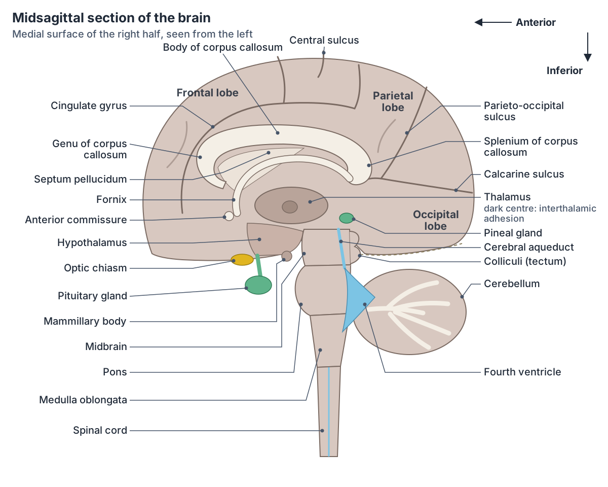
A midsagittal cut of the brain passes through the longitudinal fissure, so it shows the medial surface of one hemisphere and splits the midline structures in half. The most obvious is the corpus callosum, a C-shaped band about 10 cm long. From front to back its parts are the rostrum, genu, body (trunk) and splenium. (Radiopaedia: Corpus callosum.)
The cingulate gyrus lies on the medial surface of the hemisphere and follows the upper curve of the corpus callosum, separated from it by the callosal sulcus. The cingulate sulcus marks its upper edge. It forms a major part of the limbic system. (Radiopaedia: Cingulate gyrus.)
Under the corpus callosum is the septum pellucidum, a thin, see-through membrane between the front parts of the two lateral ventricles. It is attached above to the body of the corpus callosum, in front to the genu and below to the rostrum, and extends back to the fornix. (Radiopaedia: Septum pellucidum.)
The fornix is a C-shaped bundle that carries fibres from the hippocampus. It arches under the corpus callosum and its columns curve down past the anterior commissure towards the mammillary bodies. (Radiopaedia: Fornix.)
On the medial surface of the occipital lobe, the calcarine sulcus runs forward to join the parieto-occipital sulcus. The cuneus lies above it and the lingual gyrus below, and the primary visual cortex lines its two banks. (Radiopaedia: Calcarine sulcus.)
Below the fornix the cut shows the medial surface of the thalamus, which forms the lateral wall of the third ventricle. In most people a small interthalamic adhesion joins the two thalami across the ventricle, so it appears as a cut oval on the thalamus. (Radiopaedia: Thalamus.)
The hypothalamus lies below and in front of the thalamus. The pituitary gland hangs from it on a stalk (the infundibulum) and sits in the sella turcica, a hollow in the sphenoid bone. (OpenStax Anatomy and Physiology 2e, 17.3: The Pituitary Gland and Hypothalamus.)
The pineal gland sits in the midline at the back of the third ventricle, below the splenium of the corpus callosum and above the superior colliculi of the midbrain. It produces melatonin. (Radiopaedia: Pineal gland.)
Further down the cut passes through the midbrain, with the cerebral aqueduct in it, then the pons, the medulla oblongata and the start of the spinal cord. Behind the pons and medulla is the fourth ventricle, and behind that the cerebellum. (OpenStax Anatomy and Physiology 2e, 13.1: The Embryologic Perspective.)
Coronal section of the brain
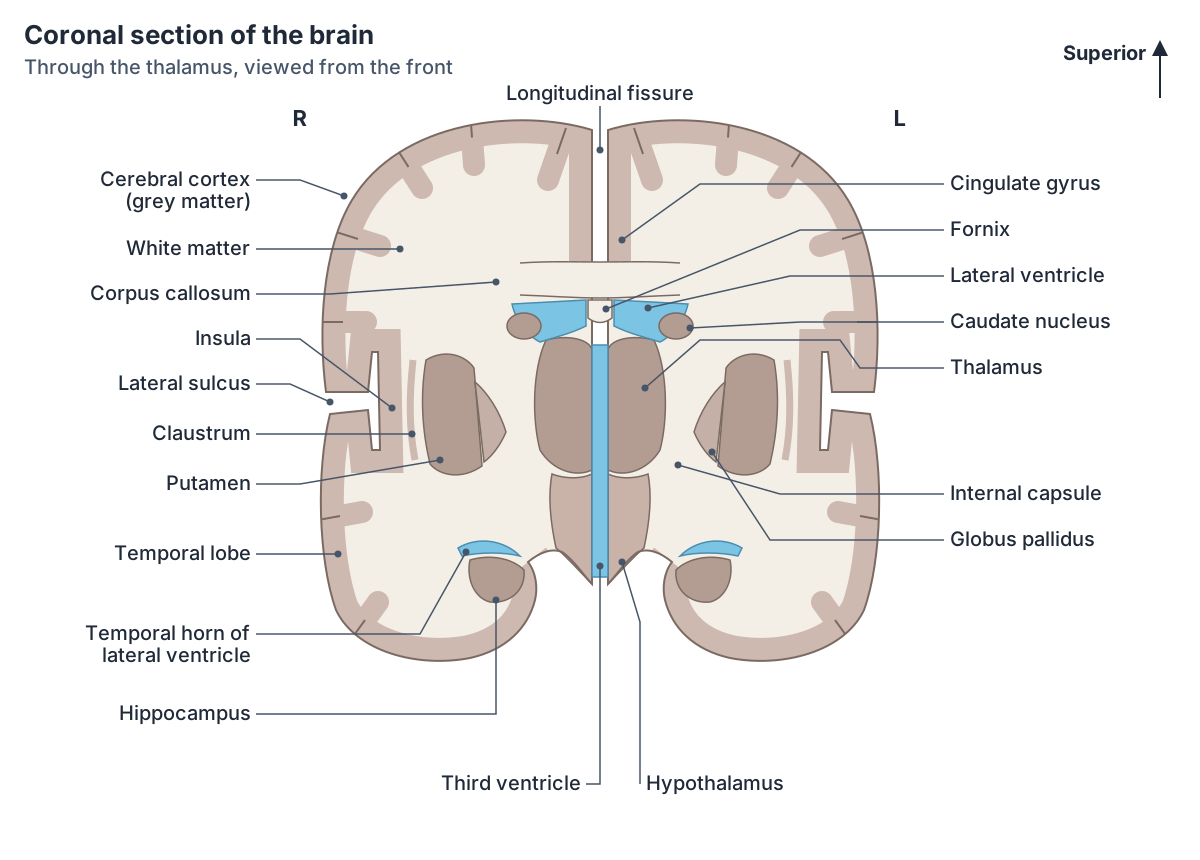
A frontal or coronal plane divides the body or an organ into a front (anterior) part and a back (posterior) part. A coronal slice of the brain is therefore a vertical cut from ear to ear. (OpenStax Anatomy and Physiology 2e, 1.6: Anatomical Terminology.)
A coronal cut through the thalamus shows grey matter on the outside (the cortex), white matter beneath it, and islands of deep grey matter. The corpus callosum crosses the midline under the longitudinal fissure. Below it lie the bodies of the lateral ventricles, with the caudate nucleus forming their lateral wall. (TeachMeAnatomy: The Basal Ganglia.)
The internal capsule is a thick sheet of white matter that carries fibres between the cortex and the thalamus, brainstem and spinal cord. Its anterior limb lies between the head of the caudate nucleus and the lentiform nucleus; its posterior limb lies between the thalamus and the lentiform nucleus. Corticospinal (motor) fibres run in the front two-thirds of the posterior limb. (Radiopaedia: Internal capsule.)
The lentiform nucleus is made of the putamen laterally and the globus pallidus medially; the caudate nucleus and putamen together form the striatum. (TeachMeAnatomy: The Basal Ganglia.)
Below the lateral sulcus is the temporal lobe. In its medial part the hippocampus, seahorse-shaped in coronal section, lies just below the floor of the temporal horn of the lateral ventricle. (Radiopaedia: Hippocampus.)
Corpus callosum
The corpus callosum is the largest commissure of the brain, a white matter tract that links the cortex of the two hemispheres. Fibres from the genu (forceps minor) connect the frontal lobes, and fibres from the splenium (forceps major) connect the occipital lobes. (Radiopaedia: Corpus callosum.)
Cutting the corpus callosum (split-brain surgery) helped spread the idea of a strict left brain and right brain. In reality the left hemisphere is mainly associated with language and the right with spatial and non-verbal reasoning, but neither side works alone. (OpenStax Anatomy and Physiology 2e, 13.2: The Central Nervous System.)
The corpus callosum forms between about weeks 12 and 20 of pregnancy. It has a rich blood supply, mainly from the pericallosal branches of the anterior cerebral arteries, so infarcts of it are uncommon. (Radiopaedia: Corpus callosum.)
Diencephalon: thalamus and hypothalamus
The diencephalon (its name means through brain) lies between the cerebrum and the brainstem and forms the walls of the third ventricle. Its main parts are the thalamus and hypothalamus; the epithalamus (with the pineal gland) and subthalamus are also included. (OpenStax Anatomy and Physiology 2e, 13.2: The Central Nervous System.)
The thalamus relays information between the cortex and the rest of the nervous system. All sensory information except smell passes through it before reaching the cortex, and it also takes part in motor control with the cerebellum and basal nuclei. The hypothalamus, below and in front of the thalamus, controls homeostasis and is the executive centre for the autonomic and endocrine systems. (OpenStax Anatomy and Physiology 2e, 13.2: The Central Nervous System.)
Each thalamus is an egg-shaped mass. The two are usually joined across the third ventricle by a small interthalamic adhesion, and the lateral surface of each touches the posterior limb of the internal capsule. (Radiopaedia: Thalamus.)
Hippocampus and limbic structures
The hippocampus lies in the medial part of the temporal lobe. It has a head, which lies behind the amygdala, a body and a tail that follows the curve of the lateral ventricle. In cross-section it looks like a rolled sheet made of the cornu Ammonis (fields CA1 to CA3) and the dentate gyrus. (Radiopaedia: Hippocampus.)
The hippocampus is needed to consolidate memories, for episodic memory and for spatial navigation. Its main output runs through the fornix. It is involved in Alzheimer disease, and it is the site of mesial temporal sclerosis. (Radiopaedia: Hippocampus.)
The hippocampus and amygdala work with nearby cortex in forming long-term memories and in emotional responses. (OpenStax Anatomy and Physiology 2e, 13.2: The Central Nervous System.)
What does the brainstem do?
The brainstem links the spinal cord to the higher brain centres. It houses the cranial nerve nuclei and coordinates vital autonomic functions. (TeachMeAnatomy: The Brainstem.)
Radiopaedia lists its roles as giving the main motor and sensory nerve supply to the face and neck through the cranial nerves, connecting the cerebrum, cerebellum and spinal cord, regulating heart and breathing, and maintaining consciousness and the sleep cycle. (Radiopaedia: Brainstem.)
The midbrain, the highest and smallest part, is about 2 cm long. Its roof (tectum) carries four colliculi: the superior colliculi deal with visual reflexes and the inferior colliculi with auditory ones. (TeachMeAnatomy: The Midbrain.)
The pons is the largest part of the brainstem. Its front bulge is made of transverse fibres that carry signals from the cerebrum to the cerebellum, and it is joined to the cerebellum by the middle cerebellar peduncles. (TeachMeAnatomy: The Pons.)
The medulla oblongata is about 3 cm long and continues as the spinal cord at the foramen magnum. On its front surface are the pyramids, and the olives. In the decussation of the pyramids about 75% of the motor fibres in the pyramids cross to the opposite side. (TeachMeAnatomy: The Medulla Oblongata.)
Major ascending and descending pathways pass through the brainstem, and the pons and medulla help regulate cardiovascular and respiratory function. The reticular formation, a diffuse network of grey matter running through the brainstem, is linked to sleep, wakefulness and attention. (OpenStax Anatomy and Physiology 2e, 13.2: The Central Nervous System.)
Parts of the brainstem
| Part | Position | Cranial nerves attached | Main roles |
|---|---|---|---|
| Midbrain | Highest part, between the diencephalon and the pons; passes through the opening in the tentorium cerebelli | Oculomotor (III), trochlear (IV) | Visual and auditory reflexes (colliculi), eye movements, motor control (substantia nigra, red nucleus) |
| Pons | Middle and largest part, in front of the cerebellum and fourth ventricle | Trigeminal (V); abducens (VI), facial (VII) and vestibulocochlear (VIII) at the pontomedullary junction | Relays signals from the cerebrum to the cerebellum; with the medulla, helps regulate breathing and heart function |
| Medulla oblongata | Lowest part, continuous with the spinal cord at the foramen magnum | Glossopharyngeal (IX), vagus (X), accessory (XI), hypoglossal (XII) | Pyramidal decussation of motor fibres; cardiovascular and respiratory regulation |
Compiled from OpenStax 13.2 and TeachMeAnatomy (see references).
Cerebellum
The cerebellum (little brain) lies in the posterior cranial fossa, below the occipital and temporal lobes, separated from them by the tentorium cerebelli. It sits behind the pons, with the fourth ventricle between them. (TeachMeAnatomy: The Cerebellum.)
It has two hemispheres joined by a narrow midline vermis. The primary and posterolateral fissures divide it into anterior, posterior and flocculonodular lobes. Four deep nuclei sit in its white matter: dentate, emboliform, globose and fastigial. (TeachMeAnatomy: The Cerebellum.)
Three pairs of cerebellar peduncles attach it to the brainstem: the superior peduncles to the midbrain, the middle peduncles to the pons and the inferior peduncles to the medulla. (Radiopaedia: Cerebellum.)
The cerebellum makes up about 10% of the brain's mass. It compares the motor commands sent by the cerebrum with sensory feedback from the body and sends correcting signals when movement is not coordinated. (OpenStax Anatomy and Physiology 2e, 13.2: The Central Nervous System.)
Ventricles of the brain
Each lateral ventricle lies within its cerebral hemisphere and has horns that reach into the frontal, occipital and temporal lobes. Each connects to the third ventricle, between the two thalami, through an interventricular foramen (foramen of Monro). The cerebral aqueduct carries cerebrospinal fluid (CSF) from the third ventricle to the fourth ventricle at the junction of the pons and medulla. (TeachMeAnatomy: The Ventricles of the Brain.)
CSF is made by the choroid plexus, where epithelial cells filter plasma from the blood. The narrow cerebral aqueduct is the most common site of blockage in non-communicating hydrocephalus. (TeachMeAnatomy: The Ventricles of the Brain.)
Brain anatomy in the 3D model
- Amygdala
- The amygdala is an almond-shaped nuclear complex in the medial temporal lobe that is central to processing emotionally significant stimuli.
- Angular gyrus
- The angular gyrus is a cortical region of the inferior parietal lobule that arches over the end of the superior temporal sulcus.
- Anterior part of superior temporal gyrus
- This is the anterior portion of the superior temporal gyrus, the uppermost gyrus on the lateral surface of the temporal lobe.
- Caudate nucleus
- The caudate nucleus is a C-shaped nucleus of the basal ganglia that, with the putamen, forms the striatum.
- Cerebellum
- The cerebellum is the large structure at the back of the brain that coordinates movement, posture and balance and takes part in motor learning.
- Cingulate gyrus
- The cingulate gyrus is a long arching gyrus on the medial surface of the cerebral hemisphere above the corpus callosum and a major part of the limbic lobe.
- Corpus callosum
- The corpus callosum is the largest white matter commissure, connecting the left and right cerebral hemispheres.
- Fornix of forebrain
- The fornix is a C-shaped white matter tract that carries the main output of the hippocampus.
- Fourth ventricle
- The fourth ventricle is a diamond-shaped, CSF-filled cavity of the hindbrain.
- Fusiform gyrus
- The fusiform gyrus is a long gyrus on the underside of the temporal and occipital lobes.
- Globus pallidus
- The globus pallidus is a basal ganglia nucleus that provides major inhibitory output to the thalamus.
- Hippocampus
- The hippocampus is a curved cortical structure in the medial temporal lobe that is essential for forming new long-term memories.
- Hypothalamus
- The hypothalamus is a small diencephalic region that regulates homeostasis, autonomic function and endocrine control.
- Inferior frontal gyrus
- The inferior frontal gyrus is the lowest of the three frontal gyri on the lateral frontal lobe, and contains Broca's area on the dominant side.
- Inferior temporal gyrus
- The inferior temporal gyrus is the lowest gyrus on the lateral surface of the temporal lobe.
- Insula
- The insula is a cortical region buried deep within the lateral sulcus, covered by the frontal, parietal and temporal opercula.
- Internal capsule
- The internal capsule is a V-shaped band of white matter carrying fibres between the cerebral cortex and the brainstem and spinal cord.
- Lateral ventricle
- The lateral ventricle is a C-shaped CSF-filled cavity within each cerebral hemisphere.
- Mammillary body
- The mammillary bodies are paired round bumps on the underside of the hypothalamus that are part of the memory circuit.
- Medulla oblongata
- The medulla oblongata is the lowest part of the brainstem, continuous with the spinal cord.
- Midbrain
- The midbrain is the smallest part of the brainstem and links the pons with the diencephalon.
- Middle frontal gyrus
- The middle frontal gyrus is the central of the three frontal gyri on the lateral surface of the frontal lobe.
- Middle temporal gyrus
- The middle temporal gyrus is the central gyrus on the lateral surface of the temporal lobe.
- Occipital lobe
- The occipital lobe is the posterior lobe of the cerebral hemisphere, and the primary visual centre.
- Optic chiasm
- The optic chiasm is the X-shaped junction where fibres from the two optic nerves partly cross.
- Orbital gyrus
- The orbital gyri are the convolutions on the orbital (inferior) surface of the frontal lobe, resting on the roof of the orbit.
- Parahippocampal gyrus
- The parahippocampal gyrus is a gyrus on the medial temporal lobe, adjoining the hippocampus.
- Pineal body
- The pineal body (gland) is a small pinecone-shaped endocrine gland in the brain.
- Pituitary gland
- The pituitary gland (hypophysis) is a pea-sized endocrine gland that regulates many other glands.
- Pons
- The pons is the part of the brainstem between the midbrain and medulla that relays signals between the cerebrum and cerebellum.
- Postcentral gyrus
- The postcentral gyrus is the primary somatosensory cortex of the parietal lobe.
- Posterior part of superior temporal gyrus
- This is the posterior portion of the superior temporal gyrus, important for hearing and language.
- Precentral gyrus
- The precentral gyrus is the primary motor cortex of the frontal lobe.
- Putamen
- The putamen is the large lateral nucleus of the basal ganglia and part of the striatum.
- Superior frontal gyrus
- The superior frontal gyrus is the uppermost gyrus of the frontal lobe, extending onto its medial surface.
- Superior parietal lobule
- The superior parietal lobule is a parietal association area above the intraparietal sulcus.
- Supramarginal gyrus
- The supramarginal gyrus is part of the inferior parietal lobule that arches over the end of the lateral sulcus.
- Thalamus
- The thalamus is a paired diencephalic mass that relays sensory and motor information to the cerebral cortex.
- Third ventricle
- The third ventricle is a narrow midline CSF-filled cavity of the diencephalon.
Frequently asked questions
What are the main parts of the brain?
The brain has four main parts. The cerebrum is the two large hemispheres with the folded cortex. The diencephalon, between the hemispheres, contains the thalamus and hypothalamus. The brainstem (midbrain, pons and medulla oblongata) connects the brain to the spinal cord. The cerebellum sits at the back, below the occipital lobes, and coordinates movement.
What are gyri?
Gyri are the ridges on the surface of the cerebral cortex, and sulci are the grooves between them. Folding lets a large sheet of grey matter fit inside the skull. Important gyri include the precentral gyrus (primary motor cortex) in front of the central sulcus and the postcentral gyrus (primary somatosensory cortex) behind it.
What is the insula?
The insula is a region of cerebral cortex folded deep inside the lateral sulcus, hidden by the frontal, parietal and temporal opercula. It is surrounded by a circular sulcus and has short gyri in front and long gyri behind. It is the primary taste cortex and is involved in autonomic control and other functions.
What does a sagittal section of the brain show?
A midsagittal section cuts the brain into right and left halves through the longitudinal fissure. It shows the medial surface of a hemisphere with the cingulate gyrus and calcarine sulcus, the C-shaped corpus callosum, the fornix, thalamus and hypothalamus, the pituitary and pineal glands, the brainstem, the fourth ventricle and the cerebellum.
What does a coronal section of the brain show?
A coronal section is a vertical cut from side to side that divides the brain into front and back parts. Through the thalamus it shows the cortex and white matter, the corpus callosum, lateral and third ventricles, caudate nucleus, internal capsule, putamen and globus pallidus, the insula in the lateral sulcus, and the hippocampus in the temporal lobe.
What does the brainstem do?
The brainstem connects the cerebrum and cerebellum with the spinal cord. All major sensory and motor pathways pass through it, and it contains the nuclei of most cranial nerves. Centres in the pons and medulla help control breathing and heart function, and the reticular formation helps regulate sleep, wakefulness and attention.
Where is the hippocampus?
The hippocampus is in the medial part of each temporal lobe, just below the floor of the temporal horn of the lateral ventricle. Its head lies behind the amygdala and its tail curves up with the ventricle. It is needed for forming new long-term memories and for spatial navigation, and its output runs through the fornix.
References
- OpenStax. Anatomy and Physiology 2e, §13.2 The Central Nervous System. Rice University, 2022.
- OpenStax. Anatomy and Physiology 2e, §13.1 The Embryologic Perspective. Rice University, 2022.
- OpenStax. Anatomy and Physiology 2e, §1.6 Anatomical Terminology. Rice University, 2022.
- OpenStax. Anatomy and Physiology 2e, §17.3 The Pituitary Gland and Hypothalamus. Rice University, 2022.
- TeachMeAnatomy. The Cerebrum.
- TeachMeAnatomy. The Brainstem.
- TeachMeAnatomy. The Midbrain.
- TeachMeAnatomy. The Pons.
- TeachMeAnatomy. The Medulla Oblongata.
- TeachMeAnatomy. The Cerebellum.
- TeachMeAnatomy. The Basal Ganglia.
- TeachMeAnatomy. The Ventricles of the Brain.
- Radiopaedia. Insula.
- Radiopaedia. Corpus callosum.
- Radiopaedia. Hippocampus.
- Radiopaedia. Internal capsule.
- Radiopaedia. Frontal lobe.
- Radiopaedia. Parietal lobe.
- Radiopaedia. Temporal lobe.
- Radiopaedia. Cingulate gyrus.
- Radiopaedia. Calcarine sulcus.
- Radiopaedia. Septum pellucidum.
- Radiopaedia. Fornix.
- Radiopaedia. Cerebellum.
- Radiopaedia. Brainstem.
- Radiopaedia. Pineal gland.
- Radiopaedia. Thalamus.
- Mitsuhashi N. et al. (2009). BodyParts3D: 3D structure database for anatomical concepts. Nucleic Acids Research 37, D782–D785. Data © The Database Center for Life Science, CC BY 4.0.
Related anatomy
- MidbrainMidbrain anatomy explained: where it is, its parts (tectum, tegmentum, cerebral peduncles), colliculi, substantia…
- ThalamusThalamus location and anatomy: what the thalami are, their nuclei (VPL, VPM, geniculate bodies), blood supply…
- Brain ventriclesVentricles of the brain explained: lateral, third and fourth ventricles, cerebral aqueduct, choroid plexus and the CSF…
- Basal gangliaBasal ganglia (basal nuclei) explained: caudate nucleus, putamen, globus pallidus, where they are located, their…
- Cranial nervesThe 12 cranial nerves in order with a labelled diagram: type, skull foramen, functions and test for CN I to CN XII…
- Spinal cordSpinal cord anatomy with labelled diagrams: cross-section, grey matter horns, spinothalamic and dorsal column tracts…
Explore the whole body in the interactive 3D human anatomy model, or browse the A–Z list of human structures.