

Central vs peripheral nervous system
The nervous system is divided anatomically into the central nervous system (CNS), which is the brain and spinal cord, and the peripheral nervous system (PNS), which is everything else. The boundary is not perfectly sharp: some peripheral structures lie inside the skull or the vertebral canal. (OpenStax Anatomy and Physiology 2e, 12.1: Basic Structure and Function of the Nervous System.)
The same kinds of structure have different names on each side of the boundary. A cluster of neuron cell bodies is a nucleus in the CNS but a ganglion in the PNS, and a bundle of axons is a tract in the CNS but a nerve in the PNS. The same axons can change name where they cross the line, as the optic nerve becomes the optic tract at the optic chiasm. (OpenStax Anatomy and Physiology 2e, 12.1: Basic Structure and Function of the Nervous System.)
The nervous system is also divided by function. The somatic nervous system handles conscious perception and voluntary control of skeletal muscle. The autonomic nervous system controls smooth muscle, cardiac muscle and glands involuntarily, mainly for homeostasis. The enteric nervous system controls the digestive tract and can work largely on its own, though it overlaps heavily with the autonomic system. (OpenStax Anatomy and Physiology 2e, 12.1: Basic Structure and Function of the Nervous System.)
What is the autonomic nervous system?
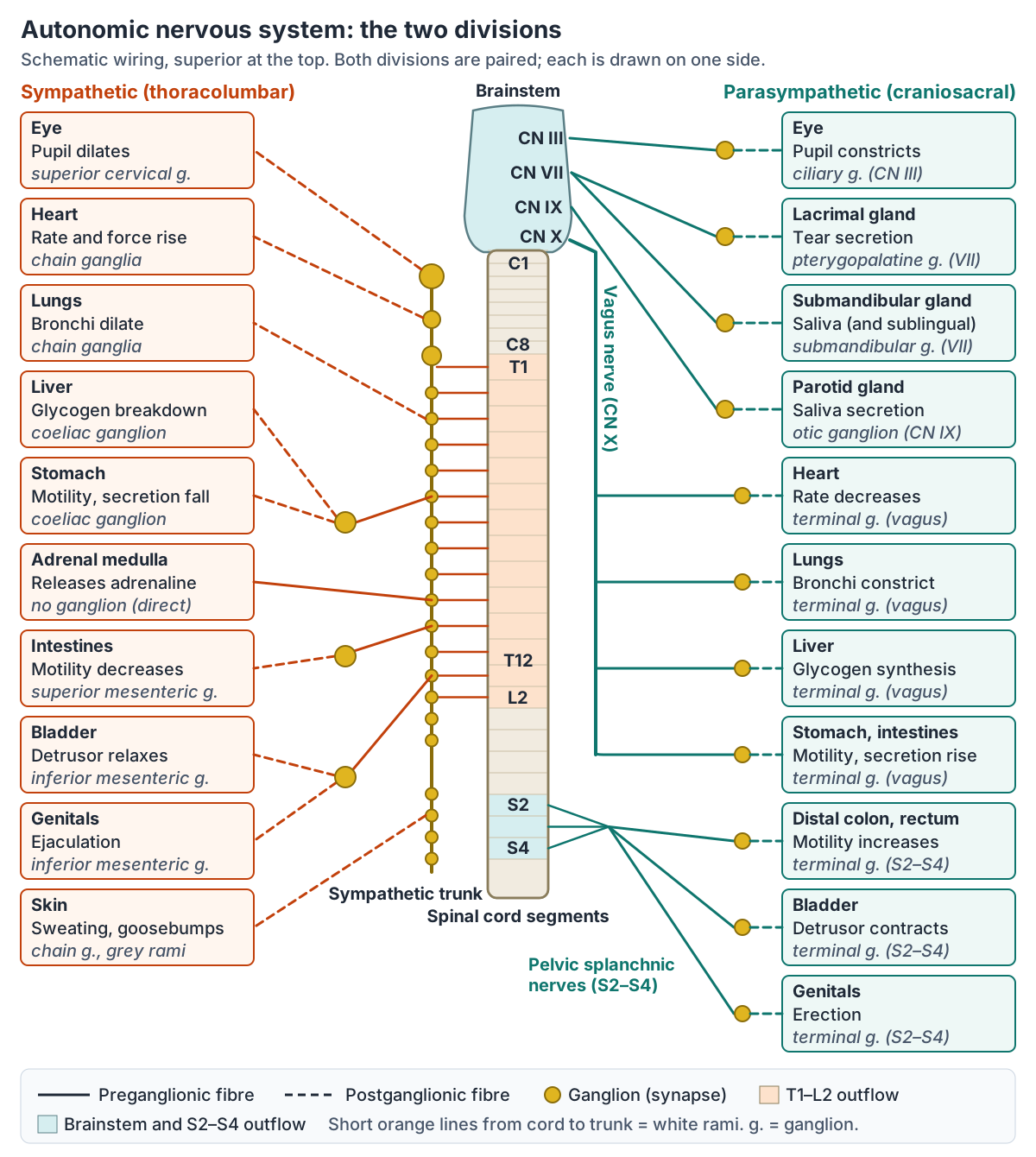
The autonomic nervous system is a functional division with parts in both the CNS and the PNS. It controls the smooth muscle, cardiac muscle and glands of the internal organs without conscious control, and it has two branches: the sympathetic nervous system and the parasympathetic nervous system. (Kenhub: Autonomic nervous system.)
An autonomic motor pathway normally has two neurons in series. A preganglionic fibre runs from a neuron in the CNS to an autonomic ganglion, and a postganglionic fibre runs from the ganglion neuron to the target organ. The endings of postganglionic fibres are usually not compact synapses but chains of swellings along the fibre, called varicosities, which release transmitter onto the target tissue. (OpenStax Anatomy and Physiology 2e, 15.1: Divisions of the Autonomic Nervous System.)
Most organs receive fibres from both divisions, a pattern called dual innervation, and the two divisions usually push activity in opposite directions. At rest one division tends to dominate a given organ; this resting balance is called autonomic tone. (OpenStax Anatomy and Physiology 2e, 15.2: Autonomic Reflexes and Homeostasis.)
Sympathetic nervous system: thoracolumbar outflow
The sympathetic division is called the thoracolumbar system because its preganglionic neurons sit in the lateral horn of the thoracic and upper lumbar spinal cord. Their axons leave through the ventral roots and reach ganglia beside the vertebral column. (OpenStax Anatomy and Physiology 2e, 15.1: Divisions of the Autonomic Nervous System.)
The preganglionic cell bodies form the intermediolateral nucleus, and their axons leave between the T1 and L2 spinal segments. Kenhub gives the range as T1 to L2 or L3, so some textbooks extend it by one segment. (Radiopaedia: Sympathetic trunk.)
Sympathetic preganglionic fibres are short and myelinated; postganglionic fibres are long and unmyelinated. One preganglionic neuron may branch to 10 to 20 targets, which is why sympathetic activation spreads widely through the body at once. (OpenStax Anatomy and Physiology 2e, 15.1: Divisions of the Autonomic Nervous System.)
The sympathetic system is associated with the "fight or flight" response: heart rate and breathing increase, blood is shifted towards skeletal muscle, digestion slows and sweating increases. Because it also responds to emotional states, OpenStax notes that the phrase is being widened to "fight, flight or fright". (OpenStax Anatomy and Physiology 2e, 15.1: Divisions of the Autonomic Nervous System.)
The sympathetic trunk (sympathetic chain)
The sympathetic trunk is a chain of ganglia joined by nerve fibres, running on each side of the vertebral bodies from the upper neck to the coccyx. Radiopaedia counts 23 paired paravertebral ganglia plus one unpaired ganglion in front of the coccyx, the ganglion impar. (Radiopaedia: Sympathetic trunk.)
The number of ganglia varies between sources. TeachMeAnatomy gives 22 pairs: 3 cervical, 11 or 12 thoracic, 4 lumbar and 4 or 5 sacral, plus the single ganglion impar. OpenStax describes typically 23 ganglia in each chain. (TeachMeAnatomy: The Sympathetic Nervous System.)
There are three cervical ganglia: superior, middle and inferior. The superior cervical ganglion is formed by the fused ganglia of C1 to C4. In most people the inferior cervical ganglion fuses with the first thoracic ganglion to form the stellate (cervicothoracic) ganglion. (Radiopaedia: Sympathetic trunk.)
Preganglionic fibres reach the trunk through white rami communicantes, which are white because their fibres are myelinated. White rami exist only from T1 to L2; above and below these levels the spinal nerves are joined to the trunk only by grey rami communicantes, which carry unmyelinated postganglionic fibres back out to the spinal nerves. (Radiopaedia: Sympathetic trunk.)
Grey rami join all 31 pairs of spinal nerves, so sympathetic fibres reach the sweat glands, arrector pili muscles and blood vessels of the whole body wall and limbs. (Kenhub: Autonomic nervous system.)
Once in the trunk, a preganglionic fibre can do one of three things: synapse in the ganglion at its own level, travel up or down the trunk to synapse at another level, or pass through the trunk without synapsing and continue as a splanchnic nerve. (TeachMeAnatomy: The Sympathetic Nervous System.)
OpenStax gives two examples. A fibre from spinal nerve T1 can synapse in the T1 chain ganglion to supply the trachea, while the T1 fibres that control the eye climb the trunk to synapse in the superior cervical ganglion, whose postganglionic fibres then reach the eye. (OpenStax Anatomy and Physiology 2e, 15.1: Divisions of the Autonomic Nervous System.)
Splanchnic nerves, prevertebral ganglia and the adrenal medulla
Fibres that pass through the trunk without synapsing form the splanchnic nerves. They end in collateral (prevertebral) ganglia that lie in front of the vertebral column in the abdomen: the coeliac (celiac), superior mesenteric and inferior mesenteric ganglia. OpenStax notes that these ganglia are also considered part of the enteric nervous system. (OpenStax Anatomy and Physiology 2e, 15.1: Divisions of the Autonomic Nervous System.)
TeachMeAnatomy describes the greater splanchnic nerve (T5 to T9) going to the coeliac ganglion, which supplies the liver, gallbladder, stomach, spleen and pancreas; the lesser splanchnic nerve (T10 to T11) going to the superior mesenteric ganglion for the small intestine and colon; and the least splanchnic nerve (T12) going to the inferior mesenteric ganglion for the kidney, bladder, colon and reproductive organs. (TeachMeAnatomy: The Sympathetic Nervous System.)
Radiopaedia gives the greater splanchnic nerve as T5 to T10 and also describes lumbar splanchnic nerves from L1 to L2, so the exact levels differ slightly between sources. (Radiopaedia: Sympathetic trunk.)
The adrenal medulla is an exception to the two-neuron rule. Preganglionic fibres pass through the trunk and the coeliac ganglion without synapsing and end directly on its chromaffin cells, which act like postganglionic neurons and release adrenaline (epinephrine) and noradrenaline (norepinephrine) into the blood as hormones. TeachMeAnatomy therefore describes the adrenal medulla as a modified sympathetic ganglion. (TeachMeAnatomy: The Sympathetic Nervous System.)
Chromaffin cells develop from the same neural crest tissue as the sympathetic ganglia, and their hormones spread the sympathetic effect throughout the body through the bloodstream. (OpenStax Anatomy and Physiology 2e, 15.1: Divisions of the Autonomic Nervous System.)
Parasympathetic nervous system: craniosacral outflow
The parasympathetic division is called the craniosacral system because its preganglionic neurons lie in nuclei of the brainstem and in the lateral horn of the sacral spinal cord. Its preganglionic fibres are long and its postganglionic fibres short: the ganglia (terminal ganglia) lie close to or inside the target organ, and those in the organ wall are called intramural ganglia. (OpenStax Anatomy and Physiology 2e, 15.1: Divisions of the Autonomic Nervous System.)
Four cranial nerves carry parasympathetic fibres. The oculomotor nerve (CN III) carries fibres from the Edinger–Westphal nucleus to the ciliary ganglion, which supplies the sphincter pupillae and ciliary muscle. The facial nerve (CN VII) carries fibres from the superior salivatory nucleus to the pterygopalatine ganglion (lacrimal gland and glands of the nose, nasopharynx and palate) and the submandibular ganglion (submandibular and sublingual glands). (TeachMeAnatomy: Parasympathetic Innervation of the Head and Neck.)
The glossopharyngeal nerve (CN IX) carries fibres from the inferior salivatory nucleus through the lesser petrosal nerve to the otic ganglion, whose postganglionic fibres travel with the auriculotemporal nerve to the parotid gland. (TeachMeAnatomy: Parasympathetic Innervation of the Head and Neck.)
The vagus nerve (CN X) carries about 75% of all parasympathetic outflow. It supplies the thoracic organs and most of the gastrointestinal tract, synapsing in ganglia in plexuses near the target organs. (TeachMeAnatomy: The Parasympathetic Nervous System.)
Vagal parasympathetic fibres reach the oesophagus (esophagus), stomach and bowel as far as the splenic flexure of the colon, and slow the resting heart rate through the sinoatrial and atrioventricular nodes. (TeachMeAnatomy: The Vagus Nerve (CN X).)
The sacral outflow comes from the anterior rami of S2 to S4 as the pelvic splanchnic nerves. They supply the distal colon, rectum, bladder and reproductive organs, including the erectile tissue of the penis and clitoris. (TeachMeAnatomy: The Parasympathetic Nervous System.)
Sympathetic vs parasympathetic effects on organs
The table below compares the two divisions organ by organ. In the eye, sympathetic fibres dilate the pupil (mydriasis) and parasympathetic fibres constrict it (miosis). The sympathetic system increases heart rate and force, dilates the bronchi, slows peristalsis, breaks down liver glycogen and causes sweating and goosebumps; the parasympathetic system does the reverse where it has an effect. (Kenhub: Autonomic nervous system.)
The heart shows autonomic tone clearly. Without any parasympathetic input it would beat at about 100 beats per minute; vagal tone slows it to a resting 60 to 80, and sympathetic drive can raise it to around 120 to 140 during exercise. (OpenStax Anatomy and Physiology 2e, 15.2: Autonomic Reflexes and Homeostasis.)
Blood vessels show the opposite pattern. Most systemic vessels receive only sympathetic fibres, so sympathetic tone alone sets their diameter and noradrenaline constricts them to raise blood pressure. Vessels in skeletal muscle tend to dilate during sympathetic activity, and the only vessels with a parasympathetic supply are those of erectile tissue. (OpenStax Anatomy and Physiology 2e, 15.2: Autonomic Reflexes and Homeostasis.)
For the bladder, sympathetic fibres relax the detrusor muscle and contract the internal urethral sphincter, which helps store urine; parasympathetic fibres contract the detrusor and relax the internal sphincter to empty it. (TeachMeAnatomy: The Sympathetic Nervous System.)
Sympathetic vs parasympathetic effects on organs
| Organ or tissue | Sympathetic (fight or flight) | Parasympathetic (rest and digest) |
|---|---|---|
| Pupil (iris) | Dilates (mydriasis) | Constricts (miosis); ciliary muscle focuses for near vision |
| Heart | Rate and force of contraction increase | Rate decreases |
| Airways (bronchi) | Bronchodilation | Bronchoconstriction; more bronchial secretion |
| Most blood vessels | Constrict (skeletal muscle vessels dilate) | No significant input, except dilation in erectile tissue |
| Sweat glands | Secretion (sympathetic fibres release acetylcholine here) | No innervation |
| Arrector pili muscles | Contract (goosebumps) | No effect |
| Stomach and intestines | Motility and secretion decrease; sphincters contract | Motility and secretion increase; sphincters relax |
| Liver | Glycogen breakdown (glycogenolysis) | Glycogen synthesis |
| Pancreas | Insulin and exocrine secretion decrease | Insulin and exocrine secretion increase |
| Adrenal medulla | Releases adrenaline and noradrenaline into the blood | No effect |
| Urinary bladder | Detrusor relaxes; internal urethral sphincter contracts | Detrusor contracts; internal urethral sphincter relaxes |
| Male genitals | Ejaculation | Erection |
Compiled from Kenhub, TeachMeAnatomy and OpenStax (see references). Most organs receive both divisions (dual innervation); sweat glands, arrector pili and most blood vessels receive sympathetic fibres only.
Neurotransmitters and receptors
All preganglionic fibres, sympathetic and parasympathetic, release acetylcholine (ACh), which acts on nicotinic receptors on the ganglion neurons. Parasympathetic postganglionic fibres also release ACh, acting on muscarinic receptors. Sympathetic postganglionic fibres release noradrenaline onto α- or β-adrenergic receptors, except for the fibres to sweat glands and to blood vessels of skeletal muscle, which release ACh onto muscarinic receptors. (OpenStax Anatomy and Physiology 2e, 15.1: Divisions of the Autonomic Nervous System.)
Nicotinic receptors are ligand-gated ion channels that depolarise the cell when ACh binds. Muscarinic and adrenergic receptors are G protein-coupled receptors, whose effect depends on the subtype. Five adrenergic subtypes are recognised: α1, α2, β1, β2 and β3. (OpenStax Anatomy and Physiology 2e, 15.1: Divisions of the Autonomic Nervous System.)
Drugs show how the receptors work. Atropine blocks muscarinic receptors and dilates the pupil; phenylephrine is an α1-agonist used to dilate the pupil and raise blood pressure; and β-blockers such as metoprolol, which selectively blocks β1 receptors, keep the heart rate from rising in heart disease. (OpenStax Anatomy and Physiology 2e, 15.4: Drugs that Affect the Autonomic System.)
In the lungs, relaxation of airway smooth muscle is mediated solely by β2 receptors, which is why inhaled β2-agonists are the main quick-relief bronchodilators in asthma. (Barisione G et al. Beta-Adrenergic Agonists. Pharmaceuticals (Basel). 2010.)
Autonomic neurotransmitters and receptors
| Receptor | Activated by | Where it is found | Drug example |
|---|---|---|---|
| Nicotinic (ligand-gated ion channel) | Acetylcholine | Postganglionic neurons in all autonomic ganglia, sympathetic and parasympathetic | Nicotine (agonist at both divisions' ganglia) |
| Muscarinic (M1 to M5, G protein-coupled) | Acetylcholine | Targets of parasympathetic postganglionic fibres; sympathetic targets in sweat glands and skeletal muscle blood vessels | Atropine and scopolamine block it; pilocarpine stimulates it |
| α1-adrenergic | Noradrenaline, adrenaline | For example, the radial (dilator) smooth muscle of the iris | Phenylephrine (agonist: dilates the pupil, raises blood pressure) |
| β1-adrenergic | Noradrenaline, adrenaline | Heart (receptors that raise heart rate) | Metoprolol (selective β1-blocker) |
| β2-adrenergic | Adrenaline, noradrenaline | Airway smooth muscle (relaxation, bronchodilation) | Inhaled β2-agonists for asthma |
Adrenergic receptors (α1, α2, β1, β2 and β3) are all G protein-coupled. Sources: OpenStax §15.1 and §15.4; Barisione et al. 2010 for β2 receptors in the airways.
The enteric nervous system
The enteric nervous system is the network of neurons in the wall of the digestive tract, from the oesophagus to the anus, with about 100 million motor, sensory and interneurons. Its myenteric (Auerbach's) plexus in the muscular layer controls the rhythm and force of contractions, and its submucosal (Meissner's) plexus regulates secretions in response to food. (OpenStax Anatomy and Physiology 2e, 23.1: Overview of the Digestive System.)
The autonomic system acts on the enteric system from outside. Sympathetic input suppresses enteric neurons and reduces secretion and motility, while parasympathetic input stimulates them and increases both. (OpenStax Anatomy and Physiology 2e, 23.1: Overview of the Digestive System.)
The enteric system can also run short reflexes without the brain or spinal cord. For example, stretch of the stomach wall activates a local neuron that increases smooth muscle motility; that neuron is a postganglionic parasympathetic neuron that the vagus nerve can influence. (OpenStax Anatomy and Physiology 2e, 15.2: Autonomic Reflexes and Homeostasis.)
How the brain controls the autonomic nervous system
The hypothalamus is the main control centre for autonomic function and also for the endocrine system. Its output reaches the brainstem parasympathetic nuclei and the thoracolumbar preganglionic neurons mainly through two pathways, the medial forebrain bundle and the dorsal longitudinal fasciculus. The amygdala signals the hypothalamus during fear and anxiety, triggering the sympathetic response. (OpenStax Anatomy and Physiology 2e, 15.3: Central Control.)
In the medulla, the cardiovascular centre controls heart rate and vessel diameter. The solitary nucleus receives blood pressure and cardiac information from the glossopharyngeal and vagus nerves, and the dorsal motor nucleus of the vagus sends parasympathetic output that slows the heart, constricts the bronchi and supports digestion. (OpenStax Anatomy and Physiology 2e, 15.3: Central Control.)
Autonomic reflexes keep variables in range. In the baroreceptor reflex, stretch receptors in the carotid sinuses and the wall of the aorta signal through the glossopharyngeal and vagus nerves. When you stand up, a sympathetic reflex raises heart rate and constricts vessels so the brain stays perfused; if it fails, for example with low blood volume or autonomic damage from diabetes, the result is orthostatic hypotension (dizziness on standing). (OpenStax Anatomy and Physiology 2e, 15.2: Autonomic Reflexes and Homeostasis.)
Autonomic dysreflexia shows what happens when the brain loses control. In people with a spinal cord injury, usually at or above T6, a stimulus below the injury such as a full bladder triggers widespread sympathetic vasoconstriction and dangerous hypertension, because descending signals that would normally damp it down cannot pass the injury. It is a medical emergency. (TeachMeAnatomy: The Sympathetic Nervous System.)
Frequently asked questions
What is the autonomic nervous system?
The autonomic nervous system is the part of the nervous system that controls cardiac muscle, smooth muscle and glands without conscious effort. It regulates heart rate, blood pressure, breathing passages, digestion, sweating, pupil size and bladder function. It has a sympathetic division (fight or flight) and a parasympathetic division (rest and digest), and works closely with the enteric nervous system of the gut.
What is the difference between the sympathetic and parasympathetic nervous systems?
The sympathetic division leaves the spinal cord from T1 to L2, has short preganglionic and long postganglionic fibres, and prepares the body for action. The parasympathetic division leaves the brainstem in cranial nerves III, VII, IX and X and the sacral cord at S2 to S4, has long preganglionic and short postganglionic fibres, and promotes rest and digestion.
What is the sympathetic trunk?
The sympathetic trunk (sympathetic chain) is a pair of nerve cords running beside the vertebral column from the upper neck to the coccyx, each carrying a series of ganglia. Preganglionic fibres enter it through white rami at T1 to L2, then synapse at the same level, travel up or down, or pass through as splanchnic nerves.
What is the difference between the central and peripheral nervous system?
The central nervous system is the brain and spinal cord. The peripheral nervous system is everything outside them: the 12 pairs of cranial nerves, the 31 pairs of spinal nerves, and the ganglia. Cell body clusters are called nuclei in the CNS and ganglia in the PNS, and axon bundles are tracts in the CNS and nerves in the PNS.
Which neurotransmitters does the autonomic nervous system use?
All preganglionic neurons release acetylcholine onto nicotinic receptors. Parasympathetic postganglionic neurons release acetylcholine onto muscarinic receptors. Most sympathetic postganglionic neurons release noradrenaline onto alpha or beta adrenergic receptors; the exceptions, to sweat glands and skeletal muscle blood vessels, release acetylcholine. The adrenal medulla releases adrenaline and noradrenaline into the blood.
Is the enteric nervous system part of the autonomic nervous system?
It is often treated as a third division. The enteric nervous system lies in the wall of the gut, contains about 100 million neurons in the myenteric and submucosal plexuses, and can run digestive reflexes on its own. The sympathetic and parasympathetic divisions modify its activity, so the two systems overlap heavily.
References
- OpenStax. Anatomy and Physiology 2e, §12.1 Basic Structure and Function of the Nervous System. Rice University, 2022.
- OpenStax. Anatomy and Physiology 2e, §15.1 Divisions of the Autonomic Nervous System. Rice University, 2022.
- OpenStax. Anatomy and Physiology 2e, §15.2 Autonomic Reflexes and Homeostasis. Rice University, 2022.
- OpenStax. Anatomy and Physiology 2e, §15.3 Central Control. Rice University, 2022.
- OpenStax. Anatomy and Physiology 2e, §15.4 Drugs that Affect the Autonomic System. Rice University, 2022.
- OpenStax. Anatomy and Physiology 2e, §23.1 Overview of the Digestive System. Rice University, 2022.
- TeachMeAnatomy. The Sympathetic Nervous System.
- TeachMeAnatomy. The Parasympathetic Nervous System.
- TeachMeAnatomy. Parasympathetic Innervation of the Head and Neck.
- TeachMeAnatomy. The Vagus Nerve (CN X).
- Kenhub. Autonomic nervous system.
- Radiopaedia. Sympathetic trunk.
- Barisione G, Baroffio M, Crimi E, Brusasco V. Beta-Adrenergic Agonists. Pharmaceuticals (Basel). 2010;3(4):1016–1044. doi:10.3390/ph3041016
Related anatomy
- Cranial nervesThe 12 cranial nerves in order with a labelled diagram: type, skull foramen, functions and test for CN I to CN XII…
- Spinal cordSpinal cord anatomy with labelled diagrams: cross-section, grey matter horns, spinothalamic and dorsal column tracts…
- Brain anatomyBrain anatomy explained: cerebrum, lobes, gyri, insula, corpus callosum, thalamus, hippocampus, brainstem and…
- Eye anatomyEyeball anatomy with a labeled 3D eye model: sclera, cornea, choroid, iris, lens, retina and chambers, the 3 layers…
Explore the whole body in the interactive 3D human anatomy model, or browse the A–Z list of human structures.