

What are the cranial nerves?
The peripheral nervous system is everything that is not the brain or spinal cord. Its nerves are grouped into two sets: the cranial nerves, which connect to the brain, and the spinal nerves, which connect to the spinal cord. There are 31 pairs of spinal nerves and 12 pairs of cranial nerves, designated CN I to CN XII with Roman numerals. (OpenStax Anatomy and Physiology 2e, 13.4: The Peripheral Nervous System.)
The cranial nerves carry three kinds of information. Some are strictly sensory (smell, vision, hearing and balance), some are strictly motor (eye movement, tongue and neck muscles), and some are mixed, carrying sensory and motor fibres together. Four of them (CN III, VII, IX and X) also carry autonomic fibres: the oculomotor nerve constricts the pupil, the facial and glossopharyngeal nerves drive salivation, and the vagus nerve supplies autonomic ganglia in the thoracic and upper abdominal cavities. (OpenStax Anatomy and Physiology 2e, 13.4: The Peripheral Nervous System.)
The cranial nerves leave the brain at different levels. CN I comes from the telencephalon (the cerebral hemispheres) and CN II from the diencephalon. CN III and IV arise from the midbrain, the middle four (CN V to VIII) from the pons, and the last four (CN IX to XII) from the medulla oblongata. (Radiopaedia: Cranial nerves.)
Cranial nerves list in order (with mnemonics)
In order, the 12 cranial nerves are: I olfactory, II optic, III oculomotor, IV trochlear, V trigeminal, VI abducens, VII facial, VIII vestibulocochlear, IX glossopharyngeal, X vagus, XI accessory and XII hypoglossal. Clinicians often write them as CN 1 to CN 12 or CN I to CN XII; "CN 8", "CN VIII" and "cranial nerve 8" all mean the vestibulocochlear nerve. (TeachMeAnatomy: Summary of the Cranial Nerves.)
A classic mnemonic for the names is "On Old Olympus's Towering Top, A Finn And German Viewed Some Hops". A version that fits the modern names is "Ooh, Ooh, Ooh, To Touch And Feel Very Good Velvet. Such Heaven!" (Radiopaedia: Cranial nerves (mnemonic).)
For type, the mnemonic "Some Say Marry Money But My Brother Says Big Brains Matter More" gives S (sensory), M (motor) or B (both) in order: I, II and VIII are sensory; III, IV, VI, XI and XII are motor; V, VII, IX and X are both. (Radiopaedia: Cranial nerves (mnemonic).)
Different textbooks word these mnemonics slightly differently. OpenStax, for example, uses "Some Say Marry Money But My Brother Says Brains Beauty Matter More", which gives the same S, M and B sequence. (OpenStax Anatomy and Physiology 2e, 13.4: The Peripheral Nervous System.)
Cranial nerves table: type, foramen, functions and test
| Nerve | Type | Leaves the skull through | Main functions | Quick bedside test |
|---|---|---|---|---|
| CN I Olfactory | Sensory | Cribriform plate of the ethmoid | Smell | Identify a familiar smell (coffee, mint) with each nostril |
| CN II Optic | Sensory | Optic canal | Vision | Snellen chart for acuity; visual fields; light reflex (afferent limb) |
| CN III Oculomotor | Motor | Superior orbital fissure | Four extraocular muscles, lifts upper eyelid, constricts pupil, accommodation | Follow a pen with the eyes; pupil response to light |
| CN IV Trochlear | Motor | Superior orbital fissure | Superior oblique muscle | Follow a pen with the eyes, including looking down and in |
| CN V Trigeminal | Both | V1 superior orbital fissure; V2 foramen rotundum; V3 foramen ovale | Sensation of the face, muscles of mastication | Light touch and sharp pain on the face; jaw-jerk reflex |
| CN VI Abducens | Motor | Superior orbital fissure | Lateral rectus (abducts the eye) | Follow a pen with the eyes, including looking outward |
| CN VII Facial | Both | Internal acoustic meatus, then stylomastoid foramen | Facial expression, taste from front two-thirds of tongue, tears and saliva | Squeeze eyes shut against resistance; taste on each side of the tongue |
| CN VIII Vestibulocochlear | Sensory | Internal acoustic meatus (does not leave the skull) | Hearing and balance | Rinne and Weber tuning-fork tests |
| CN IX Glossopharyngeal | Both | Jugular foramen | Taste and sensation from back third of tongue, carotid sinus and body, parotid secretion, stylopharyngeus | Gag reflex (sensory limb); taste at the back of the tongue |
| CN X Vagus | Both | Jugular foramen | Pharynx and larynx muscles, parasympathetic supply to thoracic and abdominal organs, visceral sensation | Gag reflex (motor limb); watch the soft palate while saying "lah-kah-pah" |
| CN XI Accessory | Motor | Jugular foramen (spinal root enters through foramen magnum) | Sternocleidomastoid and trapezius | Shrug shoulders and turn the head against resistance |
| CN XII Hypoglossal | Motor | Hypoglossal canal | All tongue muscles except palatoglossus | Stick out the tongue; it deviates towards a damaged side |
Types and foramina follow TeachMeAnatomy's summary; tests follow the OpenStax cranial nerve exam and TeachMeAnatomy. "Both" means the nerve carries sensory and motor fibres.
Cranial nerves labelled: where each nerve leaves the brain
Seen from below, the olfactory bulbs and olfactory tracts lie on the underside of the frontal lobes, one on each side of the midline. (TeachMeAnatomy: The Olfactory Nerve (CN I).)
Behind them the two optic nerves meet at the optic chiasm, which lies close to the pituitary gland; behind the chiasm the fibres continue as the optic tracts. (TeachMeAnatomy: The Optic Nerve (CN II).)
The oculomotor nerve leaves the anterior surface of the brainstem between the midbrain and the pons. (Radiopaedia: Oculomotor nerve.)
The trochlear nerve is the only cranial nerve that leaves the back (posterior) surface of the brainstem, so on the underside of the brain it is seen only after it has curved around the side of the midbrain. (TeachMeAnatomy: The Trochlear Nerve (CN IV).)
The trigeminal nerve attaches to the anterolateral pons as a large sensory root, with one or more smaller motor roots just above and medial to it. (Radiopaedia: Trigeminal nerve.)
The abducens nerve emerges at the junction of the pons and medulla (the pontomedullary junction), close to the midline. (TeachMeAnatomy: The Abducens Nerve (CN VI).)
The facial nerve emerges from the lower lateral pons at the pontomedullary sulcus, lateral to the abducens nerve and medial to the vestibulocochlear nerve, and crosses the cerebellopontine angle cistern to the internal acoustic meatus. (Radiopaedia: Facial nerve.)
On the medulla, the glossopharyngeal nerve leaves the postolivary sulcus, behind the olive, just above the vagus nerve. (Radiopaedia: Glossopharyngeal nerve.)
The hypoglossal rootlets leave through the anterolateral sulcus of the medulla, between the pyramid and the olive, in front of the olive, whereas the glossopharyngeal and vagus rootlets leave behind it. (Radiopaedia: Hypoglossal nerve.)
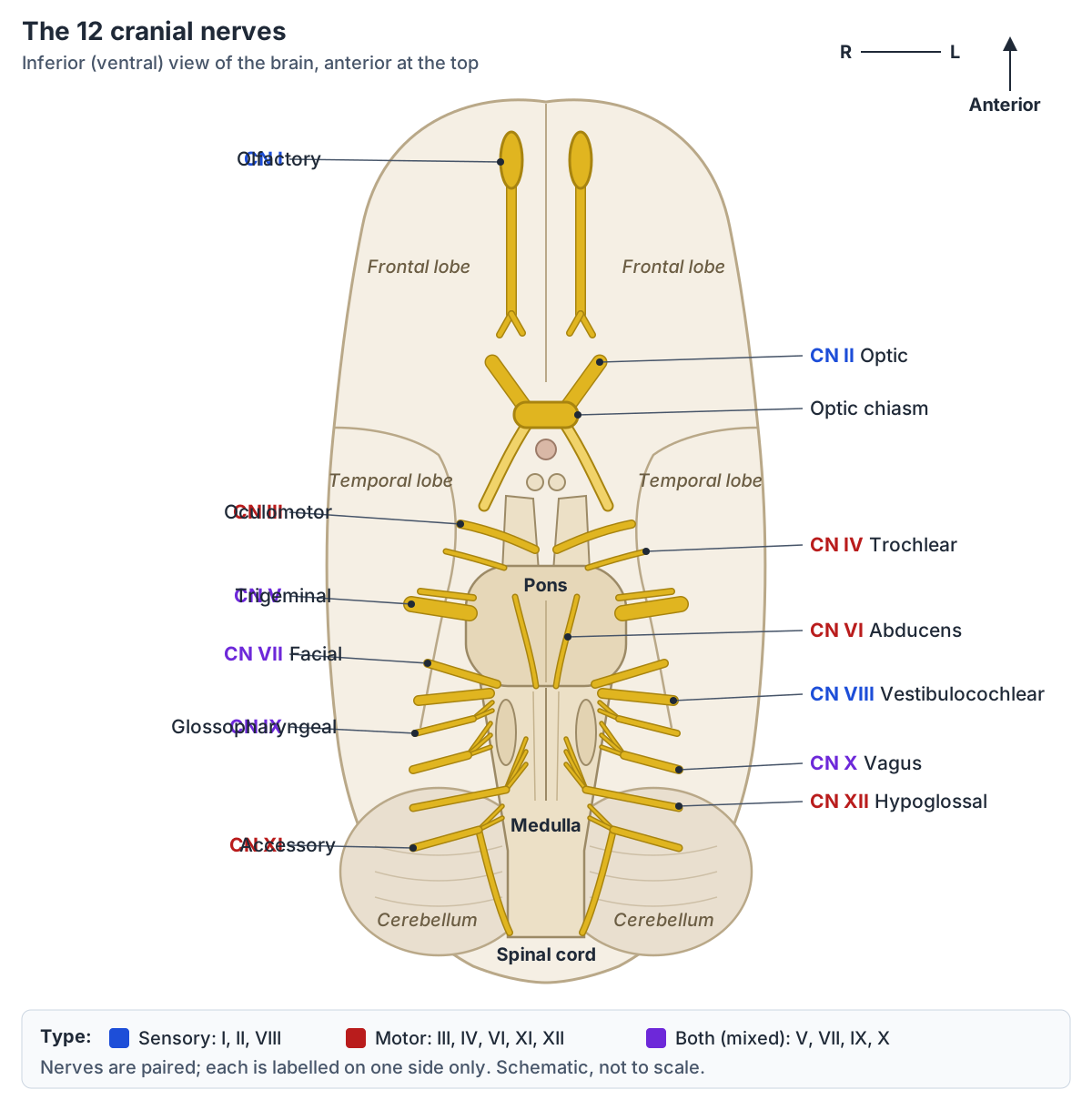
CN I: olfactory nerve and olfactory tract
The olfactory nerve (CN I) is the first and shortest cranial nerve and is purely sensory, for smell. Its receptor cells are bipolar neurons in the olfactory mucosa in the roof of the nasal cavity. Their axons gather into bundles that pass up through the small holes of the cribriform plate of the ethmoid bone. (TeachMeAnatomy: The Olfactory Nerve (CN I).)
The fibres end in the olfactory bulb, which lies in the anterior cranial fossa, where they synapse with mitral cells. The olfactory tract then runs back along the underside of the frontal lobe and divides into lateral and medial striae. The lateral stria carries fibres to the primary olfactory cortex near the uncus of the temporal lobe. (TeachMeAnatomy: The Olfactory Nerve (CN I).)
Loss of smell is called anosmia. It can be temporary (a cold), permanent (head injury, or a tumour such as a meningioma in the olfactory groove) or progressive, as in Parkinson's and Alzheimer's disease, where it may come before the movement or memory symptoms. (TeachMeAnatomy: The Olfactory Nerve (CN I).)
CN II: optic nerve
The optic nerve (CN II) is formed by the axons of retinal ganglion cells. It leaves the orbit through the optic canal of the sphenoid bone. At the optic chiasm, fibres from the nasal (medial) half of each retina cross to the other side, while fibres from the temporal half stay on the same side. Behind the chiasm the fibres continue as the optic tract to the lateral geniculate nucleus of the thalamus, and from there to the visual cortex. (TeachMeAnatomy: The Optic Nerve (CN II).)
Because the chiasm lies close to the pituitary gland, a pituitary adenoma can press on the crossing nasal fibres and cause bitemporal hemianopia: loss of the outer (temporal) half of the visual field in both eyes. (TeachMeAnatomy: The Optic Nerve (CN II).)
CN III: oculomotor nerve
The oculomotor nerve (CN III) arises from the oculomotor nucleus in the midbrain, runs through the lateral wall of the cavernous sinus and enters the orbit through the superior orbital fissure. Its superior division supplies superior rectus and levator palpebrae superioris; its inferior division supplies inferior rectus, medial rectus and inferior oblique. (TeachMeAnatomy: The Oculomotor Nerve (CN III).)
CN III also carries parasympathetic fibres from the Edinger–Westphal nucleus. They synapse in the ciliary ganglion, and the short ciliary nerves then supply the sphincter pupillae (pupil constriction) and the ciliary muscle (focusing the lens for near vision). (TeachMeAnatomy: Parasympathetic Innervation of the Head and Neck.)
A complete oculomotor palsy leaves the eye "down and out", because only lateral rectus (CN VI) and superior oblique (CN IV) still work, with a drooping upper eyelid (ptosis) and a dilated pupil. Causes include raised intracranial pressure and an aneurysm of the posterior communicating artery. (TeachMeAnatomy: The Oculomotor Nerve (CN III).)
CN IV: trochlear nerve and its function
The trochlear nerve (CN IV) is the smallest cranial nerve by number of axons but has the longest course inside the skull. It is the only cranial nerve to leave the posterior (dorsal) surface of the brainstem, and its fibres cross in the midbrain, so each nerve supplies the superior oblique muscle of the opposite side. It enters the orbit through the superior orbital fissure. (TeachMeAnatomy: The Trochlear Nerve (CN IV).)
Trochlear nerve function is purely motor: the superior oblique mainly depresses and intorts (rotates inward) the eyeball. A trochlear palsy causes vertical double vision that is worst when looking down and in, for example when reading or walking downstairs, and patients often tilt the head away from the affected side. (TeachMeAnatomy: The Trochlear Nerve (CN IV).)
CN V (CN5): trigeminal nerve and the mandibular nerve
The trigeminal nerve (CN V) is the main sensory nerve of the face and the motor nerve of chewing. It has four nuclei, three sensory and one motor, and attaches to the pons as a large sensory root and a small motor root. The sensory root swells into the trigeminal ganglion in a depression of the middle cranial fossa, lateral to the cavernous sinus. (TeachMeAnatomy: The Trigeminal Nerve (CN V).)
From the ganglion come three divisions. The ophthalmic nerve (V1) leaves through the superior orbital fissure and supplies the forehead, scalp, upper eyelid, cornea and back of the nose. The maxillary nerve (V2) leaves through the foramen rotundum and supplies the lower eyelid, cheek, upper lip, upper teeth, palate and nasal cavity. (TeachMeAnatomy: The Trigeminal Nerve (CN V).)
The mandibular nerve (V3) leaves through the foramen ovale into the infratemporal fossa. It gives sensation to the lower lip, chin, lower teeth, part of the external ear, the floor of the mouth and general sensation (not taste) to the anterior two-thirds of the tongue. Its main sensory branches are the buccal, inferior alveolar, auriculotemporal and lingual nerves. V3 is the only division with motor fibres: it supplies masseter, temporalis, the medial and lateral pterygoids, the anterior belly of digastric, mylohyoid, tensor veli palatini and tensor tympani. (TeachMeAnatomy: The Trigeminal Nerve (CN V).)
Compression of the trigeminal root by a nearby blood vessel at the point where it enters the pons is linked with trigeminal neuralgia. (Radiopaedia: Trigeminal nerve.)
CN VI: abducens nerve
The abducens nerve (CN VI) arises from the abducens nucleus in the pons and leaves the brainstem at the pontomedullary junction. It pierces the dura, runs through Dorello's canal, passes over the tip of the petrous temporal bone into the cavernous sinus, and enters the orbit through the superior orbital fissure. It supplies only lateral rectus, which abducts the eye (turns it outward). (TeachMeAnatomy: The Abducens Nerve (CN VI).)
In an abducens palsy the affected eye rests turned inward, because medial rectus is unopposed, and cannot look outward, causing double vision. Its long course inside the skull makes it vulnerable to stretching when the brainstem is pushed downward, for example by a space-occupying lesion. (TeachMeAnatomy: The Abducens Nerve (CN VI).)
CN VII (CN 7): facial nerve and facial nerve branches
The facial nerve (CN VII) has a large motor root and a small sensory root, the nervus intermedius. Both enter the internal acoustic meatus of the petrous temporal bone. In the facial canal they fuse and form the geniculate ganglion, and the nerve finally leaves the skull through the stylomastoid foramen, just behind the styloid process. (TeachMeAnatomy: The Facial Nerve (CN VII).)
Inside the facial canal it gives three branches: the greater petrosal nerve (parasympathetic fibres to the lacrimal gland and mucous glands), the nerve to stapedius, and the chorda tympani, which carries taste from the anterior two-thirds of the tongue and parasympathetic fibres to the submandibular and sublingual glands. After the stylomastoid foramen it gives the posterior auricular nerve and the nerves to the posterior belly of digastric and to stylohyoid. (TeachMeAnatomy: The Facial Nerve (CN VII).)
The nerve then enters the parotid gland and divides into five terminal branches for the muscles of facial expression. From top to bottom they are the temporal, zygomatic, buccal, marginal mandibular and cervical branches. Two common mnemonics are "Two Zebras Bit My Coccyx" and "To Zanzibar By Motor Car". (Radiopaedia: Facial nerve branches (mnemonic).)
The marginal mandibular branch usually runs along the lower border of the mandible, but in about one person in five it lies up to 2 cm below it. (Radiopaedia: Facial nerve.)
Damage to the facial nerve itself (a lower motor neuron lesion) weakens the whole of one side of the face. Bell palsy, the most common cause, is an idiopathic lower motor neuron facial palsy. A lesion higher up, in the motor pathway from the cerebral cortex (an upper motor neuron lesion), typically spares the forehead on the affected side, so the patient can still wrinkle the brow and close the eye. The traditional explanation is that the upper face receives input from both cerebral hemispheres, although Radiopaedia notes this may not be fully supported by the literature. (Radiopaedia: Facial palsy.)
Facial nerve (CN VII) branches
| Branch | Where it arises | What it supplies |
|---|---|---|
| Greater petrosal nerve | Facial canal (geniculate ganglion) | Parasympathetic fibres to the lacrimal gland and mucous glands of the nose and palate |
| Nerve to stapedius | Facial canal | Stapedius muscle in the middle ear |
| Chorda tympani | Facial canal | Taste from the anterior two-thirds of the tongue; parasympathetic fibres to the submandibular and sublingual glands |
| Posterior auricular nerve | Just below the stylomastoid foramen | Muscles around the ear and the occipital belly of occipitofrontalis |
| Nerve to posterior belly of digastric | Just below the stylomastoid foramen | Posterior belly of digastric |
| Nerve to stylohyoid | Just below the stylomastoid foramen | Stylohyoid |
| Temporal, zygomatic, buccal, marginal mandibular and cervical branches | Within the parotid gland | Muscles of facial expression; the cervical branch supplies platysma |
CN VIII (cranial nerve 8): vestibulocochlear nerve
The vestibulocochlear nerve (CN VIII) is purely sensory. It leaves the brainstem at the cerebellopontine angle with the facial nerve and enters the internal acoustic meatus of the temporal bone, where it divides into a cochlear nerve for hearing and a vestibular nerve for balance. (TeachMeAnatomy: The Vestibulocochlear Nerve (CN VIII).)
The cochlear part carries signals from hair cells in the cochlea, where loudness and pitch are coded, to the cochlear nuclei; its cell bodies form the spiral ganglion. The vestibular part carries signals from the otolith organs (linear acceleration) and the semicircular canals (rotation) to the vestibular nuclei, which coordinate balance and keep the gaze steady. (TeachMeAnatomy: The Vestibulocochlear Nerve (CN VIII).)
Inflammation of the vestibular part (vestibular neuritis) causes vertigo, nystagmus, imbalance and nausea. In benign paroxysmal positional vertigo (BPPV), calcium crystals in a semicircular canal cause brief vertigo when the head changes position. (TeachMeAnatomy: The Vestibulocochlear Nerve (CN VIII).)
CN IX (CN 9): glossopharyngeal nerve
The glossopharyngeal nerve (CN IX) leaves the medulla from the postolivary sulcus and exits the skull through the jugular foramen, where its superior and inferior ganglia hold its sensory cell bodies. Its tympanic branch supplies the middle ear, and its carotid sinus nerve carries signals from the carotid sinus (blood pressure) and carotid body (blood oxygen). (TeachMeAnatomy: The Glossopharyngeal Nerve (CN IX).)
CN IX supplies only one muscle, stylopharyngeus, which raises the pharynx and larynx during swallowing. Its pharyngeal, tonsillar and lingual branches give sensation to the oropharynx and palatine tonsil, and both general sensation and taste to the posterior third of the tongue. (TeachMeAnatomy: The Glossopharyngeal Nerve (CN IX).)
Its parasympathetic fibres start in the inferior salivatory nucleus, travel in the tympanic and lesser petrosal nerves to the otic ganglion, and then ride on the auriculotemporal nerve (a branch of V3) to the parotid gland. In the gag reflex, CN IX is the sensory (afferent) limb and the vagus nerve is the motor (efferent) limb. (TeachMeAnatomy: The Glossopharyngeal Nerve (CN IX).)
CN X: vagus nerve
The vagus nerve (CN X) takes its name from the Latin for "wandering", because it travels from the brainstem down into the abdomen. It leaves the medulla, exits the skull through the jugular foramen with CN IX and CN XI, and descends in the carotid sheath with the internal jugular vein and common carotid artery. (TeachMeAnatomy: The Vagus Nerve (CN X).)
In the neck it gives pharyngeal branches to most muscles of the pharynx and soft palate, and the superior laryngeal nerve. The recurrent laryngeal nerves run back up to the larynx: the right hooks under the right subclavian artery and the left under the arch of the aorta. Damage to a recurrent laryngeal nerve causes hoarseness. The vagus also supplies palatoglossus, the one tongue muscle not supplied by CN XII. (TeachMeAnatomy: The Vagus Nerve (CN X).)
In the thorax and abdomen the vagus is the main parasympathetic nerve: it slows the heart and supplies the airways, oesophagus (esophagus), stomach and bowel as far as the splenic flexure of the colon. It also carries visceral sensation from these organs back to the brainstem. (TeachMeAnatomy: The Vagus Nerve (CN X).)
CN XI: spinal accessory nerve (nervus accessorius)
The accessory nerve (CN XI, spinal accessory nerve, Latin nervus accessorius) is unusual because its main part starts in the spinal cord. The spinal root arises from neurons of the upper cervical cord (about C1 to C5 or C6), climbs into the skull through the foramen magnum, and leaves again through the jugular foramen. A small cranial root from the medulla joins the vagus and is often treated as part of CN X. (TeachMeAnatomy: The Accessory Nerve (CN XI).)
Outside the skull, CN XI supplies sternocleidomastoid and then crosses the posterior triangle of the neck to trapezius. Because it runs superficially there, it can be injured during lymph node biopsy, which weakens shoulder shrugging and head turning. It is tested by asking the patient to shrug and to turn the head against resistance. (TeachMeAnatomy: The Accessory Nerve (CN XI).)
CN XII (cranial nerve 12): hypoglossal nerve and the hypoglossal canal
The hypoglossal nerve (CN XII, often written "hypoglossal n.") arises from the hypoglossal nucleus in the medulla and leaves the skull through the hypoglossal canal of the occipital bone, the nerve that gives the canal its name. It then descends below the angle of the mandible, crosses the internal and external carotid arteries and enters the tongue. (TeachMeAnatomy: The Hypoglossal Nerve (CN XII).)
It is purely motor and supplies all the intrinsic tongue muscles and the extrinsic muscles genioglossus, hyoglossus and styloglossus; palatoglossus is supplied by the vagus. Fibres from the cervical plexus travel in its sheath without mixing with its own fibres, and leave it to supply geniohyoid, thyrohyoid and, through the ansa cervicalis, the other infrahyoid muscles. (TeachMeAnatomy: The Hypoglossal Nerve (CN XII).)
If one hypoglossal nerve is damaged, the protruded tongue deviates towards the damaged side, and that side may waste and twitch (fasciculate). (TeachMeAnatomy: The Hypoglossal Nerve (CN XII).)
How are the cranial nerves tested?
The cranial nerve exam is part of the neurological examination. Smell is tested with familiar odours, vision with a Snellen chart, visual fields and an ophthalmoscope, and the three eye-movement nerves (III, IV and VI) by asking the patient to follow a pen with the eyes. Pupil constriction to light tests the optic nerve (input) and the oculomotor nerve (output). (OpenStax Anatomy and Physiology 2e, 16.3: The Cranial Nerve Exam.)
The trigeminal nerve is tested by asking the patient to tell light touch from a sharp stimulus on the face, and the facial nerve by asking the patient to squeeze the eyes shut while the examiner tries to open them, and by taste. The Rinne and Weber tests check hearing. Touching the back of the throat tests the gag reflex (IX and X), and the patient says "lah-kah-pah" while the soft palate is watched. Shrugging against resistance tests XI, and sticking out the tongue tests XII. (OpenStax Anatomy and Physiology 2e, 16.3: The Cranial Nerve Exam.)
The 3D model on EasyAnatomyLab includes the skull, so you can find the openings these nerves pass through on the skull foramina page, but the nerves themselves are not part of the BodyParts3D data set.
Frequently asked questions
What are the 12 cranial nerves in order?
In order from front to back they are: I olfactory, II optic, III oculomotor, IV trochlear, V trigeminal, VI abducens, VII facial, VIII vestibulocochlear, IX glossopharyngeal, X vagus, XI accessory and XII hypoglossal. A common mnemonic is "Ooh, Ooh, Ooh, To Touch And Feel Very Good Velvet. Such Heaven!".
Which cranial nerves are sensory, motor or both?
CN I (smell), II (vision) and VIII (hearing and balance) are sensory. CN III, IV, VI, XI and XII are motor. CN V, VII, IX and X are mixed, carrying both sensory and motor fibres. The mnemonic "Some Say Marry Money But My Brother Says Big Brains Matter More" gives the sequence S, S, M, M, B, M, B, S, B, B, M, M.
What does cranial nerve 8 (CN VIII) do?
Cranial nerve 8, the vestibulocochlear nerve, is a purely sensory nerve for hearing and balance. Its cochlear part carries sound signals from the cochlea, and its vestibular part carries head-movement signals from the semicircular canals and otolith organs. It runs through the internal acoustic meatus with the facial nerve and does not leave the skull.
What are the branches of the facial nerve?
Inside the temporal bone the facial nerve gives the greater petrosal nerve, the nerve to stapedius and the chorda tympani. After the stylomastoid foramen it gives the posterior auricular nerve and nerves to digastric and stylohyoid. In the parotid gland it ends in five branches: temporal, zygomatic, buccal, marginal mandibular and cervical ("To Zanzibar By Motor Car").
Which nerve passes through the hypoglossal canal?
The hypoglossal nerve (CN XII) passes through the hypoglossal canal in the occipital bone. It is the motor nerve of the tongue, supplying every tongue muscle except palatoglossus. When one hypoglossal nerve is damaged, the tongue points towards the injured side when it is stuck out.
Which cranial nerves pass through the jugular foramen?
Three cranial nerves leave the skull through the jugular foramen: the glossopharyngeal nerve (CN IX), the vagus nerve (CN X) and the accessory nerve (CN XI). The spinal root of the accessory nerve first enters the skull through the foramen magnum before leaving with the other two.
Which cranial nerves carry parasympathetic fibres?
Four do: the oculomotor nerve (CN III) to the ciliary ganglion for pupil constriction and focusing, the facial nerve (CN VII) to the pterygopalatine and submandibular ganglia for tears and saliva, the glossopharyngeal nerve (CN IX) to the otic ganglion for the parotid gland, and the vagus nerve (CN X) to the heart, lungs and most of the gut.
References
- OpenStax. Anatomy and Physiology 2e, §13.4 The Peripheral Nervous System. Rice University, 2022.
- OpenStax. Anatomy and Physiology 2e, §16.3 The Cranial Nerve Exam. Rice University, 2022.
- TeachMeAnatomy. Summary of the Cranial Nerves.
- TeachMeAnatomy. The Olfactory Nerve (CN I).
- TeachMeAnatomy. The Optic Nerve (CN II).
- TeachMeAnatomy. The Oculomotor Nerve (CN III).
- TeachMeAnatomy. The Trochlear Nerve (CN IV).
- TeachMeAnatomy. The Trigeminal Nerve (CN V).
- TeachMeAnatomy. The Abducens Nerve (CN VI).
- TeachMeAnatomy. The Facial Nerve (CN VII).
- TeachMeAnatomy. The Vestibulocochlear Nerve (CN VIII).
- TeachMeAnatomy. The Glossopharyngeal Nerve (CN IX).
- TeachMeAnatomy. The Vagus Nerve (CN X).
- TeachMeAnatomy. The Accessory Nerve (CN XI).
- TeachMeAnatomy. The Hypoglossal Nerve (CN XII).
- TeachMeAnatomy. Parasympathetic Innervation of the Head and Neck.
- Kenhub. The 12 cranial nerves.
- Radiopaedia. Cranial nerves.
- Radiopaedia. Cranial nerves (mnemonic).
- Radiopaedia. Oculomotor nerve.
- Radiopaedia. Trigeminal nerve.
- Radiopaedia. Facial nerve.
- Radiopaedia. Facial nerve branches (mnemonic).
- Radiopaedia. Facial palsy.
- Radiopaedia. Glossopharyngeal nerve.
- Radiopaedia. Hypoglossal nerve.
Related anatomy
- Skull foraminaSkull foramina explained: the three cranial fossae, foramen magnum, jugular foramen and every other opening, with a…
- Brain anatomyBrain anatomy explained: cerebrum, lobes, gyri, insula, corpus callosum, thalamus, hippocampus, brainstem and…
- Autonomic nervous systemThe autonomic nervous system explained: sympathetic vs parasympathetic outflow diagram, sympathetic trunk, effects on…
- Eye musclesEye muscles explained: the six extraocular muscles, levator palpebrae superioris and superior tarsal muscle, with the…
- Facial musclesFace muscles labeled: labelled diagrams and a table of every muscle of facial expression, from procerus and…
- TongueTongue anatomy explained: parts and papillae of the dorsum, the underside with the frenulum and sublingual folds…
Explore the whole body in the interactive 3D human anatomy model, or browse the A–Z list of human structures.