

What are the basal ganglia (basal nuclei)?
The basal ganglia are a group of grey matter nuclei located deep within the brain, connected to the cerebral cortex, the thalami and the brainstem. Strictly, anatomists count only the caudate nucleus, the putamen and the globus pallidus, which together form the corpus striatum. Functionally, the subthalamic nucleus and the substantia nigra are also included. (Radiopaedia: Basal ganglia.)
The name "basal ganglia" is a misnomer. A ganglion is a collection of nerve cell bodies outside the central nervous system, and these nuclei are inside the brain. The official Terminologia Anatomica term is nuclei basales, translated as "basal nuclei". Both names are in common use, and "basal nucleus" is sometimes used for a single one of them. (Radiopaedia: Basal ganglia.)
OpenStax describes the basal nuclei as a set of cerebral nuclei that compare cortical processing with the general state of activity in the body, which changes how likely a movement is. For example, they help stop a student sitting in a lecture from suddenly jumping up and shouting. The major structures that control movement are the caudate, the putamen and the globus pallidus. (OpenStax Anatomy and Physiology 2e, 13.2: The Central Nervous System.)
Where are the basal ganglia located?
The basal ganglia sit deep in the white matter of each cerebral hemisphere, below the cortex. The caudate nucleus lies at the superomedial side of the internal capsule, the band of white matter carrying fibres to and from the cortex, while the putamen and globus pallidus lie at its inferolateral side. (Radiopaedia: Basal ganglia.)
The caudate nucleus forms the lateral wall of the lateral ventricle. The fibres of the internal capsule separate it from the putamen. Lateral to the putamen is a thin sheet of white matter, the external capsule, then a thin layer of grey matter, the claustrum, then the extreme capsule and the insula. The claustrum was once thought to be part of the basal ganglia but is no longer counted. (TeachMeAnatomy: The Basal Ganglia.)
In OpenStax's figure of the basal nuclei, the putamen lies inferior to the caudate, separated from it by the large white-matter structure called the internal capsule. (OpenStax Anatomy and Physiology 2e, 13.2: The Central Nervous System.)
In the lateral ventricle, the head of the caudate nucleus forms part of the floor of the frontal horn, and the tail of the caudate runs along the floor of the body of the ventricle, inferolaterally, as it curves back and down. (Kenhub: Ventricular system of the brain.)
Below the thalamus lies the subthalamic nucleus, directly above the substantia nigra of the midbrain. The substantia nigra looks dark in brain slices because the cells of its pars compacta contain neuromelanin. (TeachMeAnatomy: The Basal Ganglia.)
The nuclei of the basal ganglia: caudate nucleus, putamen and globus pallidus
Kenhub lists five paired nuclei: the caudate nucleus, putamen, globus pallidus, subthalamic nucleus and substantia nigra. The putamen and globus pallidus together are called the lentiform (lenticular) nucleus. The globus pallidus has an internal segment (GPi) and an external segment (GPe), and the substantia nigra has a pars compacta (SNc) and a pars reticulata (SNr). (Kenhub: Basal ganglia.)
The caudate nucleus and putamen together form the striatum. Where they join at the front is the nucleus accumbens, part of the ventral striatum. Although the putamen and globus pallidus are grouped as the lentiform nucleus because they sit together, TeachMeAnatomy notes that they share no functional relationship: the putamen belongs with the caudate as an input nucleus. (TeachMeAnatomy: The Basal Ganglia.)
From lateral to medial, the order is putamen, GPe, then GPi, with thin sheets of white matter (medullary laminae) between them. The caudate is C-shaped when fully developed, following the curve of the lateral ventricle. (TeachMeAnatomy: The Basal Ganglia.)
TeachMeAnatomy groups the nuclei by role. Input nuclei: the caudate and putamen. Intrinsic nuclei: GPe, the subthalamic nucleus and SNc. Output nuclei: GPi and SNr. This grouping makes the pathways easier to follow. (TeachMeAnatomy: The Basal Ganglia.)
Nuclei of the basal ganglia
| Nucleus | Group | Where it is | Role in the circuit |
|---|---|---|---|
| Caudate nucleus | Striatum (input) | C-shaped; forms the lateral wall of the lateral ventricle; head in the floor of the frontal horn, tail reaching the temporal horn | Receives cortical input, mainly from association areas; projects in loops back to prefrontal cortex |
| Putamen | Striatum (input); with the globus pallidus forms the lentiform nucleus | Lateral to the globus pallidus; separated from the caudate by the internal capsule; external capsule on its lateral side | Receives cortical input; main part of the motor loop |
| Globus pallidus, external segment (GPe) | Intrinsic nucleus; lentiform nucleus | Medial to the putamen | Relay in the indirect pathway; GABAergic, inhibits the subthalamic nucleus |
| Globus pallidus, internal segment (GPi) | Output nucleus; lentiform nucleus | Medial to the GPe | GABAergic output that inhibits the thalamus |
| Subthalamic nucleus (STN) | Intrinsic nucleus | Below the thalamus, directly above the substantia nigra | Glutamatergic; excites GPi and SNr |
| Substantia nigra pars compacta (SNc) | Intrinsic nucleus | Midbrain; dark because of neuromelanin | Dopamine to the striatum (nigrostriatal pathway) |
| Substantia nigra pars reticulata (SNr) | Output nucleus | Midbrain | GABAergic output to the thalamus, like GPi |
Grouping follows TeachMeAnatomy and Kenhub; neurotransmitters from Kenhub and Lanciego et al. 2012.
Basal ganglia function
The basal ganglia fine-tune voluntary movements. Kenhub also lists planning and modulation of movement, memory, eye movements, reward processing and motivation among their functions. (Kenhub: Basal ganglia.)
They work as a feedback system for the cortex. Information passes from the cortex into the basal ganglia and returns to the cortex through the thalamus, mainly reducing excitatory input to the cortex, which helps prevent excessive movement. (TeachMeAnatomy: The Basal Ganglia.)
Different parts run different loops. The putamen is linked to the motor loop. The caudate receives input from association areas of the cortex and projects back to prefrontal areas, which matters for planning and cognition. The ventral striatum receives limbic input and is involved in emotion. (TeachMeAnatomy: The Basal Ganglia.)
The primary output of the basal nuclei is to the thalamus, which relays it to the cerebral cortex. (OpenStax Anatomy and Physiology 2e, 13.2: The Central Nervous System.)
Direct and indirect pathways explained
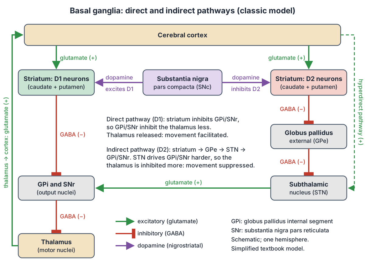
Start with the resting state. The output nuclei, GPi and SNr, send inhibitory GABA signals to the thalamus, which holds it back. The thalamus excites the cortex with glutamate. Whether a movement happens depends on whether the basal ganglia release or tighten this brake on the thalamus. (Kenhub: Basal ganglia.)
Direct pathway: the cortex excites the striatum with glutamate. Striatal neurons of the direct pathway send inhibitory GABA projections straight to GPi and SNr. Inhibiting an inhibitor is called disinhibition: GPi and SNr fall quiet, the thalamus is released, and thalamocortical neurons can excite the cortex. The net effect is to allow movement to start. (Kenhub: Basal ganglia.)
Indirect pathway: striatal neurons of the indirect pathway inhibit GPe. GPe normally inhibits the subthalamic nucleus, so when GPe is quiet the subthalamic nucleus is released. The subthalamic nucleus then excites GPi and SNr with glutamate, they inhibit the thalamus more strongly, and movement is suppressed. (Kenhub: Basal ganglia.)
OpenStax summarises the result: the direct pathway causes disinhibition of the thalamus, whereas the indirect pathway causes, or reinforces, the normal inhibition of the thalamus. The indirect pathway has extra connections, including disinhibition of the subthalamic nucleus. (OpenStax Anatomy and Physiology 2e, 13.2: The Central Nervous System.)
The two pathways start from different striatal neurons (medium spiny neurons). Those projecting directly to GPi and SNr carry dopamine D1 receptors and contain substance P and dynorphin. Those projecting to GPe carry D2 receptors and contain enkephalin. A third route, the hyperdirect pathway, runs from the cortex straight to the subthalamic nucleus, skipping the striatum. (Lanciego JL, Luquin N, Obeso JA. Functional neuroanatomy of the basal ganglia. Cold Spring Harb Perspect Med. 2012.)
This is a simplified textbook model. Lanciego and colleagues point out that it has been modified as new data appeared, that it oversimplified the connections of the subthalamic nucleus, and that it does not explain some findings, for example that surgical lesions of the globus pallidus (pallidotomy) reduce rather than cause involuntary movements. (Lanciego JL, Luquin N, Obeso JA. Functional neuroanatomy of the basal ganglia. Cold Spring Harb Perspect Med. 2012.)
Dopamine and the substantia nigra
The substantia nigra pars compacta projects to the striatum and acts as the switch between the two pathways. Dopamine receptors are either excitatory (D1-type) or inhibitory (D2-type), so dopamine activates the direct pathway and inhibits the indirect pathway. Both effects push the circuit towards movement. (OpenStax Anatomy and Physiology 2e, 13.2: The Central Nervous System.)
This dopamine supply is called the nigrostriatal pathway. The striatum is the main input of the basal ganglia: it receives excitatory glutamate input from the cortex, and its own projection neurons are inhibitory and use GABA. (Kenhub: Basal ganglia.)
Blood supply of the basal ganglia
The main supply is the middle cerebral artery through its lenticulostriate (perforating) branches. The anterior cerebral and anterior choroidal arteries supply anterior parts such as the head of the caudate and the nucleus accumbens. The substantia nigra and subthalamic nucleus receive branches of the posterior cerebral and posterior communicating arteries. Veins drain through striate branches of the internal cerebral vein into the great cerebral vein. (TeachMeAnatomy: The Basal Ganglia.)
Radiopaedia gives the same main sources: perforating branches of the middle cerebral artery, branches of the anterior cerebral artery and the terminal internal carotid artery. (Radiopaedia: Basal ganglia.)
Basal ganglia disorders: Parkinson disease, Huntington disease and more
Parkinson disease is the result of neurons in the substantia nigra pars compacta dying. Without dopamine, the basal ganglia are stuck in the indirect pathway, and the increased indirect-pathway activity causes a hypokinetic (too little movement) disorder. (OpenStax Anatomy and Physiology 2e, 13.2: The Central Nervous System.)
In the classic model, dopamine loss leads to excessive activity of the subthalamic nucleus, which overstimulates GPi and SNr and increases their inhibition of the thalamus. The same model explains chorea and ballism (hyperkinetic, too much movement) as reduced activity in the subthalamic-to-GPi projection. (Lanciego JL, Luquin N, Obeso JA. Functional neuroanatomy of the basal ganglia. Cold Spring Harb Perspect Med. 2012.)
Kenhub lists the main features of Parkinson disease as bradykinesia (slowness of movement), resting tremor, rigidity and postural instability. Huntington disease is an autosomal dominant condition in which striatal GABAergic neurons degenerate, causing atrophy of the head of the caudate nucleus, with chorea, dementia and psychiatric problems. Hemiballismus, a hyperkinetic disorder affecting one side of the body, typically follows a lesion such as a stroke near the subthalamic nucleus. In Wilson disease, an autosomal recessive copper transport disorder, copper builds up in the basal ganglia, especially the putamen. (Kenhub: Basal ganglia.)
Radiopaedia also lists multiple system atrophy, corticobasal degeneration, basal ganglia calcification and Hallervorden-Spatz syndrome among conditions that affect this region on imaging. The globus pallidus normally contains more myelin and calcium than the putamen. (Radiopaedia: Basal ganglia.)
Finding the basal ganglia in the 3D model
In the anterior view, look for the caudate nucleus hugging the lateral ventricle on each side, the putamen as the large lens-shaped mass further out, and the globus pallidus tucked medial to the putamen. This arrangement matches the description of the caudate at the superomedial side of the internal capsule and the lentiform nucleus at its inferolateral side. (Radiopaedia: Basal ganglia.)
Basal ganglia in the 3D model
- Caudate nucleus
- The caudate nucleus is a C-shaped nucleus of the basal ganglia that, with the putamen, forms the striatum.
- Globus pallidus
- The globus pallidus is a basal ganglia nucleus that provides major inhibitory output to the thalamus.
- Putamen
- The putamen is the large lateral nucleus of the basal ganglia and part of the striatum.
- Thalamus
- The thalamus is a paired diencephalic mass that relays sensory and motor information to the cerebral cortex.
Frequently asked questions
What are the basal ganglia made of?
The core nuclei are the caudate nucleus, putamen and globus pallidus. The caudate and putamen form the striatum, and the putamen with the globus pallidus forms the lentiform nucleus. Functionally the subthalamic nucleus and the substantia nigra are included too, because they share the same circuits. The claustrum, a thin sheet of grey matter beside the putamen, was once counted too but is no longer included.
Where are the basal ganglia located?
Deep inside each cerebral hemisphere, in the white matter below the cortex. The caudate nucleus lies along the lateral wall of the lateral ventricle, on the medial side of the internal capsule. The putamen and globus pallidus lie on the lateral side of the internal capsule. The subthalamic nucleus is below the thalamus and the substantia nigra is in the midbrain.
Are they called basal ganglia or basal nuclei?
Both names are used. "Basal nuclei" is the official Terminologia Anatomica term and is technically more correct, because a ganglion is a group of nerve cell bodies outside the central nervous system. "Basal ganglia" remains the usual term in medicine and research, so you will see both in textbooks and exams.
What is the main function of the basal ganglia?
Their best-known role is controlling movement: they help select and start the movements you want and suppress unwanted ones, by adjusting how strongly the thalamus is inhibited. They also take part in planning, memory, eye movements, reward and motivation, through separate loops with the prefrontal cortex and limbic areas.
What is the difference between the direct and indirect pathway?
Both start in the striatum. The direct pathway inhibits GPi and SNr, which releases the thalamus and facilitates movement. The indirect pathway inhibits GPe, which frees the subthalamic nucleus to excite GPi and SNr, so the thalamus is inhibited more and movement is suppressed. Dopamine excites direct-pathway (D1) neurons and inhibits indirect-pathway (D2) neurons.
Why does Parkinson disease affect the basal ganglia?
Parkinson disease destroys dopamine-producing neurons in the substantia nigra pars compacta. Without dopamine the direct pathway loses its boost and the indirect pathway is no longer held back, so the output nuclei inhibit the thalamus too much. The result is a hypokinetic disorder with slowness of movement, rigidity and tremor.
References
- OpenStax. Anatomy and Physiology 2e, §13.2 The Central Nervous System. Rice University, 2022.
- TeachMeAnatomy. The Basal Ganglia.
- Kenhub. Basal ganglia: gross anatomy and function.
- Radiopaedia. Basal ganglia.
- Kenhub. Ventricular system of the brain.
- Lanciego JL, Luquin N, Obeso JA. Functional neuroanatomy of the basal ganglia. Cold Spring Harb Perspect Med. 2012;2(12):a009621. doi:10.1101/cshperspect.a009621
- Mitsuhashi N. et al. (2009). BodyParts3D: 3D structure database for anatomical concepts. Nucleic Acids Research 37, D782–D785. Data © The Database Center for Life Science, CC BY 4.0.
Related anatomy
- Brain anatomyBrain anatomy explained: cerebrum, lobes, gyri, insula, corpus callosum, thalamus, hippocampus, brainstem and…
- ThalamusThalamus location and anatomy: what the thalami are, their nuclei (VPL, VPM, geniculate bodies), blood supply…
- MidbrainMidbrain anatomy explained: where it is, its parts (tectum, tegmentum, cerebral peduncles), colliculi, substantia…
- Brain ventriclesVentricles of the brain explained: lateral, third and fourth ventricles, cerebral aqueduct, choroid plexus and the CSF…
- Brain arteriesCircle of Willis and the arteries of the brain: anterior, middle and posterior cerebral, communicating, basilar and…
Explore the whole body in the interactive 3D human anatomy model, or browse the A–Z list of human structures.