

Blood supply of the brain: carotid and vertebrobasilar systems
The brain receives blood from two systems. The anterior (carotid) system is formed by the internal carotid arteries, which enter the skull through the carotid canal of the temporal bone. The posterior (vertebrobasilar) system is formed by the vertebral arteries, which run up through the transverse foramina of the cervical vertebrae, enter the skull through the foramen magnum and merge into the basilar artery. (OpenStax Anatomy and Physiology 2e, 13.3: Circulation and the Central Nervous System.)
The paired vertebral arteries join to form the large basilar artery, which sends branches to the cerebellum and brainstem and divides into the posterior cerebral arteries. The internal carotid artery gives rise to the ophthalmic artery to the eye, then the anterior and middle cerebral arteries. (OpenStax Anatomy and Physiology 2e, 20.5: Circulatory Pathways.)
Inside the skull, each internal carotid artery passes through the cavernous sinus and gives off the ophthalmic, posterior communicating, anterior choroidal and anterior cerebral arteries before continuing as the middle cerebral artery. (TeachMeAnatomy: Arterial Supply to the Brain.)
What is the circle of Willis?
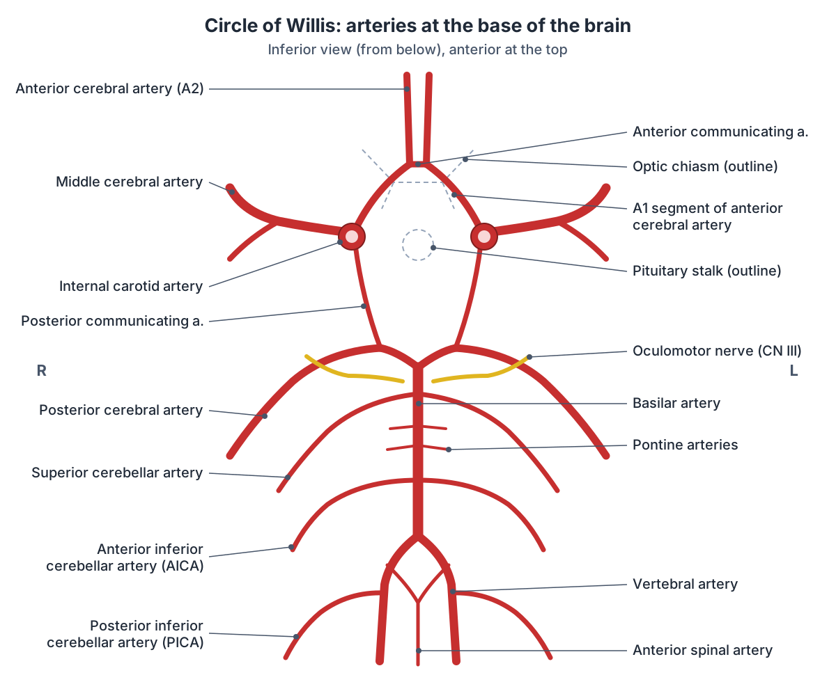
The circle of Willis (circulus arteriosus cerebri) is a ring of arteries, strictly a seven-sided polygon, formed where the internal carotid and vertebrobasilar systems join at the base of the brain. It lies in the suprasellar cistern around the optic chiasm and the pituitary stalk. (Radiopaedia: Circle of Willis.)
Its front half is made of the two internal carotid arteries, the A1 segments of the two anterior cerebral arteries and the single anterior communicating artery. Its back half is made of the two posterior communicating arteries, the P1 segments of the two posterior cerebral arteries and the top of the basilar artery. (Radiopaedia: Circle of Willis.)
Because the ring joins the left and right sides and the front and back systems, it allows blood flow to be balanced between the two sides of the brain and provides a bypass if one of the supplying arteries is narrowed or blocked. (Radiopaedia: Circle of Willis.)
The textbook complete circle is present in only about 20 to 25% of people. Common variants include a hypoplastic posterior communicating artery (about 30%), a "fetal" posterior cerebral artery that arises from the internal carotid artery (about 20%), a hypoplastic or absent A1 segment (about 15%) and an absent or fenestrated anterior communicating artery (about 12.5%). (Radiopaedia: Circle of Willis.)
Anterior cerebral artery (arteria cerebri anterior)
The anterior cerebral artery (Latin arteria cerebri anterior, a. cerebri anterior) is one of the two terminal branches of the internal carotid artery; the other is the middle cerebral artery. It runs forwards and medially to the midline, then arches up and back around the genu of the corpus callosum. (Radiopaedia: Anterior cerebral artery.)
It is divided into five segments. A1 (horizontal, pre-communicating) runs from the carotid termination to the anterior communicating artery and is about 14 mm long. A2 (post-communicating) runs up in front of the corpus callosum, A3 curves around the genu, and A4 and A5 run over the body of the corpus callosum in front of and behind the coronal suture plane. The pericallosal artery continues from A2 or A3, and the callosomarginal artery runs in the cingulate sulcus. (Radiopaedia: Anterior cerebral artery.)
The ACA supplies the medial surface of the cerebral hemisphere back to the parietal lobe, most of the corpus callosum except the splenium, the septum pellucidum, the anterior putamen, the head of the caudate nucleus and part of the internal capsule. Small medial lenticulostriate arteries arise from A1, and the recurrent artery of Heubner usually arises from A2. (Radiopaedia: Anterior cerebral artery.)
In OpenStax terms, the anterior cerebral artery supplies blood to the frontal lobe of the cerebrum. (OpenStax Anatomy and Physiology 2e, 20.5: Circulatory Pathways.)
Anterior communicating artery
The anterior communicating artery (ACom) is a short vessel, about 4 mm long, that joins the left and right anterior cerebral arteries and marks the junction of their A1 and A2 segments. It gives small branches to the optic chiasm, lamina terminalis, hypothalamus, anterior columns of the fornix and cingulate gyrus. (Radiopaedia: Anterior communicating artery.)
It is one of the most variable parts of the circle: Radiopaedia reports duplication in about 18% of people, fenestration in about 15% and absence in about 5%. (Radiopaedia: Anterior communicating artery.)
Middle cerebral artery
The middle cerebral artery (MCA) is the larger of the two terminal branches of the internal carotid artery. It is sometimes searched as the "medial cerebral artery", but there is no artery by that name; the middle cerebral artery actually runs laterally, into the lateral (Sylvian) sulcus. (Radiopaedia: Middle cerebral artery.)
It has four segments. M1 (sphenoidal or horizontal) runs laterally parallel to the sphenoid ridge and gives the lateral lenticulostriate arteries to the deep grey matter. M2 (insular) runs up and back over the insula, usually splitting into superior and inferior divisions. M3 (opercular) runs out along the frontoparietal operculum, and M4 (cortical) spreads over the lateral surface. (Radiopaedia: Middle cerebral artery.)
The MCA supplies most of the lateral surface of the cerebral hemisphere, as well as part of the internal capsule and basal ganglia. (Radiopaedia: Middle cerebral artery.)
OpenStax notes that the middle cerebral artery supplies the temporal and parietal lobes, which are the most common sites of strokes (cerebrovascular accidents). (OpenStax Anatomy and Physiology 2e, 20.5: Circulatory Pathways.)
Posterior communicating artery
The posterior communicating artery (PCom) arises from the back of the C7 (communicating) segment of the internal carotid artery and runs backwards and medially to join the posterior cerebral artery on the same side. It gives many fine perforating branches; the largest is the premammillary (anterior thalamoperforating) artery. (Radiopaedia: Posterior communicating artery.)
It is often small: Radiopaedia gives hypoplasia in about 35% of people, while a fetal origin of the posterior cerebral artery, where the PCom is large and the P1 segment small, is common. (Radiopaedia: Posterior communicating artery.)
Posterior cerebral artery
The posterior cerebral arteries (PCA) are the terminal branches of the basilar artery. Each is divided into segments: P1 runs from the basilar tip to the posterior communicating artery, P2 curls around the cerebral peduncle of the midbrain, P3 passes through the quadrigeminal cistern and P4 lies in the sulci of the occipital lobe. (Radiopaedia: Posterior cerebral artery.)
The PCA supplies the occipital lobes and the posteromedial temporal lobes. Its small perforating branches, together with those of the PCom, supply the thalamus and midbrain. Its cortical branches include the calcarine and parieto-occipital arteries. (Radiopaedia: Posterior cerebral artery.)
A fetal posterior cerebral artery, in which the PCA is supplied mainly from the internal carotid artery, is found on one side in about 13 to 15% of people and on both sides in about 0.5%. (Radiopaedia: Posterior cerebral artery.)
Vertebral and basilar arteries
The intracranial (V4) segment of each vertebral artery runs up in front of the hypoglossal nerve roots. The two V4 segments join at the lower border of the pons to form the basilar artery. Inside the skull the vertebral artery gives the posterior inferior cerebellar artery, the anterior and posterior spinal arteries and small medullary branches. The two vertebral arteries are frequently unequal in size. (Radiopaedia: Vertebral artery.)
The basilar artery lies in front of the pons in the pontine cistern and runs up the central groove of the pons. Along the way it gives the anterior inferior cerebellar arteries, the pontine arteries and the superior cerebellar arteries, and at the upper border of the pons it divides into the two posterior cerebral arteries. (Radiopaedia: Basilar artery.)
Branches from the two vertebral arteries merge into the anterior spinal artery, which supplies the front of the spinal cord. The basilar artery supplies the cerebellum and pons. (OpenStax Anatomy and Physiology 2e, 13.3: Circulation and the Central Nervous System.)
Cerebellar arteries: PICA, AICA and superior cerebellar artery
The posterior inferior cerebellar artery (PICA) arises from the V4 segment of the vertebral artery, although in about 20% of people it starts below the foramen magnum. It winds around the medulla and the cerebellar tonsil and supplies the posteroinferior cerebellar hemisphere, the inferior vermis, the inferior cerebellar peduncle and, in about half of people, the lower medulla. Its territory is larger when the AICA is small, and the reverse. (Radiopaedia: Posterior inferior cerebellar artery.)
The anterior inferior cerebellar artery (AICA) arises from the basilar artery in about 99% of people, usually from its lower third. It supplies the middle cerebellar peduncle, the inferolateral pons, the flocculus and the anteroinferior surface of the cerebellum, and it supplies the facial and vestibulocochlear nerves. Its internal auditory branch, the labyrinthine artery, enters the internal acoustic meatus to supply the inner ear. (Radiopaedia: Anterior inferior cerebellar artery.)
The superior cerebellar artery (SCA) arises from the top of the basilar artery just below the posterior cerebral artery. Near its origin it runs beside the oculomotor nerve, which separates it from the first segment of the PCA, and further back it runs beside the trochlear nerve. It supplies the whole upper surface of the cerebellum, the superior vermis, the dentate nucleus, most of the cerebellar white matter and parts of the midbrain. (Radiopaedia: Superior cerebellar artery.)
Brain arteries and the territories they supply
| Artery | Arises from | Main territory | Key branches or notes |
|---|---|---|---|
| Anterior cerebral artery (ACA) | Internal carotid artery (terminal branch) | Medial surface of the frontal and parietal lobes, most of the corpus callosum, caudate head and part of the internal capsule | Recurrent artery of Heubner, pericallosal and callosomarginal arteries |
| Middle cerebral artery (MCA) | Internal carotid artery (larger terminal branch) | Most of the lateral surface of the hemisphere, plus part of the internal capsule and basal ganglia | Lateral lenticulostriate arteries; superior and inferior divisions |
| Posterior cerebral artery (PCA) | Basilar artery (terminal branch) | Occipital lobe, posteromedial temporal lobe, thalamus and midbrain | Thalamoperforators, posterior choroidal, calcarine and parieto-occipital arteries |
| Anterior communicating artery | Joins the two ACAs | Small branches to the optic chiasm, lamina terminalis, hypothalamus and fornix | About 4 mm long |
| Posterior communicating artery | Internal carotid artery (C7 segment) | Fine perforating branches, the largest being the premammillary (anterior thalamoperforating) artery; with the PCA, supplies the thalamus | Joins the ICA to the PCA; often hypoplastic |
| Superior cerebellar artery (SCA) | Distal basilar artery, just below the PCA | Superior surface of the cerebellum, superior vermis, dentate nucleus, parts of the midbrain | Runs beside the oculomotor and trochlear nerves |
| Anterior inferior cerebellar artery (AICA) | Lower basilar artery | Anteroinferior cerebellum, flocculus, middle cerebellar peduncle, inferolateral pons | Usually gives the labyrinthine artery to the inner ear |
| Posterior inferior cerebellar artery (PICA) | Vertebral artery (V4) | Posteroinferior cerebellum, inferior vermis, lower medulla | Largest branch of the vertebral artery |
Compiled from Radiopaedia articles on each artery (see references). Territories vary between people and overlap at their borders.
Clinical notes: aneurysms and strokes
Saccular (berry) aneurysms are rounded outpouchings of the arteries at the base of the brain and the main cause of non-traumatic subarachnoid haemorrhage. About 90% arise from the anterior circulation: 30 to 40% at the anterior cerebral and anterior communicating artery complex, about 30% at the supraclinoid internal carotid artery including the junction with the posterior communicating artery, and 20 to 30% at the middle cerebral artery bifurcation. About 10% arise in the posterior circulation, for example at the basilar tip. (Radiopaedia: Saccular cerebral aneurysm.)
A rapidly enlarging posterior communicating artery aneurysm is the most common cause of an oculomotor nerve palsy at this level. Compression by an aneurysm usually affects the pupil, whereas palsies caused by poor blood supply to the nerve usually spare it. (Radiopaedia: Oculomotor nerve palsy.)
Stroke can be caused by thrombosis, embolism, hypoperfusion or haemorrhage; embolism is the most common, often from atherosclerotic plaque in the arteries of the neck. Aneurysms most often occur in the vessels of the circle of Willis and usually cause few symptoms until they rupture. (TeachMeAnatomy: Arterial Supply to the Brain.)
Blockage in the posterior circulation causes characteristic syndromes. Lateral medullary (Wallenberg) syndrome is caused by ischaemia of the lateral medulla, most often from disease of the vertebral artery, or less often of the PICA. It produces vertigo and limb incoordination, a Horner syndrome and difficulty swallowing on the same side, and loss of pain and temperature sensation on the same side of the face and the opposite side of the body. (Radiopaedia: Lateral medullary syndrome.)
Brain arteries in the 3D model
- Anterior cerebral artery
- The anterior cerebral artery is one of the two terminal branches of the internal carotid artery and supplies the medial surface of the cerebral hemisphere.
- Anterior choroidal artery
- The anterior choroidal artery is a small branch of the internal carotid artery that supplies deep brain structures and the choroid plexus.
- Anterior communicating artery
- The anterior communicating artery is a short vessel that links the two anterior cerebral arteries.
- Anterior inferior cerebellar artery
- The anterior inferior cerebellar artery (AICA) is a branch of the basilar artery supplying the underside of the cerebellum and part of the pons.
- Anterior spinal artery
- The anterior spinal artery is a single midline artery that supplies the front of the spinal cord.
- Basilar artery
- The basilar artery is a midline artery of the brainstem formed by the two vertebral arteries.
- Callosomarginal artery
- The callosomarginal artery is a branch of the anterior cerebral artery that runs in the cingulate sulcus.
- Insular part of middle cerebral artery
- The insular part (M2 segment) of the middle cerebral artery lies on the insula.
- Internal carotid artery
- The internal carotid artery supplies the brain and the eye.
- Ophthalmic artery
- A branch of the internal carotid artery supplying the eye and orbit.
- Pericallosal artery
- The distal continuation of the anterior cerebral artery along the corpus callosum.
- Postcommunicating part of posterior cerebral artery
- The P2 segment of the posterior cerebral artery, running beyond the posterior communicating artery.
- Posterior communicating artery
- An artery of the circle of Willis connecting the internal carotid and posterior cerebral arteries.
- Posterior inferior cerebellar artery
- The largest branch of the vertebral artery, supplying the underside of the cerebellum and lateral medulla.
- Precommunicating part of posterior cerebral artery
- The P1 segment of the posterior cerebral artery.
- Sphenoid part of middle cerebral artery
- The M1 segment of the middle cerebral artery.
- Superior cerebellar artery
- An artery of the posterior circulation supplying the upper cerebellum.
Frequently asked questions
What arteries make up the circle of Willis?
The two internal carotid arteries, the A1 segments of the two anterior cerebral arteries, the anterior communicating artery, the two posterior communicating arteries, the P1 segments of the two posterior cerebral arteries and the top of the basilar artery. The middle cerebral arteries arise from it but are not part of the ring itself.
What does the middle cerebral artery supply?
Most of the lateral surface of the cerebral hemisphere, including much of the frontal, parietal and temporal lobes, together with part of the internal capsule and basal ganglia through its lenticulostriate branches. It is the larger terminal branch of the internal carotid artery. There is no "medial cerebral artery"; that search usually means the middle cerebral artery.
What does the anterior cerebral artery supply?
The medial surface of the frontal and parietal lobes, most of the corpus callosum apart from the splenium, the septum pellucidum, the head of the caudate nucleus and part of the internal capsule. It is the smaller terminal branch of the internal carotid artery, and the two anterior cerebral arteries are joined by the anterior communicating artery.
What is PICA?
PICA is the posterior inferior cerebellar artery, the largest branch of the vertebral artery. It winds around the medulla and supplies the lower back part of the cerebellum, the inferior vermis and part of the lower medulla. Blockage of the vertebral artery or PICA can cause lateral medullary (Wallenberg) syndrome.
How is the basilar artery formed?
The two vertebral arteries join at the lower border of the pons to form the single basilar artery. It runs up in front of the pons, giving the anterior inferior cerebellar, pontine and superior cerebellar arteries, and divides at the upper border of the pons into the two posterior cerebral arteries.
Is the circle of Willis complete in everyone?
No. A complete circle with no missing or very small segments is found in only about 20 to 25% of people. The posterior communicating arteries are often hypoplastic, about one person in five has a fetal posterior cerebral artery, and the A1 segment or the anterior communicating artery can be small, absent or duplicated.
References
- OpenStax. Anatomy and Physiology 2e, §20.5 Circulatory Pathways. Rice University, 2022.
- OpenStax. Anatomy and Physiology 2e, §13.3 Circulation and the Central Nervous System. Rice University, 2022.
- TeachMeAnatomy. Arterial Supply to the Brain.
- Radiopaedia. Circle of Willis.
- Radiopaedia. Internal carotid artery.
- Radiopaedia. Anterior cerebral artery.
- Radiopaedia. Anterior communicating artery.
- Radiopaedia. Middle cerebral artery.
- Radiopaedia. Posterior communicating artery.
- Radiopaedia. Posterior cerebral artery.
- Radiopaedia. Basilar artery.
- Radiopaedia. Vertebral artery.
- Radiopaedia. Posterior inferior cerebellar artery.
- Radiopaedia. Anterior inferior cerebellar artery.
- Radiopaedia. Superior cerebellar artery.
- Radiopaedia. Saccular cerebral aneurysm.
- Radiopaedia. Lateral medullary syndrome.
- Radiopaedia. Oculomotor nerve palsy.
- Mitsuhashi N. et al. (2009). BodyParts3D: 3D structure database for anatomical concepts. Nucleic Acids Research 37, D782–D785. Data © The Database Center for Life Science, CC BY 4.0.
Related anatomy
- Carotid arteriesCarotid arteries explained: common carotid, bifurcation, internal carotid, the 8 external carotid branches with a…
- Brain anatomyBrain anatomy explained: cerebrum, lobes, gyri, insula, corpus callosum, thalamus, hippocampus, brainstem and…
- Dural venous sinusesDural venous sinuses explained: superior sagittal, straight, transverse, sigmoid, occipital and cavernous sinus…
- Cranial nervesThe 12 cranial nerves in order with a labelled diagram: type, skull foramen, functions and test for CN I to CN XII…
- Basilar arteryBasilar artery: The basilar artery is a midline artery of the brainstem formed by the two vertebral arteries. See…
- Anterior cerebral arteryAnterior cerebral artery: The anterior cerebral artery is one of the two terminal branches of the internal carotid…
Explore the whole body in the interactive 3D human anatomy model, or browse the A–Z list of human structures.