

What are the ventricles of the brain?
There are four ventricles in the brain. The first two are the lateral ventricles, deep within the cerebrum. They connect to the third ventricle, the space between the left and right sides of the diencephalon, through two openings called the interventricular foramina. The third ventricle opens into the cerebral aqueduct, which passes through the midbrain and opens into the fourth ventricle, the space between the cerebellum and the pons and upper medulla. The fourth ventricle then narrows into the central canal of the spinal cord. (OpenStax Anatomy and Physiology 2e, 13.3: Circulation and the Central Nervous System.)
The ventricles are filled with cerebrospinal fluid (CSF), the clear liquid that surrounds and bathes the brain and spinal cord. CSF is made inside the ventricles, flows through them, and then leaves into the subarachnoid space around the outside of the brain. (Radiopaedia: Cerebrospinal fluid.)
The words used for this topic vary. "Cerebral ventricles", "ventricles of the cerebrum", "brain ventricles" and "ventricular system of the brain" all refer to the same set of cavities. Strictly, only the two lateral ventricles lie in the cerebrum (telencephalon); the third ventricle belongs to the diencephalon and the fourth to the hindbrain. (OpenStax Anatomy and Physiology 2e, 13.1: The Embryologic Perspective.)
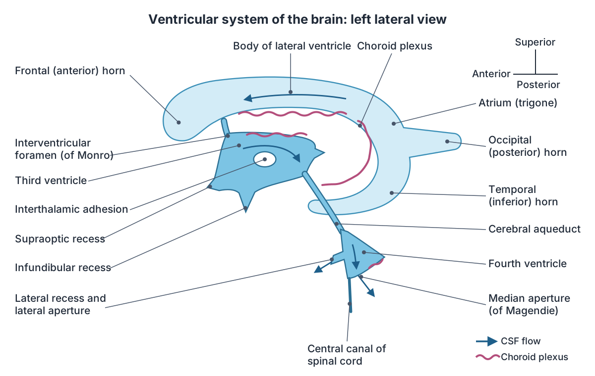
Lateral ventricles: horns, body and walls
Each cerebral hemisphere contains one lateral ventricle. Each has five divisions: the anterior (frontal) horn, the body, the atrium (trigone), the posterior (occipital) horn and the inferior (temporal) horn. The three horns project into the lobes they are named after. (Radiopaedia: Lateral ventricle.)
The walls of the lateral ventricle are formed by important structures. The corpus callosum forms the roof of the central part (body). The floor of the body includes the thalamus and, inferolaterally, the tail of the caudate nucleus, with the fornix inferomedially. The head of the caudate nucleus bulges into the floor of the frontal horn, and the septum pellucidum separates the two frontal horns in the midline. The hippocampus lies in the floor of the temporal horn, and the calcar avis in the floor of the occipital horn. (Kenhub: Ventricular system of the brain.)
The lateral ventricles drain through the interventricular foramen (of Monro) into the third ventricle. The two sides are often slightly asymmetrical, especially the occipital horns, and this is a normal finding. The volume of the lateral ventricles increases with age as the brain shrinks (cerebral involution). (Radiopaedia: Lateral ventricle.)
Third ventricle: boundaries and recesses
The third cerebral ventricle is a narrow midline cavity of the diencephalon. Its lateral walls are formed by the thalamus, the hypothalamic sulcus and the hypothalamus. From top to bottom, its anterior wall is formed by the columns of the fornix, the anterior commissure and the lamina terminalis. Its posterior wall is formed by the habenular commissure, the pineal gland and the posterior commissure. The roof is formed by the choroidal fissure and the bodies of the fornix, and the floor runs back from the optic chiasm and the infundibulum of the pituitary gland. (Radiopaedia: Third ventricle.)
The third ventricle has small extensions called recesses. In front, the supraoptic recess lies above the optic chiasm and the infundibular recess lies above the pituitary stalk. Behind, there are the pineal and suprapineal recesses. Many people have an interthalamic adhesion (massa intermedia), a small bridge between the two thalami that crosses the ventricle; Radiopaedia notes it is present more often than not. (Radiopaedia: Third ventricle.)
The third ventricle receives CSF from the lateral ventricles through the foramina of Monro, which lie just behind the columns of the fornix, and drains it through the cerebral aqueduct, which begins just below the posterior commissure. Its width normally ranges from 2 to 10 mm and increases with age; more than 10 mm is considered dilated. (Radiopaedia: Third ventricle.)
Cerebral aqueduct (aqueduct of Sylvius)
The cerebral aqueduct, also called the aqueduct of Sylvius, is the narrow CSF-filled channel that joins the third ventricle to the fourth. It runs through the midbrain, surrounded by periaqueductal grey matter, with the tectum behind it and the tegmentum in front. It is roughly 1 to 2 mm in diameter. (Radiopaedia: Cerebral aqueduct.)
The aqueduct opens just inferior to the epithalamus, the part of the diencephalon that contains the pineal gland, and passes through the midbrain to reach the fourth ventricle. (OpenStax Anatomy and Physiology 2e, 13.3: Circulation and the Central Nervous System.)
Obstruction of the aqueduct causes hydrocephalus, with enlargement of the lateral and third ventricles that lie upstream of the block. On T2-weighted MRI, the pulsing CSF normally produces a dark "flow void" in the aqueduct; its absence can point to disease. (Radiopaedia: Cerebral aqueduct.)
The aqueduct is the most common site of obstruction in non-communicating (obstructive) hydrocephalus. (TeachMeAnatomy: The Ventricles of the Brain.)
Fourth ventricle and its apertures
The fourth ventricle is tent-shaped or triangular when seen from the side and diamond-shaped in a coronal section. Its floor, in front, is the diamond-shaped rhomboid fossa on the back of the pons and upper medulla. Its roof is formed by the superior and inferior medullary vela, which overlie the cerebellum, and its side walls by the vela and the cerebellar peduncles. (Radiopaedia: Fourth ventricle.)
On each side of the floor there is a small swelling, the facial colliculus, made by fibres of the facial nerve looping around the abducens nucleus. The lowest tip of the ventricle is the obex, where the cavity continues into the central canal of the spinal cord. (Radiopaedia: Fourth ventricle.)
CSF leaves the fourth ventricle through three openings. The single median aperture (foramen of Magendie) opens into the cisterna magna, and the two lateral apertures (foramina of Luschka) open into the cerebellopontine cisterns. (Radiopaedia: Fourth ventricle.)
The four ventricles and the channels that join them
| Part | Where it is | Connects to | Key features |
|---|---|---|---|
| Lateral ventricles (2) | Inside each cerebral hemisphere | Third ventricle, through the interventricular foramen (of Monro) | C-shaped; frontal, occipital and temporal horns, body and atrium; separated in front by the septum pellucidum |
| Third ventricle | Midline, between the left and right thalamus and hypothalamus (diencephalon) | Lateral ventricles in front and above; fourth ventricle through the cerebral aqueduct | Narrow slit; supraoptic, infundibular, pineal and suprapineal recesses; choroid plexus in its roof |
| Cerebral aqueduct (of Sylvius) | Midbrain, between the tectum behind and the tegmentum in front | Third ventricle above, fourth ventricle below | About 1 to 2 mm in diameter; common site of obstruction |
| Fourth ventricle | Between the pons and upper medulla in front and the cerebellum behind | Aqueduct above; central canal below; subarachnoid space through one median and two lateral apertures | Floor is the rhomboid fossa; tent-shaped in side view; choroid plexus in its roof |
Compiled from OpenStax, Kenhub and Radiopaedia (see references).
Choroid plexus: where CSF is made
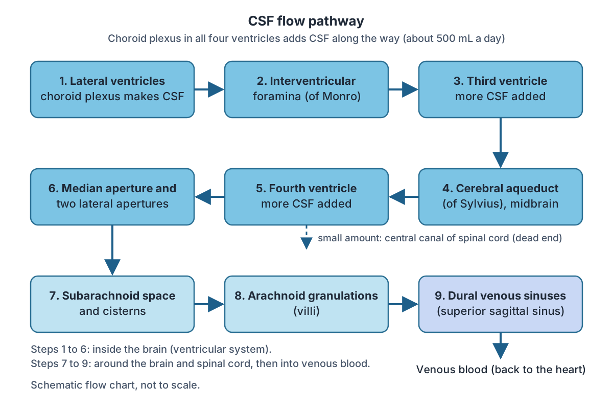
CSF is produced inside the ventricles by a specialised membrane called the choroid plexus. Ependymal cells surround blood capillaries and filter the blood to make CSF, a clear solution with a limited amount of the constituents of blood. Choroid plexuses are found in all four ventricles, and in total they make about 500 mL of CSF a day. (OpenStax Anatomy and Physiology 2e, 13.3: Circulation and the Central Nervous System.)
The choroid plexus is made of choroidal epithelial cells, loose connective tissue (the tela choroidea) and permeable capillaries. In the lateral ventricle it lies in the floor of the body and the roof of the temporal horn and runs through the foramen of Monro into the roof of the third ventricle; it typically does not extend into the frontal or occipital horns. In the fourth ventricle it lies in the lower roof and can extend out through the lateral apertures. (Radiopaedia: Choroid plexus.)
Kenhub notes that the choroid plexus of the lateral ventricles produces the most CSF, followed by that of the third ventricle and then the fourth. (Kenhub: Ventricular system of the brain.)
At any one time there is only about 150 to 160 mL of CSF in the ventricles and subarachnoid space together, but about 500 mL is made each day, so the whole volume is renewed roughly three times a day. The choroid plexus is thought to make most of it, with a smaller part filtering from capillaries in the brain tissue itself. (Radiopaedia: Cerebrospinal fluid.)
CSF flow pathway step by step
From the lateral ventricles, CSF flows through the interventricular foramina into the third ventricle, where more CSF is produced, and then through the cerebral aqueduct into the fourth ventricle, where even more is produced. A little CSF continues down the central canal of the spinal cord, but this is essentially a dead end, so most of it leaves the ventricular system through the median and lateral apertures into the subarachnoid space. (OpenStax Anatomy and Physiology 2e, 13.3: Circulation and the Central Nervous System.)
Within the subarachnoid space, CSF flows around the whole brain and spinal cord. It returns to the blood through the arachnoid granulations, outpocketings of the arachnoid mater into the dural venous sinuses, especially the superior sagittal sinus, which runs in the longitudinal fissure. Its flow is tied to the pulse, because CSF is made from blood. (OpenStax Anatomy and Physiology 2e, 13.3: Circulation and the Central Nervous System.)
The classic teaching is that the arachnoid villi absorb most CSF into the venous system. Radiopaedia notes that a substantial share also drains into lymphatics along the sheaths of cranial and spinal nerves and around the olfactory bulb. (Radiopaedia: Cerebrospinal fluid.)
A short way to remember the sequence: lateral ventricles, foramina of Monro, third ventricle, aqueduct of Sylvius, fourth ventricle, foramina of Magendie and Luschka, subarachnoid space, arachnoid granulations, superior sagittal sinus.
What does CSF do?
CSF cushions the brain and spinal cord and protects them against rapid acceleration and deceleration. It gives the brain buoyancy, helps maintain intracranial pressure, and carries away waste products. (Radiopaedia: Cerebrospinal fluid.)
Because CSF is reabsorbed into the dural sinuses along with metabolic wastes, its continuous production and drainage also act as a clearance system for the nervous tissue. (OpenStax Anatomy and Physiology 2e, 13.3: Circulation and the Central Nervous System.)
How the ventricles develop
The ventricles are the remnant of the hollow centre of the embryonic neural tube. Each brain vesicle keeps part of this cavity: the telencephalon (cerebrum) has the lateral ventricles, the diencephalon has the third ventricle, the mesencephalon (midbrain) has the cerebral aqueduct, and the metencephalon and myelencephalon (pons, cerebellum and medulla) share the fourth ventricle. The posterior neural tube becomes the spinal cord with its central canal. (OpenStax Anatomy and Physiology 2e, 13.1: The Embryologic Perspective.)
Hydrocephalus and other clinical notes
Hydrocephalus is an abnormal collection of CSF within the ventricles of the brain. In communicating (non-obstructive) hydrocephalus there is no blockage within the ventricles. In non-communicating (obstructive) hydrocephalus, flow is blocked inside the ventricular system, most often at the cerebral aqueduct. (TeachMeAnatomy: The Ventricles of the Brain.)
Kenhub adds that blockage at the cisterns or dural sinuses causes communicating hydrocephalus. If hydrocephalus develops in an infant before the fontanelles close, the head enlarges; after the skull bones fuse, raised pressure is more likely to push brain tissue out of place (herniation). (Kenhub: Ventricular system of the brain.)
On imaging, the size of the ventricles must be judged against age: the lateral ventricles enlarge as the brain shrinks with age, and a third ventricle wider than 10 mm is considered dilated. (Radiopaedia: Third ventricle.)
The choroid plexus can calcify. A fully calcified, non-enhancing plexus can mimic other lesions, such as tuberculoma or neurocysticercosis, on scans. (Radiopaedia: Choroid plexus.)
Brain ventricles in the 3D model
- Cerebral aqueduct
- The cerebral aqueduct (of Sylvius) is a narrow channel carrying cerebrospinal fluid from the third to the fourth ventricle.
- Choroid plexus of cerebral hemisphere
- The choroid plexus is a vascular, fringe-like tissue in the cerebral ventricles that produces cerebrospinal fluid.
- Fourth ventricle
- The fourth ventricle is a diamond-shaped, CSF-filled cavity of the hindbrain.
- Interventricular foramen
- The interventricular foramen (of Monro) is the opening connecting each lateral ventricle to the third ventricle.
- Lateral ventricle
- The lateral ventricle is a C-shaped CSF-filled cavity within each cerebral hemisphere.
- Third ventricle
- The third ventricle is a narrow midline CSF-filled cavity of the diencephalon.
Frequently asked questions
How many ventricles are in the brain?
There are four: the right and left lateral ventricles, one in each cerebral hemisphere, the midline third ventricle in the diencephalon, and the fourth ventricle between the pons and medulla in front and the cerebellum behind. The lateral ventricles drain into the third through the interventricular foramina, and the cerebral aqueduct joins the third to the fourth.
Where is CSF produced?
Most CSF is made by the choroid plexus, a specialised membrane of capillaries covered by ependymal cells, found in all four ventricles. The lateral ventricles produce the most. In total about 500 mL is made each day, although only about 150 mL is present at any one time, so the fluid is renewed roughly three times a day.
What is the correct order of CSF flow?
Lateral ventricles, interventricular foramina (of Monro), third ventricle, cerebral aqueduct (of Sylvius), fourth ventricle, then out through the median aperture (Magendie) and two lateral apertures (Luschka) into the subarachnoid space. From there CSF is absorbed through arachnoid granulations into the dural venous sinuses, mainly the superior sagittal sinus. Some also drains along nerve sheaths into lymphatics.
What connects the third and fourth ventricles?
The cerebral aqueduct, also called the aqueduct of Sylvius. It is a narrow channel, about 1 to 2 mm wide, running through the midbrain with the tectum behind and the tegmentum in front. It is the most common site of blockage in obstructive hydrocephalus; a block here enlarges the lateral and third ventricles upstream of it.
What separates the two lateral ventricles?
The septum pellucidum, a thin midline sheet that separates the frontal horns of the right and left lateral ventricles. The two lateral ventricles do not open into each other directly; each drains separately through its own interventricular foramen into the single midline third ventricle.
Which ventricle is in the midbrain?
None of the four ventricles lies in the midbrain. The midbrain contains the cerebral aqueduct, the channel that links the third ventricle in the diencephalon to the fourth ventricle in the hindbrain. In development, the aqueduct is the part of the neural tube cavity that belongs to the mesencephalon.
References
- OpenStax. Anatomy and Physiology 2e, §13.3 Circulation and the Central Nervous System. Rice University, 2022.
- OpenStax. Anatomy and Physiology 2e, §13.1 The Embryologic Perspective. Rice University, 2022.
- TeachMeAnatomy. The Ventricles of the Brain.
- Kenhub. Ventricular system of the brain.
- Radiopaedia. Lateral ventricle.
- Radiopaedia. Third ventricle.
- Radiopaedia. Fourth ventricle.
- Radiopaedia. Cerebral aqueduct.
- Radiopaedia. Choroid plexus.
- Radiopaedia. Cerebrospinal fluid.
- Mitsuhashi N. et al. (2009). BodyParts3D: 3D structure database for anatomical concepts. Nucleic Acids Research 37, D782–D785. Data © The Database Center for Life Science, CC BY 4.0.
Related anatomy
- Brain anatomyBrain anatomy explained: cerebrum, lobes, gyri, insula, corpus callosum, thalamus, hippocampus, brainstem and…
- MidbrainMidbrain anatomy explained: where it is, its parts (tectum, tegmentum, cerebral peduncles), colliculi, substantia…
- ThalamusThalamus location and anatomy: what the thalami are, their nuclei (VPL, VPM, geniculate bodies), blood supply…
- Pineal bodyPineal gland (pineal body) explained: where it is situated, its size, relations, nerve and blood supply, melatonin…
- Dural venous sinusesDural venous sinuses explained: superior sagittal, straight, transverse, sigmoid, occipital and cavernous sinus…
- Spinal cordSpinal cord anatomy with labelled diagrams: cross-section, grey matter horns, spinothalamic and dorsal column tracts…
Explore the whole body in the interactive 3D human anatomy model, or browse the A–Z list of human structures.