

What are the dural venous sinuses?
Dural venous sinuses are venous channels inside the skull, located between the two layers of the dura mater. They are valveless, so blood can flow in either direction between them and the veins that join them, and they form the main drainage route from the brain, mostly to the internal jugular veins. (Radiopaedia: Dural venous sinuses.)
The sinuses drain the cerebrum, cerebellum and brainstem, as well as the diploic veins of the skull bones and the emissary veins that connect them to veins outside the skull, and they empty into the internal jugular vein. (TeachMeAnatomy: The Dural Venous Sinuses.)
Radiopaedia lists five unpaired sinuses (superior sagittal, inferior sagittal, straight, occipital and intercavernous) and seven paired channels (transverse, sigmoid, superior petrosal, inferior petrosal, cavernous and sphenoparietal sinuses and the basilar venous plexus). (Radiopaedia: Dural venous sinuses.)
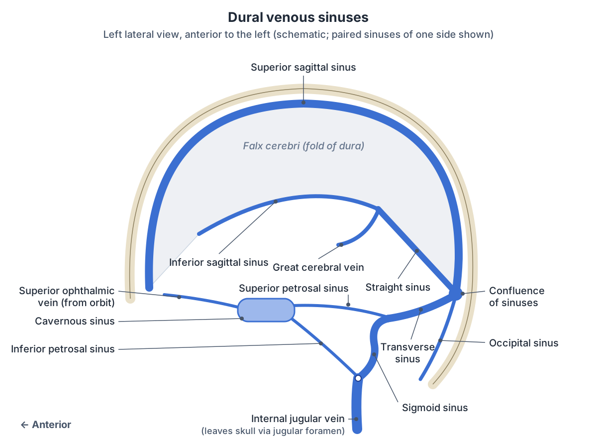
Dura mater: the layers and folds that hold the sinuses
The cranial dura mater has two layers. The outer periosteal layer lines the inner surface of the skull bones; the inner meningeal layer is continuous with the dura around the spinal cord. The sinuses lie between these two layers. (TeachMeAnatomy: The Meninges.)
The meningeal layer folds inwards to form partitions. The falx cerebri hangs down between the right and left cerebral hemispheres. The tentorium cerebelli separates the occipital lobes from the cerebellum. The falx cerebelli separates the two cerebellar hemispheres, and the diaphragma sellae covers the pituitary fossa. Several sinuses run along the edges of these folds. (TeachMeAnatomy: The Meninges.)
Superior sagittal sinus
The superior sagittal sinus is the largest dural venous sinus. It runs in the midline along the upper, attached border of the falx cerebri, from the frontal crest at the front of the skull to the internal occipital protuberance at the back, where it joins the confluence of sinuses. (TeachMeAnatomy: The Dural Venous Sinuses.)
It starts in front at the foramen caecum and receives the superficial cortical veins of the cerebral hemispheres. At its back end it usually turns to the right and drains into the right transverse sinus. Variations are common, especially in its front part, which can be duplicated or underdeveloped. (Radiopaedia: Superior sagittal sinus.)
The superior sagittal sinus also returns cerebrospinal fluid to the blood. The arachnoid mater pushes into the dural sinuses as the arachnoid granulations, where CSF is filtered back into the venous blood. (OpenStax Anatomy and Physiology 2e, 13.3: Circulation and the Central Nervous System.)
Inferior sagittal sinus and straight sinus
The inferior sagittal sinus is a smaller sinus that runs from front to back along the lower, free edge of the falx cerebri. It receives veins from the falx and small veins from the medial surfaces of the cerebral hemispheres and drains into the straight sinus. In one MR venography study it could not be seen in 14% of 204 patients. (Radiopaedia: Inferior sagittal sinus.)
The straight sinus lies at the junction of the falx cerebri and the tentorium cerebelli and is triangular in cross-section. Its front end receives the inferior sagittal sinus and the great cerebral vein (vein of Galen), which drains the deep parts of the brain, and it also receives some superior cerebellar veins. (Radiopaedia: Straight sinus.)
The straight sinus runs back and down towards the confluence of sinuses, but its exact ending varies: Radiopaedia gives the confluence in 56% of people, the left transverse sinus in 21% and the right transverse sinus in 13%. (Radiopaedia: Straight sinus.)
Confluence of sinuses and occipital sinus
The confluence of sinuses (torcular Herophili) lies at the internal occipital protuberance. It is where the superior sagittal, straight and occipital sinuses meet, and from it the two transverse sinuses run out to each side. (TeachMeAnatomy: The Dural Venous Sinuses.)
The occipital sinus is one of the smallest dural sinuses. It lies on the inner surface of the occipital bone, in the attached margin of the falx cerebelli. It begins from small tributaries around the foramen magnum (the marginal sinus), some of which connect with the sigmoid sinus and the vertebral venous plexus, and it drains upwards into the confluence. It can be large or lie off the midline, which matters to surgeons planning an operation in the posterior cranial fossa. (Radiopaedia: Occipital sinus.)
Transverse sinus and sigmoid sinus
Each transverse sinus runs out from the confluence in the lateral border of the tentorium cerebelli, grooving the occipital bone and the squamous temporal bone. In its front part it receives the inferior anastomotic vein (of Labbé). It ends by becoming the sigmoid sinus at the point where the superior petrosal sinus joins. (Radiopaedia: Transverse sinus.)
The two transverse sinuses are often unequal. In the study quoted by Radiopaedia, the left was hypoplastic in 39% of people and absent in 20%, while the right was hypoplastic in only 6% and absent in 4%; both were symmetrical in 31%. This variation can make it hard to diagnose thrombosis on scans. (Radiopaedia: Transverse sinus.)
The sigmoid sinus continues from the transverse sinus where the tentorium ends. It runs down in an S-shaped groove behind and medial to the mastoid air cells and ends in the jugular bulb in the posterior part of the jugular foramen. It is connected to veins outside the skull by the mastoid and condylar emissary veins. (Radiopaedia: Sigmoid sinus.)
In OpenStax's summary, the transverse sinuses lie near the lambdoid suture and flow into the sigmoid sinuses, which then flow into the internal jugular veins. (OpenStax Anatomy and Physiology 2e, 20.5: Circulatory Pathways.)
Cavernous sinus: location, contents and connections
The cavernous sinuses lie on either side of the pituitary fossa and the body of the sphenoid bone, extending from the apex of the orbit in front to the apex of the petrous temporal bone behind. The two sides are joined by the intercavernous sinuses. (Radiopaedia: Cavernous sinus.)
The oculomotor nerve (CN III), trochlear nerve (CN IV) and the ophthalmic (V1) and maxillary (V2) divisions of the trigeminal nerve run in the lateral wall, in that order from top to bottom. The internal carotid artery passes through the sinus itself, surrounded by a plexus of sympathetic nerves, and the abducens nerve (CN VI) runs through the sinus on the inferolateral side of the artery. (Radiopaedia: Cavernous sinus.)
The cavernous sinus receives the superior and inferior ophthalmic veins from the orbit, the superficial middle cerebral vein, the sphenoparietal sinus and the intercavernous sinuses. It drains backwards through the superior petrosal sinus to the transverse sinus and through the inferior petrosal sinus to the jugular bulb. Emissary veins through the foramen ovale and nearby openings connect it with the pterygoid venous plexus outside the skull. (Radiopaedia: Cavernous sinus.)
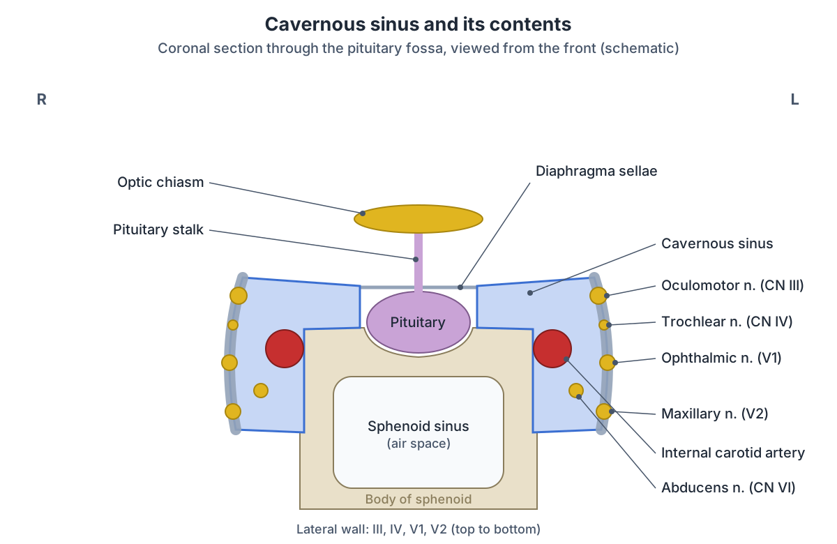
Contents of the cavernous sinus
| Structure | Position in the sinus |
|---|---|
| Oculomotor nerve (CN III) | Lateral wall, highest |
| Trochlear nerve (CN IV) | Lateral wall, below CN III |
| Ophthalmic nerve (V1) | Lateral wall, below CN IV |
| Maxillary nerve (V2) | Lowest, inferolateral part of the lateral wall |
| Internal carotid artery (cavernous segment, C4) | Passes through the sinus, forming the carotid siphon; gives the meningohypophyseal and inferolateral trunks |
| Sympathetic plexus (from the superior cervical ganglion) | Around the internal carotid artery |
| Abducens nerve (CN VI) | Passes through the sinus, inferolateral to the artery; the nerve most often affected in cavernous sinus thrombosis |
Sources: Radiopaedia, Cavernous sinus; TeachMeAnatomy, The Dural Venous Sinuses.
How blood drains from the brain to the internal jugular vein
The overall route is: cerebral and cerebellar veins, then the dural venous sinuses, then the internal jugular vein and the general circulation. The superior and inferior sagittal and straight sinuses lead to the confluence; the transverse sinuses carry blood from the confluence to the sigmoid sinuses, which leave the skull through the jugular foramen as the internal jugular veins. The inferior petrosal sinus joins the internal jugular vein at the jugular foramen. (TeachMeAnatomy: The Dural Venous Sinuses.)
OpenStax describes the same pathway: blood from the brain drains through the superior sagittal sinus to the confluence of sinuses, then through the transverse and sigmoid sinuses into the jugular veins, which return it towards the heart. (OpenStax Anatomy and Physiology 2e, 13.3: Circulation and the Central Nervous System.)
Radiopaedia describes the internal jugular vein as formed by the union of the sigmoid sinus and the inferior petrosal sinus in or just below the jugular foramen, at the jugular bulb. (Radiopaedia: Internal jugular vein.)
The main dural venous sinuses at a glance
| Sinus | Paired? | Where it runs | Drains into |
|---|---|---|---|
| Superior sagittal | No | Attached upper edge of the falx cerebri, from the foramen caecum to the internal occipital protuberance | Confluence of sinuses, usually towards the right transverse sinus |
| Inferior sagittal | No | Free lower edge of the falx cerebri | Straight sinus |
| Straight | No | Junction of the falx cerebri and tentorium cerebelli | Confluence (56%) or one transverse sinus |
| Occipital | No | Attached edge of the falx cerebelli, on the inner occipital bone | Confluence of sinuses |
| Transverse | Yes | Lateral edge of the tentorium, grooving the occipital bone | Sigmoid sinus |
| Sigmoid | Yes | S-shaped groove behind the mastoid air cells | Internal jugular vein at the jugular foramen |
| Cavernous | Yes | Either side of the pituitary fossa and body of the sphenoid | Superior and inferior petrosal sinuses |
| Superior petrosal | Yes | From the cavernous sinus backwards | Transverse sinus at its junction with the sigmoid sinus |
| Inferior petrosal | Yes | From the cavernous sinus back to the jugular foramen; linked by the basilar venous plexus | Internal jugular vein |
Compiled from the Radiopaedia articles on each sinus and TeachMeAnatomy (see references).
Clinical notes: sinus thrombosis and infection
Cerebral venous thrombosis is a blockage of the veins or sinuses inside the skull. It is rare, accounting for about 0.5% of all cerebrovascular disease. Headache is the most common symptom (about 90%), and focal neurological deficits and seizures are frequent. A predisposing factor, such as hormonal causes including the oral contraceptive pill, a blood clotting disorder, or a local or systemic cause, is found in most cases (about 87.5%). On contrast CT, a clot in the superior sagittal sinus can show as the "empty delta sign". (Radiopaedia: Cerebral venous thrombosis.)
Cavernous sinus thrombosis is a clot within the cavernous sinus, most often caused by infection spreading from the orbit, the paranasal sinuses or the "danger zone" of the face through the connection between the facial vein and the superior ophthalmic vein. Features include headache, swelling around one eye, bulging of the eye (proptosis), sensitivity to light and cranial nerve palsies, with the abducens nerve (CN VI) most often affected. It can spread quickly to cause meningitis. (TeachMeAnatomy: The Dural Venous Sinuses.)
A subdural haematoma is a collection of venous blood between the dura and the arachnoid mater. It usually results from tearing of the cerebral veins where they cross to enter the dural venous sinuses. (TeachMeAnatomy: The Meninges.)
Frequently asked questions
What are the dural venous sinuses?
They are valveless venous channels between the outer periosteal and inner meningeal layers of the dura mater inside the skull. They collect blood from the brain, skull and meninges, receive CSF through the arachnoid granulations, and drain mainly into the internal jugular veins. The main ones are the superior and inferior sagittal, straight, occipital, transverse, sigmoid, cavernous and petrosal sinuses.
Where does the superior sagittal sinus drain?
The superior sagittal sinus runs back along the top of the falx cerebri to the confluence of sinuses at the internal occipital protuberance. From there blood usually passes into the right transverse sinus, then the right sigmoid sinus, and leaves the skull as the right internal jugular vein. The pattern varies between people.
What passes through the cavernous sinus?
The internal carotid artery with its sympathetic plexus and the abducens nerve (CN VI) pass through the sinus itself. The oculomotor (CN III) and trochlear (CN IV) nerves and the ophthalmic (V1) and maxillary (V2) divisions of the trigeminal nerve run in its lateral wall, from top to bottom in that order.
What is the sigmoid sinus?
The sigmoid sinus is the paired, S-shaped continuation of the transverse sinus. It begins where the tentorium cerebelli ends, runs down in a groove behind the mastoid air cells, receives the superior petrosal sinus at its start and ends in the jugular bulb at the jugular foramen, where the internal jugular vein begins.
Where is the occipital sinus?
The occipital sinus is a small sinus on the inner surface of the occipital bone, within the attached edge of the falx cerebelli. It begins from small veins around the foramen magnum and drains up into the confluence of sinuses. It can be large or off the midline, which surgeons note before operating in the posterior cranial fossa.
Why are dural sinus infections dangerous?
The sinuses and the veins that join them have no valves, so blood and infection can travel in either direction. Infection from the face, orbit or paranasal sinuses can reach the cavernous sinus through the facial and ophthalmic veins and cause cavernous sinus thrombosis, with eye swelling, cranial nerve palsies and a risk of meningitis.
References
- OpenStax. Anatomy and Physiology 2e, §20.5 Circulatory Pathways. Rice University, 2022.
- OpenStax. Anatomy and Physiology 2e, §13.3 Circulation and the Central Nervous System. Rice University, 2022.
- TeachMeAnatomy. The Dural Venous Sinuses.
- TeachMeAnatomy. The Meninges.
- Radiopaedia. Dural venous sinuses.
- Radiopaedia. Superior sagittal sinus.
- Radiopaedia. Inferior sagittal sinus.
- Radiopaedia. Straight sinus.
- Radiopaedia. Transverse sinus.
- Radiopaedia. Sigmoid sinus.
- Radiopaedia. Occipital sinus.
- Radiopaedia. Cavernous sinus.
- Radiopaedia. Cerebral venous thrombosis.
- Radiopaedia. Internal jugular vein.
Related anatomy
- Internal jugular veinInternal jugular vein (IJ vein) anatomy: jugular foramen to brachiocephalic vein, carotid sheath relations…
- Brain arteriesCircle of Willis and the arteries of the brain: anterior, middle and posterior cerebral, communicating, basilar and…
- Brain ventriclesVentricles of the brain explained: lateral, third and fourth ventricles, cerebral aqueduct, choroid plexus and the CSF…
- Skull foraminaSkull foramina explained: the three cranial fossae, foramen magnum, jugular foramen and every other opening, with a…
- Carotid arteriesCarotid arteries explained: common carotid, bifurcation, internal carotid, the 8 external carotid branches with a…
- Pituitary glandPituitary gland (hypophyseal gland) anatomy: the hypophyseal fossa and sella turcica, relations, lobes, portal blood…
Explore the whole body in the interactive 3D human anatomy model, or browse the A–Z list of human structures.