

What are the three parts of the ear?
The ear has three parts: the external ear, the middle ear and the inner ear. The inner ear houses the vestibulocochlear organ, which has two jobs: converting sound into nerve signals and maintaining balance. (TeachMeAnatomy: The Inner Ear.)
Sound is collected by the outer ear and makes the tympanic membrane (eardrum) vibrate. The middle ear bones pass this vibration to the cochlea, and the middle ear is connected to the pharynx through the Eustachian tube, which helps equalise pressure on either side of the eardrum. (OpenStax Anatomy and Physiology 2e, 14.1: Sensory Perception.)
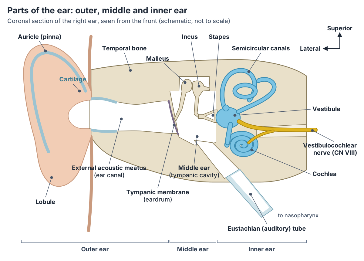
Parts of the ear at a glance
| Part | Main structures | Job |
|---|---|---|
| Outer (external) ear | Auricle (pinna), external acoustic meatus (ear canal) | Collects sound and directs it to the tympanic membrane |
| Tympanic membrane | Pars tensa, pars flaccida, umbo | Vibrates with sound; separates outer and middle ear |
| Middle ear | Tympanic cavity, ossicles, tensor tympani and stapedius, opening of Eustachian tube | Carries vibrations to the oval window; equalises pressure |
| Inner ear: cochlea | Scala vestibuli, cochlear duct with organ of Corti, scala tympani | Hearing |
| Inner ear: vestibular system | Vestibule (utricle and saccule), three semicircular canals and ducts | Balance: head position, linear and rotational movement |
The auricle (pinna)
The auricle (pinna) is the large, fleshy structure on the side of the head. It catches sound waves and directs them into the external acoustic meatus. (OpenStax Anatomy and Physiology 2e, 14.1: Sensory Perception.)
The auricle is mostly cartilage covered by skin; the lobule (earlobe) is the only part without cartilage. Its curved outer rim is the helix, and the antihelix runs inside and parallel to it. The concha is the hollow in the middle that leads into the ear canal, the tragus is the small cartilage flap just in front of the canal opening, and the antitragus lies opposite it. (TeachMeAnatomy: The External Ear.)
The skin of the auricle is supplied by the greater and lesser occipital nerves from the cervical plexus and by the auriculotemporal nerve from the mandibular nerve, with small contributions from the facial and vagus nerves. Its blood comes from branches of the external carotid artery: the posterior auricular, superficial temporal and occipital arteries. (TeachMeAnatomy: The External Ear.)
A blow to the ear can cause bleeding between the cartilage and its covering (an auricular haematoma). If not drained, the cartilage can lose its blood supply and die, leaving a "cauliflower ear". (TeachMeAnatomy: The External Ear.)
The external ear canal (external acoustic meatus)
The external ear canal, or external acoustic (auditory) meatus, runs from the concha to the tympanic membrane. It is typically about 2.5 cm long and S-shaped. Its lateral third is a fibrocartilaginous tube continuous with the auricle, and its medial two-thirds is surrounded by bone. (Radiopaedia: External auditory canal.)
In the bony part the skin lies directly on the periosteum with no subcutaneous layer in between. (Radiopaedia: External auditory canal.)
The canal enters the skull through the external auditory meatus of the temporal bone. (OpenStax Anatomy and Physiology 2e, 14.1: Sensory Perception.)
From outside in, the S-shaped canal first runs upwards and forwards, then turns slightly to run upwards and backwards, and finally runs downwards and forwards. The auricular branch of the vagus nerve supplies part of the canal, which is why cleaning the ear can trigger a cough. (TeachMeAnatomy: The External Ear.)
The tympanic membrane: what a normal eardrum looks like
The tympanic membrane separates the outer ear from the middle ear and passes sound vibrations from the ear canal to the ossicles. It is shaped like a flat cone pointing into the middle ear; its deepest point, at the centre, is the umbo. (Radiopaedia: Tympanic membrane.)
Most of the membrane is the tense pars tensa, held in a fibrocartilaginous ring (the annulus). The small pars flaccida sits at the top, where the ring is missing at the notch of Rivinus. The membrane has three layers: skin on the outside continuous with the ear canal, a fibroelastic middle layer, and mucosa on the inside continuous with the middle ear. (Radiopaedia: Tympanic membrane.)
A normal tympanic membrane is translucent, so structures in the middle ear can be seen through it during otoscopy. The handle of the malleus attaches to its inner surface at the umbo. (TeachMeAnatomy: The External Ear.)
On otoscopy a normal eardrum shows a cone of light (light reflex) in its anteroinferior part. Its outer surface is supplied mainly by the auriculotemporal nerve (CN V3), and its inner surface by the tympanic branch of the glossopharyngeal nerve (CN IX). (Radiopaedia: Tympanic membrane.)
The eardrum can be perforated by injury or infection. In middle ear infection (otitis media), pus and fluid build up behind it until it may rupture; small perforations often heal on their own. (TeachMeAnatomy: The External Ear.)
The middle ear (tympanic cavity)
The middle ear is an air-filled space in the temporal bone. It has two parts: the tympanic cavity, just medial to the tympanic membrane, which holds the ossicles, and the epitympanic recess above it, which holds parts of the malleus and incus. (TeachMeAnatomy: The Middle Ear.)
Its walls have important neighbours. A thin roof of bone separates it from the middle cranial fossa and the brain, the floor lies over the internal jugular vein, the front wall lies close to the internal carotid artery, and the back wall opens through the aditus into the mastoid antrum and mastoid air cells. The medial wall bulges where the facial nerve runs in its canal. (TeachMeAnatomy: The Middle Ear.)
Because the mastoid air cells connect with the middle ear, infection can spread into them (mastoiditis) and from there to the middle cranial fossa, where it can cause meningitis. (TeachMeAnatomy: The Middle Ear.)
The ossicles: malleus, incus and stapes
The ossicles are three small bones that form a chain across the middle ear. The malleus is attached to the tympanic membrane and articulates with the incus, and the incus articulates with the stapes, which attaches to the cochlea at the oval window. (OpenStax Anatomy and Physiology 2e, 14.1: Sensory Perception.)
The malleus is the largest and most lateral ossicle; its handle attaches to the eardrum and its head meets the incus in the epitympanic recess. The incus has a body, a short limb attached to the back wall and a long limb that joins the stapes. The stapes is the smallest; its base sits in the oval window. (TeachMeAnatomy: The Middle Ear.)
The joints between them are small synovial joints: the incudomalleolar joint between the head of the malleus and the body of the incus, and the incudostapedial joint between the lenticular process of the incus and the head of the stapes. The footplate of the stapes is held in the oval (vestibular) window by the annular ligament. (Kenhub: Auditory ossicles.)
The chain increases the force of the vibrations while reducing their amplitude. Because the footplate of the stapes is much smaller than the tympanic membrane, the force at the oval window is about 10 times greater. (Kenhub: Auditory ossicles.)
Two tiny muscles damp the chain. Tensor tympani, supplied by a branch of the mandibular nerve, pulls the handle of the malleus inwards; stapedius, supplied by the facial nerve, acts on the stapes. Both contract in response to loud noise (the acoustic reflex) to reduce the sound reaching the inner ear. (TeachMeAnatomy: The Middle Ear.)
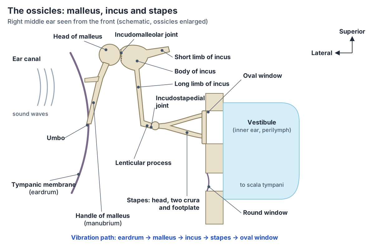
The ossicles: malleus, incus and stapes
| Bone | Parts | Connects to | Muscle attached |
|---|---|---|---|
| Malleus | Head, neck, handle (manubrium) | Handle on the tympanic membrane; head with the body of the incus (incudomalleolar joint) | Tensor tympani (CN V3) on the handle |
| Incus | Body, short limb, long limb, lenticular process | Body with the malleus; lenticular process with the head of the stapes (incudostapedial joint) | None |
| Stapes (stirrup) | Head, anterior and posterior limbs (crura), base (footplate) | Head with the incus; footplate in the oval window by the annular ligament | Stapedius (CN VII) |
Parts and joints from Kenhub; muscles and nerves from TeachMeAnatomy.
The Eustachian (auditory) tube
The Eustachian tube, also called the auditory tube, connects the middle ear to the nasopharynx. The third nearest the middle ear is bone, and the two-thirds nearest the nasopharynx is cartilage. Its pharyngeal opening lies in the nasopharynx behind the inferior nasal concha. (TeachMeAnatomy: The Eustachian Tube.)
The tube ventilates the middle ear by equalising pressure across the tympanic membrane, clears secretions from the middle ear into the nasopharynx, and protects the middle ear from secretions and sounds coming from the nasopharynx. Tensor veli palatini does most of the work of opening it. (TeachMeAnatomy: The Eustachian Tube.)
In children the tube is shorter and straighter, which is why middle ear infections are more common in childhood. Persistent tube dysfunction can lead to otitis media with effusion ("glue ear"). (TeachMeAnatomy: The Middle Ear.)
The inner ear: bony and membranous labyrinth
The inner ear lies in the petrous part of the temporal bone. The bony labyrinth is a series of cavities in the bone, lined with periosteum and filled with perilymph. Inside it lies the membranous labyrinth, a continuous system of ducts filled with endolymph. (TeachMeAnatomy: The Inner Ear.)
The bony labyrinth has three parts: the vestibule in the centre, the cochlea in front of it and the semicircular canals behind it. Two openings face the middle ear: the oval window, which separates the middle ear from the vestibule, and the round window, which separates the middle ear from the scala tympani of the cochlea. (TeachMeAnatomy: The Inner Ear.)
The vestibule and semicircular canals (balance)
The vestibule is the central part of the bony labyrinth. It contains two membranous sacs: the larger utricle, which receives the three semicircular ducts, and the globular saccule, which is connected to the cochlear duct. (TeachMeAnatomy: The Inner Ear.)
The utricle and saccule sense the position of the head; their sensory patches, the maculae, are specialised for detecting linear acceleration, such as tilting the head or moving in a straight line. (OpenStax Anatomy and Physiology 2e, 14.1: Sensory Perception.)
There are three semicircular canals, anterior, lateral and posterior, set at right angles to each other. Each has a swelling at one end called the ampulla. The semicircular ducts inside them lie in the same planes, and receptors in the ampullae detect flow of endolymph when the head moves. (TeachMeAnatomy: The Inner Ear.)
Rotational movement of the head is encoded by hair cells at the base of the semicircular canals, so together the vestibule and canals tell the brain both where the head is and how it is turning. (OpenStax Anatomy and Physiology 2e, 14.1: Sensory Perception.)
The cochlea and cochlear duct (hearing)
The cochlea is a spiral that winds two and a half to two and three-quarter times around a central column of porous bone, the modiolus. A bony shelf, the spiral lamina, divides its canal into the scala vestibuli above and the scala tympani below, with the scala media (cochlear duct) between them. The scala vestibuli and scala tympani contain perilymph; the cochlear duct contains endolymph. (Radiopaedia: Cochlea.)
The cochlear duct is triangular in cross-section. Its roof is Reissner's membrane, its floor is the basilar membrane and its outer wall is the spiral ligament. The organ of Corti, the sensory epithelium of hearing, sits on the basilar membrane. The scala vestibuli is continuous with the vestibule, and the scala tympani ends at the round window. (TeachMeAnatomy: The Inner Ear.)
The basilar membrane responds to different pitches along its length. Higher-frequency sounds move the region near the base of the cochlea, and lower-frequency sounds move the region near its tip (apex). (OpenStax Anatomy and Physiology 2e, 14.1: Sensory Perception.)
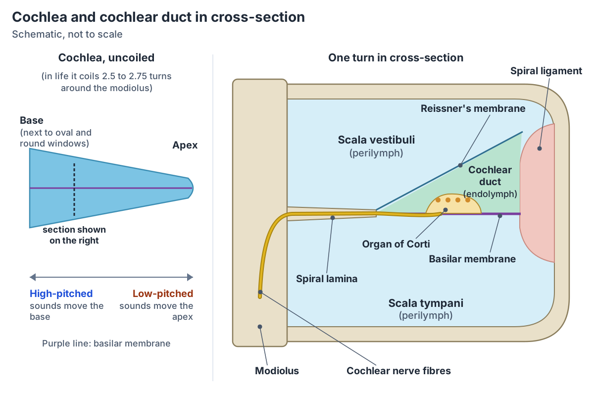
How hearing works, step by step
Sound waves funnelled by the auricle travel down the ear canal and strike the tympanic membrane, which vibrates. The malleus, incus and stapes pass the vibration to the oval window, which lies at the start of a fluid-filled tube in the cochlea, the scala vestibuli. (OpenStax Anatomy and Physiology 2e, 14.1: Sensory Perception.)
The fluid wave in the cochlea moves the basilar membrane, and the organ of Corti on top of it turns the motion into nerve signals. The other fluid-filled tube, the scala tympani, ends at the round window, which is covered by a membrane that keeps the fluid in. (OpenStax Anatomy and Physiology 2e, 14.1: Sensory Perception.)
The vestibulocochlear nerve (CN VIII) enters the internal acoustic meatus and divides into the vestibular nerve, which supplies the utricle, saccule and semicircular ducts, and the cochlear nerve, which enters the base of the modiolus and supplies the organ of Corti. The facial nerve passes through the inner ear region but does not supply it. (TeachMeAnatomy: The Inner Ear.)
Ménière's disease is thought to result from excess endolymph distending the membranous labyrinth. It causes episodes of vertigo, low-pitched tinnitus and hearing loss, showing how closely hearing and balance are linked in the inner ear. (TeachMeAnatomy: The Inner Ear.)
Ear anatomy in the 3D model
- External ear
- The external ear is the part of the ear outside the tympanic membrane that collects sound.
Frequently asked questions
What are the ossicles?
The ossicles are the three small bones of the middle ear: the malleus, incus and stapes (stirrup). They form a chain from the tympanic membrane to the oval window of the inner ear, joined by small synovial joints. The stapes is the smallest bone in the body. The chain increases the force of sound vibrations about tenfold at the oval window.
What is the order of malleus, incus and stapes?
From outside in, the order is malleus, incus, stapes. The handle of the malleus is attached to the eardrum, the head of the malleus articulates with the body of the incus, the long limb of the incus ends in the lenticular process that articulates with the head of the stapes, and the footplate of the stapes sits in the oval window.
How long is the external ear canal?
The external ear canal (external acoustic meatus) is typically about 2.5 cm long in adults. It is S-shaped, running from the concha of the auricle to the tympanic membrane. Its outer third has cartilage walls continuous with the auricle, and its inner two-thirds runs through the temporal bone, where the skin lies directly on the bone's periosteum.
What does a normal tympanic membrane look like?
A normal tympanic membrane is a thin, translucent, cone-shaped membrane, so the middle ear bones can be seen through it during otoscopy. The handle of the malleus can be seen running down to the umbo, its deepest central point, and a bright cone of light (light reflex) is seen in the anteroinferior part. The small pars flaccida sits at the top.
What is the cochlear duct?
The cochlear duct, or scala media, is the part of the membranous labyrinth that runs through the spiral of the cochlea. It is triangular in cross-section and filled with endolymph. Its roof is Reissner's membrane, its floor the basilar membrane and its outer wall the spiral ligament. The organ of Corti, which detects sound, sits on the basilar membrane.
What does the vestibule of the ear do?
The vestibule is the central chamber of the bony labyrinth, between the cochlea and the semicircular canals. It contains the utricle and saccule, whose maculae sense head position and linear acceleration, such as tilting the head or moving forwards. Together with the semicircular canals, which sense rotation, it forms the balance part of the vestibulocochlear organ.
References
- OpenStax. Anatomy and Physiology 2e, §14.1 Sensory Perception. Rice University, 2022.
- TeachMeAnatomy. The External Ear.
- TeachMeAnatomy. The Middle Ear.
- TeachMeAnatomy. The Inner Ear.
- TeachMeAnatomy. The Eustachian Tube.
- Kenhub. Auditory ossicles.
- Radiopaedia. Tympanic membrane.
- Radiopaedia. External auditory canal.
- Radiopaedia. Cochlea.
- Mitsuhashi N. et al. (2009). BodyParts3D: 3D structure database for anatomical concepts. Nucleic Acids Research 37, D782–D785. Data © The Database Center for Life Science, CC BY 4.0.
Related anatomy
- Temporal boneThe temporal bone (os temporale) explained: petrous part and inner ear, mastoid process, zygomatic and styloid…
- Cranial nervesThe 12 cranial nerves in order with a labelled diagram: type, skull foramen, functions and test for CN I to CN XII…
- Skull foraminaSkull foramina explained: the three cranial fossae, foramen magnum, jugular foramen and every other opening, with a…
- External earExternal ear: The external ear is the part of the ear outside the tympanic membrane that collects sound. See where it…
- Brain anatomyBrain anatomy explained: cerebrum, lobes, gyri, insula, corpus callosum, thalamus, hippocampus, brainstem and…
Explore the whole body in the interactive 3D human anatomy model, or browse the A–Z list of human structures.