

Esophagus at a glance
- What it is
- The esophagus is a muscular tube about 25 cm long that carries swallowed food from the pharynx to the stomach.
- Location
- It begins at C6 behind the cricoid cartilage, descends through the posterior mediastinum behind the trachea and left atrium, passes through the diaphragm at T10 and joins the stomach at about T11.
- Function
- It moves food and liquid to the stomach by peristalsis, with upper and lower esophageal sphincters preventing reflux.
- Body system
- Digestive · single, midline structure
- FMA ID
- FMA7131 (Foundational Model of Anatomy)
Key facts
- Upper third is skeletal muscle, lower third is smooth muscle, with a mixed middle third.
- The lining is non-keratinised stratified squamous epithelium.
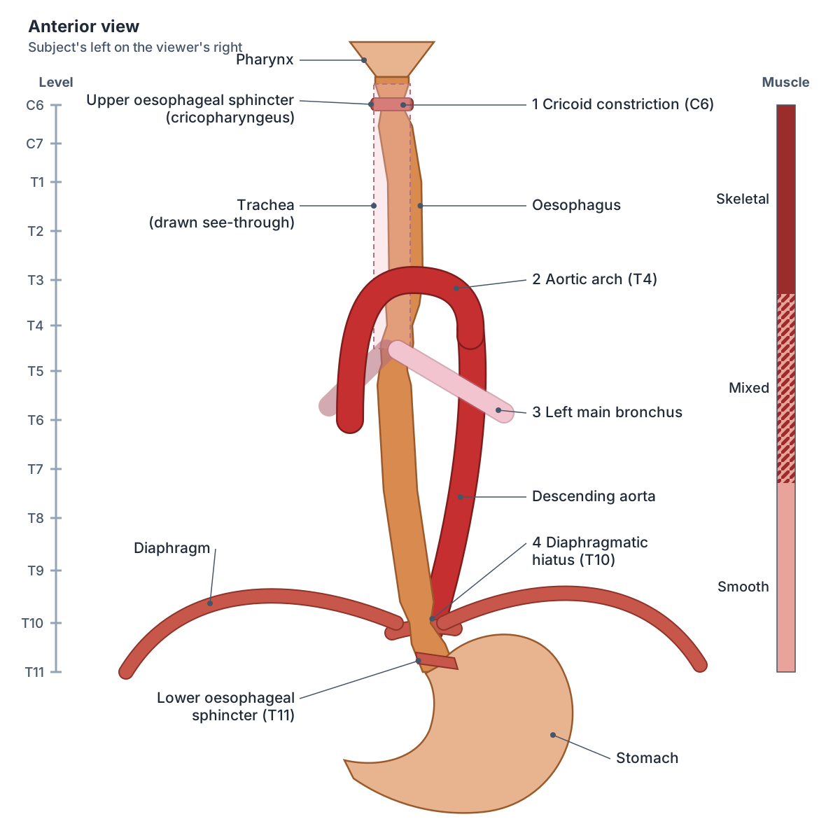
- It has three normal constrictions: at the cricoid, aortic arch/left main bronchus, and diaphragm.
- The lower oesophagus is a site of portosystemic anastomosis between left gastric and oesophageal (azygos) veins.
Clinical relevance
Gastroesophageal reflux can cause Barrett esophagus, in which the lower lining changes to columnar epithelium.
What is the esophagus?
The oesophagus is a muscular tube about 25.4 cm (10 in) long that lies behind the trachea. When you are not swallowing it is collapsed flat. It runs a mainly straight course through the mediastinum of the chest and passes through the diaphragm at the oesophageal hiatus to reach the abdomen. (OpenStax Anatomy and Physiology 2e, 23.3: The Mouth, Pharynx, and Esophagus.)
TeachMeAnatomy describes it as a fibromuscular tube about 25 cm long. It begins at the lower border of the cricoid cartilage at C6, where it continues on from the laryngopharynx, and ends at the cardiac orifice of the stomach at T11. Radiopaedia gives about 22 to 24 cm, measured between the upper and lower sphincters, and a diameter of about 1 to 2 cm. (TeachMeAnatomy: The Oesophagus.)
Radiopaedia divides it into three parts: cervical, from the cricoid cartilage (about C5/6) down to the superior thoracic aperture; thoracic, from T1 to the oesophageal hiatus at T10; and abdominal, from the hiatus to the gastro-oesophageal junction at the cardia of the stomach. (Radiopaedia: Oesophagus.)
The abdominal part is short: TeachMeAnatomy gives about 1.25 cm. The oesophagus passes through the diaphragm in the right crus at T10. (TeachMeAnatomy: The Oesophagus.)
In the 3D model the oesophagus is shown with the stomach, trachea, left main bronchus, aorta, the oesophageal arteries, the gastric vessels and the azygos vein, seen from the left side. The model is BodyParts3D (an adult male).
Course: where does the esophagus run?
The oesophagus is not perfectly straight. It descends slightly to the left of the midline through the neck and superior mediastinum, comes back to the midline at about T5, then moves left again in the posterior mediastinum and curves forwards to pass through the diaphragm. (Radiopaedia: Oesophagus.)
In the superior mediastinum it lies between the trachea in front and the bodies of vertebrae T1 to T4 behind. (TeachMeAnatomy: The Oesophagus.)
At the root of the left lung, the left main bronchus passes in front of the oesophagus on its way to the left hilum, below the aortic arch. (Radiopaedia: Left main bronchus.)
The four constrictions of the esophagus
The oesophagus has natural narrow points where swallowed objects are most likely to get stuck. TeachMeAnatomy lists four, with the mnemonic ABCD: the aortic arch, the left main (principal) bronchus, the cricoid cartilage and the diaphragmatic hiatus. (TeachMeAnatomy: The Oesophagus.)
Radiopaedia counts three physiological constrictions by treating the aortic arch and left main bronchus as one: at C5/C6 behind the cricoid cartilage (the narrowest point), at T4/5 where the aortic arch crosses, and at T10/11 at the oesophageal hiatus. Both descriptions point to the same places. (Radiopaedia: Oesophagus.)
Upper and lower esophageal sphincters
The upper oesophageal sphincter is a true anatomical sphincter of striated (skeletal) muscle at the junction of the pharynx and oesophagus. It is formed by the cricopharyngeus muscle and stays contracted at rest to stop air entering the oesophagus. (TeachMeAnatomy: The Oesophagus.)
The lower oesophageal sphincter is a physiological sphincter at the gastro-oesophageal junction, to the left of T11. TeachMeAnatomy explains that four things keep it closed: the acute angle at which the oesophagus enters the stomach; compression of the abdominal oesophagus by pressure in the abdomen; folds of mucosa that plug the lumen; and the right crus of the diaphragm acting as a pinch-valve. The junction is marked by an abrupt change from oesophageal to gastric lining. (TeachMeAnatomy: The Oesophagus.)
Radiopaedia describes the lower sphincter as a specialised thickening of the circular muscle at the lower end of the oesophagus. On a barium swallow, the 2 to 4 cm dilated segment around it is called the phrenic ampulla. Sources therefore differ on whether there is a distinct muscle; all agree the region acts as a sphincter. (Radiopaedia: Oesophagus.)
The lower sphincter, also called the gastro-oesophageal or cardiac sphincter, relaxes to let food into the stomach and closes to stop acid moving back up. If it does not close fully, stomach acid can reflux into the oesophagus and cause heartburn or gastro-oesophageal reflux disease (GERD, GORD in British usage). (OpenStax Anatomy and Physiology 2e, 23.3: The Mouth, Pharynx, and Esophagus.)
Wall and muscle layers: skeletal to smooth muscle
The oesophageal wall has four layers. From the outside in: an adventitia (not a serosa, because most of the oesophagus is not covered by peritoneum), the muscularis propria with an outer longitudinal and inner circular layer, the submucosa, and the mucosa. (Radiopaedia: Oesophagus.)
The muscle type changes along its length: the upper third is skeletal (voluntary striated) muscle, the middle third is a mix of skeletal and smooth muscle, and the lower third is smooth muscle. (OpenStax Anatomy and Physiology 2e, 23.3: The Mouth, Pharynx, and Esophagus.)
The mucosa is non-keratinised stratified squamous epithelium, which resists abrasion by food, and mucous glands lubricate the lumen. The outer adventitia is not covered by visceral peritoneum, unlike the stomach and intestines. (OpenStax Anatomy and Physiology 2e, 23.3: The Mouth, Pharynx, and Esophagus.)
At the lower end the squamous lining changes abruptly to the columnar epithelium of the stomach. (Radiopaedia: Oesophagus.)
How the esophagus moves food: swallowing and peristalsis
Swallowing (deglutition) has three phases. In the voluntary oral phase the tongue pushes the food bolus back against the palate into the oropharynx. In the involuntary pharyngeal phase the soft palate rises to close off the nasopharynx, breathing stops briefly, the larynx rises, the epiglottis folds over the glottis, the pharyngeal constrictors push the bolus down and the upper oesophageal sphincter relaxes. (OpenStax Anatomy and Physiology 2e, 23.3: The Mouth, Pharynx, and Esophagus.)
In the oesophageal phase, waves of peristalsis, contractions of the circular and longitudinal muscle, carry the bolus towards the stomach. As the bolus nears the stomach, distension briefly relaxes the lower sphincter so it can pass. Solid food takes about 4 to 8 seconds to swallow and liquids about 1 second. (OpenStax Anatomy and Physiology 2e, 23.3: The Mouth, Pharynx, and Esophagus.)
Relations: what lies around the esophagus
In front of the oesophagus are the trachea (down to T4/5), the recurrent laryngeal nerves in the groove between trachea and oesophagus, the left main bronchus and the left atrium of the heart. Behind it are the vertebral column, the descending aorta, the thoracic duct, and the hemiazygos and accessory hemiazygos veins. (Radiopaedia: Oesophagus.)
On its left lie the lung and pleura, the aorta, the left subclavian artery and the thoracic duct; on its right lie the lung, pleura and azygos vein. (Radiopaedia: Oesophagus.)
TeachMeAnatomy notes that the left recurrent laryngeal nerve lies in front of the cervical and upper thoracic oesophagus and the pericardium in front of its lower thoracic part. The abdominal oesophagus lies in front of the left vagus nerve and is related to the left crus of the diaphragm. (TeachMeAnatomy: The Oesophagus.)
Blood supply, veins and the portosystemic anastomosis
The arterial supply comes in three parts. The cervical oesophagus is supplied by the inferior thyroid artery (from the thyrocervical trunk). The thoracic oesophagus is supplied by oesophageal branches of the thoracic aorta and by bronchial arteries. The abdominal oesophagus is supplied by the left gastric artery (from the coeliac trunk) and the left inferior phrenic artery. (TeachMeAnatomy: The Oesophagus.)
The aortic oesophageal arteries are unpaired branches from the front of the aorta: most people have two, but there can be up to five. (TeachMeAnatomy: The Oesophagus.)
Veins follow the same pattern. The cervical part drains through the inferior thyroid veins to the brachiocephalic veins; the thoracic part drains to the azygos and hemiazygos veins. The abdominal part drains two ways, to the left gastric vein (portal circulation) and to the azygos vein (systemic circulation), so the lower oesophagus is a site of portosystemic anastomosis. (TeachMeAnatomy: The Oesophagus.)
When pressure in the portal vein rises (portal hypertension), blood is diverted through this anastomosis from the portal vein to the superior vena cava via the azygos vein. The submucosal veins of the lower oesophagus dilate to form oesophageal varices. Cirrhosis, most often from alcohol excess, is the commonest cause. Varices usually cause no symptoms until they bleed, which presents as vomiting blood (haematemesis) or black stools (melaena). (Radiopaedia: Oesophageal varices.)
Blood supply, venous drainage and lymph of the oesophagus by part
| Part | Arteries | Veins | Lymph nodes |
|---|---|---|---|
| Upper (cervical) | Inferior thyroid artery | Inferior thyroid veins to the brachiocephalic veins | Deep cervical |
| Middle (thoracic) | Oesophageal branches of the thoracic aorta; bronchial arteries | Azygos and hemiazygos veins (to the superior vena cava) | Superior and posterior mediastinal |
| Lower (abdominal) | Oesophageal branches of the left gastric artery; left inferior phrenic artery | Left gastric vein (to the portal vein) and azygos vein: a portosystemic anastomosis | Left gastric and coeliac |
Compiled from TeachMeAnatomy and Radiopaedia. Lymph can travel long distances inside the oesophageal wall, so drainage is not strictly by segment.
Nerve supply and lymph drainage
The oesophagus is supplied by the oesophageal plexus, formed by the vagal trunks (parasympathetic) and branches of the sympathetic trunks. The upper sphincter and the skeletal muscle of the upper oesophagus are controlled by fibres from the nucleus ambiguus; the lower sphincter and smooth muscle by fibres from the dorsal motor nucleus of the vagus. (TeachMeAnatomy: The Oesophagus.)
The upper oesophagus receives the recurrent laryngeal nerves and sympathetic fibres from the middle cervical ganglia; the middle and lower parts receive the vagal trunks, the thoracic sympathetic trunk and the greater splanchnic nerves. Pain is carried by both vagal and sympathetic pathways. (Radiopaedia: Oesophagus.)
Lymph from the upper third drains to the deep cervical nodes, the middle third to the superior and posterior mediastinal nodes, and the lower third to the left gastric and coeliac nodes. (TeachMeAnatomy: The Oesophagus.)
Clinical anatomy of the esophagus
Barrett's oesophagus is a change (metaplasia) of the lining of the lower oesophagus, usually caused by long-standing acid reflux. It is diagnosed by endoscopy and biopsy and is a premalignant condition for adenocarcinoma. (TeachMeAnatomy: The Oesophagus.)
Oesophageal cancer comes in two main types. Squamous cell carcinoma affects mainly the upper and middle thirds and is linked to smoking and alcohol. Adenocarcinoma affects the lower third and is linked to reflux and Barrett's oesophagus. Both typically cause progressively worsening difficulty swallowing (dysphagia) and weight loss. (TeachMeAnatomy: The Oesophagus.)
Achalasia is a failure of the lower sphincter to relax, with loss of normal peristalsis, thought to be caused by loss of neurons in the myenteric (Auerbach) plexus. Food stagnates and the oesophagus widens. A barium swallow shows a tapered lower end, the bird beak sign, and patients have difficulty swallowing both solids and liquids. (Radiopaedia: Achalasia.)
A Zenker diverticulum (pharyngeal pouch) is a pouch of lining that pushes out through a weak spot called Killian's dehiscence, between the cricopharyngeus and thyropharyngeus parts of the inferior constrictor, just above the upper oesophageal sphincter at about C5/6. It mostly affects people aged 60 to 80 and causes difficulty swallowing, regurgitation, bad breath and a risk of aspiration. (Radiopaedia: Zenker diverticulum.)
The oesophagus and trachea develop from the same part of the foregut and are separated by the tracheoesophageal septum. If the septum forms abnormally, a tracheoesophageal fistula connects the two tubes; babies may turn blue or bring milk back during feeds. (TeachMeAnatomy: Development of the Respiratory System.)
Structures next to the esophagus
The nearest named structures in the 3D model, measured from the centre of each part:
- Accessory hemiazygos vein · cardiovascular
- Cavity of atrium · cardiovascular
- Main bronchus proper · respiratory
- Wall of atrium · cardiovascular
- Ascending aorta · cardiovascular
- Posterior cusp of aortic valve · cardiovascular
- Anterior cusp of aortic valve · cardiovascular
- Trunk of coronary artery · cardiovascular
- Main bronchus · respiratory
- Pulmonary artery · cardiovascular
Frequently asked questions
What's the esophagus?
The esophagus (oesophagus) is the muscular food pipe that carries swallowed food and drink from the pharynx to the stomach by waves of muscle contraction called peristalsis. It is about 25 cm long, runs from C6 in the neck through the chest behind the trachea, and passes through the diaphragm at T10 to join the stomach at about T11.
Is it spelled esophagus or oesophagus?
Both are correct. Esophagus is the American spelling and oesophagus the British one, as in oedema and edema. They name the same organ. Related terms follow the same pattern: oesophageal or esophageal, gastro-oesophageal or gastroesophageal reflux (GORD or GERD).
What are the four constrictions of the oesophagus?
The four narrow points are at the cricoid cartilage (about C6, the narrowest), where the aortic arch crosses (about T4), where the left main bronchus crosses just below that, and at the diaphragmatic hiatus (about T10). The mnemonic is ABCD: arch, bronchus, cricoid, diaphragm. Some sources merge the arch and bronchus and count three.
What muscle is the esophagus made of?
The upper third of the oesophagus is skeletal (striated) muscle, the middle third is a mixture of skeletal and smooth muscle, and the lower third is smooth muscle. In all parts the muscle is arranged as an inner circular and an outer longitudinal layer, which together produce peristalsis.
Is the lower esophageal sphincter a real muscle?
The lower oesophageal sphincter is usually described as a physiological sphincter. It is kept closed by the angle at which the oesophagus enters the stomach, abdominal pressure, mucosal folds and the right crus of the diaphragm, along with a thickening of the circular muscle. The upper sphincter, formed by cricopharyngeus, is a true anatomical sphincter.
Why do oesophageal varices form?
The lower oesophagus drains both to the left gastric vein (portal system) and to the azygos vein (systemic system). When portal pressure is high, usually because of cirrhosis, blood is forced through these connections and the submucosal veins swell into varices. They can bleed heavily, causing vomiting of blood.
References
- OpenStax. Anatomy and Physiology 2e, §23.3 The Mouth, Pharynx, and Esophagus. Rice University, 2022.
- TeachMeAnatomy. The Oesophagus.
- TeachMeAnatomy. Development of the Respiratory System.
- Radiopaedia. Oesophagus.
- Radiopaedia. Oesophageal varices.
- Radiopaedia. Zenker diverticulum.
- Radiopaedia. Achalasia.
- Radiopaedia. Left main bronchus.
- Foundational Model of Anatomy: Esophagus (FMA7131). EMBL-EBI Ontology Lookup Service.
- Mitsuhashi N. et al. (2009). BodyParts3D: 3D structure database for anatomical concepts. Nucleic Acids Research 37, D782–D785. Data © The Database Center for Life Science, CC BY 4.0.
Related anatomy
- TracheaTrachea anatomy explained: where the windpipe runs, C-shaped rings, the carina, right vs left main bronchi and the…
- LarynxLarynx anatomy with 3D model and diagrams: thyroid, cricoid, arytenoid and epiglottis cartilages, vocal folds…
- StomachStomach: The stomach is a J-shaped, expandable part of the digestive tract that mixes food with acid and enzymes. See…
- Portal veinHepatic portal vein explained: how it forms, its tributaries, the porta hepatis, portal vs hepatic veins…
- AortaAorta anatomy explained: ascending aorta, aortic arch and its three branches, thoracic aorta, and every abdominal…
- DiaphragmDiaphragm: The dome-shaped muscle separating the thoracic and abdominal cavities and the main muscle of inspiration…
Explore the whole body in the interactive 3D human anatomy model, or browse the A–Z list of human structures.