

What is the larynx and where is it?
The larynx is a cartilaginous structure below the laryngopharynx that connects the pharynx to the trachea. It is part of the conducting airway, and the inner edges of its true vocal cords are free, so they can vibrate to produce sound. (OpenStax Anatomy and Physiology 2e, 22.1: Organs and Structures of the Respiratory System.)
The larynx sits in the front of the neck in the midline, in front of the oesophagus, normally at the level of vertebrae C3 to C6. It is continuous with the oropharynx above and the trachea below, and is built from nine cartilages joined by membranes, ligaments and muscles. (Kenhub: Larynx.)
Radiologists define the larynx as running from the epiglottis down to the lower edge of the cricoid cartilage, below which it continues as the cervical trachea. It develops from the laryngotracheal groove in the floor of the primitive pharynx, and its cartilages come from the 4th and 6th pharyngeal arches. (Radiopaedia: Larynx.)
In infants the larynx sits higher in the neck, which lets the epiglottis lock into the nasopharynx so a baby can breathe through the nose while feeding. This arrangement is lost between about 2 and 6 years of age. (Kenhub: Larynx.)
Laryngeal cartilages: thyroid, cricoid, epiglottis and arytenoids
The larynx has nine cartilages: three unpaired (thyroid, cricoid and epiglottis) and three pairs (arytenoid, corniculate and cuneiform). Together they form the laryngeal skeleton. The thyroid and cricoid cartilages are hyaline cartilage, while the epiglottis is elastic cartilage. (Kenhub: Larynx.)
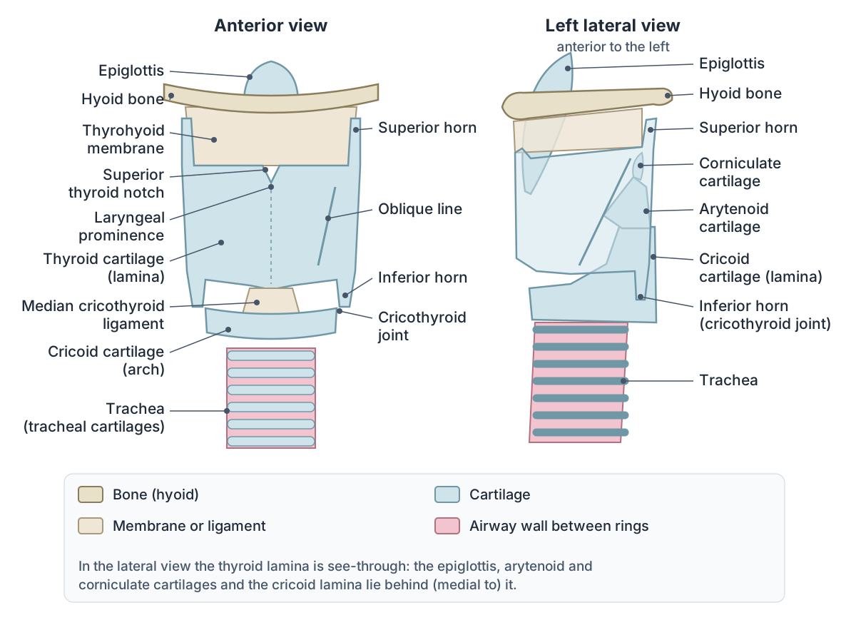
The thyroid cartilage is the largest. Its two flat laminae fuse in the front midline to form the laryngeal prominence (Adam's apple), while their back borders are free and extend up as the superior horns and down as the inferior horns. The superior horns are tied to the hyoid bone by the lateral thyrohyoid ligaments; the inferior horns articulate with the cricoid at the cricothyroid joints. An oblique line on each lamina gives attachment to the inferior pharyngeal constrictor, thyrohyoid and sternothyroid muscles. Its upper border lies at about C4. (Radiopaedia: Thyroid cartilage.)
The cricoid cartilage is a complete ring of hyaline cartilage, with a broad lamina at the back and a narrow arch at the front. It marks the lower border of the larynx at C6 and is the only complete ring of cartilage in the larynx and trachea. Pressing on it (cricoid pressure, or Sellick's manoeuvre) squeezes the oesophagus behind it, which is used during emergency intubation to reduce regurgitation of stomach contents. (TeachMeAnatomy: Laryngeal Cartilages.)
The epiglottis is a leaf-shaped flap of elastic cartilage. Its narrow stalk (petiolus) is attached to the inside of the thyroid cartilage, behind the laryngeal prominence, by the thyroepiglottic ligament, and the hyoepiglottic ligament ties its front surface to the hyoid bone. Its free upper end projects up and back over the laryngeal inlet. On the tongue side the mucosa forms glossoepiglottic folds with a shallow pit, the vallecula, on each side. (Kenhub: Epiglottis.)
The arytenoid cartilages are paired, pyramid-shaped hyaline cartilages sitting on the sloping upper border of the cricoid lamina, where they form synovial cricoarytenoid joints. Each has a vocal process pointing forwards, to which the vocal fold attaches, a muscular process pointing laterally for the posterior and lateral cricoarytenoid muscles, and an apex that articulates with the corniculate cartilage. (Radiopaedia: Arytenoid cartilages.)
The corniculate cartilages sit on the apices of the arytenoids. The cuneiform cartilages lie within the aryepiglottic folds, have no direct attachment to the other cartilages, and simply stiffen the folds. (TeachMeAnatomy: Laryngeal Cartilages.)
Laryngeal joints, membranes and ligaments
The larynx has two pairs of joints. At the cricothyroid joints the inferior horns of the thyroid cartilage articulate with the side of the cricoid. At the cricoarytenoid joints the arytenoids rotate on the upper cricoid lamina, which opens (abducts) the vocal folds during breathing and closes (adducts) them during swallowing. (Kenhub: Larynx.)
Extrinsic membranes and ligaments tie the larynx to its neighbours. The thyrohyoid membrane joins the thyroid cartilage to the hyoid bone, with median and lateral thickenings; the superior laryngeal vessels and the internal laryngeal nerve pierce it on each side. The hyoepiglottic ligament joins the hyoid to the epiglottis, and the cricotracheal ligament joins the cricoid to the trachea. (TeachMeAnatomy: Laryngeal Ligaments and Folds.)
Inside, the cricothyroid ligament (conus elasticus) rises from the cricoid cartilage. Its free upper edge, stretched between the thyroid cartilage in front and the arytenoid behind, is the vocal ligament. Higher up, the quadrangular membrane runs from the arytenoid to the side of the epiglottis, and its lower free edge is the vestibular ligament. The median cricothyroid ligament is the thick midline part of the cricothyroid membrane between the thyroid and cricoid cartilages. (TeachMeAnatomy: Laryngeal Ligaments and Folds.)
Vocal folds, vestibular folds and the inside of the larynx
The vocal folds (true vocal cords) are controlled by the muscles of phonation. From the surface inwards each is made of non-keratinised stratified squamous epithelium, Reinke's space, the vocal ligament and the vocalis muscle. They have little blood supply and look white. The gap between them is the rima glottidis. (TeachMeAnatomy: Laryngeal Ligaments and Folds.)
The vestibular folds (false vocal cords) lie just above the vocal folds. Each is the vestibular ligament covered by mucosa; they are pink, do not move, and mainly protect the larynx. (TeachMeAnatomy: Laryngeal Ligaments and Folds.)
The two pairs of folds divide the laryngeal cavity into three parts: the vestibule, from the laryngeal inlet down to the vestibular folds; a middle part between the vestibular and vocal folds, which includes the laryngeal ventricles and saccules; and the infraglottic cavity, from the vocal folds down to the trachea. The glottis is the vocal folds together with the rima glottidis. (Kenhub: Larynx.)
For cancer staging, radiologists use three subsites: the supraglottis, from the tip of the epiglottis to the laryngeal ventricle (including the aryepiglottic folds and false cords); the glottis (the true vocal cords and the anterior and posterior commissures); and the subglottis, from the underside of the true cords to the lower edge of the cricoid. (Radiopaedia: Larynx.)
Intrinsic muscles of the larynx
The intrinsic muscles lie within the larynx and control the shape of the rima glottidis and the tension of the vocal folds. All are supplied by the inferior laryngeal nerve, the terminal branch of the recurrent laryngeal nerve, except the cricothyroid, which is supplied by the external laryngeal nerve. (Radiopaedia: Intrinsic muscles of the larynx.)
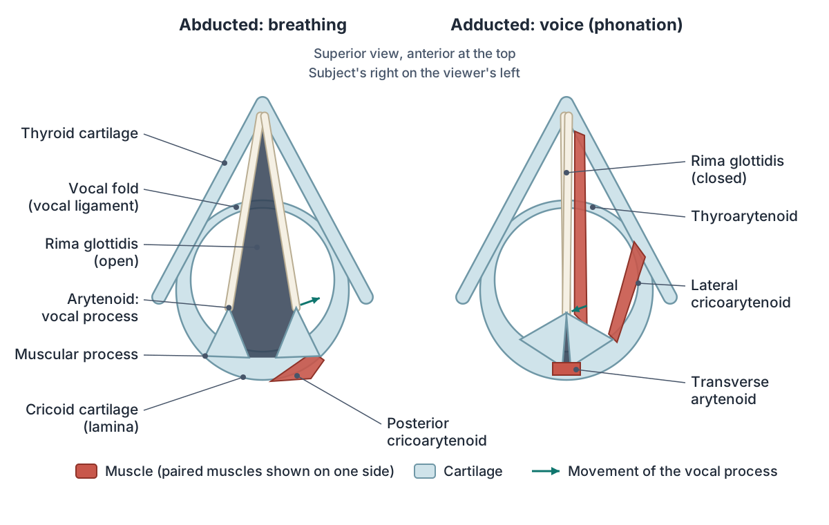
The posterior cricoarytenoid runs from the back of the cricoid lamina to the muscular process of the arytenoid. It is the only muscle that abducts the vocal folds, so it is the only muscle that opens the airway at the glottis. (TeachMeAnatomy: Posterior Cricoarytenoid.)
It works by pulling the muscular processes backwards, which rotates the vocal processes outwards. The lateral cricoarytenoid, from the upper border of the cricoid arch to the same muscular process, does the opposite: it pulls the muscular process forwards, swinging the vocal processes towards the midline. It is the principal adductor. (Radiopaedia: Intrinsic muscles of the larynx.)
The transverse arytenoid joins the back surfaces of the two arytenoids and pulls them together. The oblique arytenoid runs from the muscular process of one arytenoid to the apex of the opposite one, so the two oblique muscles cross in an X. Some of its fibres continue around the arytenoid apex into the aryepiglottic fold as the aryepiglottic muscle (aryepiglotticus). Together they act as a sphincter of the laryngeal inlet that helps prevent aspiration during swallowing and coughing. (Kenhub: Oblique arytenoid muscle.)
The cricothyroid runs from the front of the cricoid arch to the thyroid cartilage in two parts: a straight part to the lower margin of the thyroid lamina and an oblique part to the inferior horn. It rotates the thyroid cartilage forwards and down at the cricothyroid joint, moving it away from the arytenoids, so the vocal folds are stretched, tightened and thinned and the pitch rises. (Kenhub: Cricothyroid muscle.)
The thyroarytenoid (often spelled thyro-arytenoid) runs from the inner surface of the thyroid cartilage near the midline, and from the cricothyroid ligament, back to the arytenoid. It draws the arytenoids forwards, which shortens, thickens and relaxes the vocal folds. Some anatomists describe the part along the vocal ligament as a separate vocalis muscle, and a few fibres continue into the aryepiglottic fold as the thyroepiglotticus. (Kenhub: Thyroarytenoid muscle.)
Radiopaedia sums up the groups: the posterior cricoarytenoids are the only abductors; the lateral cricoarytenoids are the principal adductors, helped by the transverse and oblique arytenoids; the cricothyroids are the tensors; the thyroarytenoids are the relaxers; and the vocalis makes fine adjustments of tension when the folds are together for speech or singing. (Radiopaedia: Intrinsic muscles of the larynx.)
Intrinsic muscles of the larynx: attachments, actions and nerves
| Muscle | Attachments | Action | Nerve |
|---|---|---|---|
| Cricothyroid (straight and oblique parts) | Anterolateral arch of the cricoid to the inferior margin of the thyroid lamina (straight part) and the inferior horn (oblique part) | Tilts the thyroid cartilage forwards and down at the cricothyroid joint; lengthens and tenses the vocal folds, raising pitch | External laryngeal nerve (external branch of the superior laryngeal nerve) |
| Posterior cricoarytenoid | Posterior surface of the cricoid lamina to the muscular process of the arytenoid | The only abductor: swings the vocal processes laterally and opens the rima glottidis | Inferior laryngeal nerve (from the recurrent laryngeal nerve) |
| Lateral cricoarytenoid | Upper border of the cricoid arch to the muscular process of the arytenoid | Principal adductor: swings the vocal processes medially | Inferior laryngeal nerve |
| Transverse arytenoid | Posterior surface of one arytenoid to the other | Draws the arytenoids together; closes the back of the rima glottidis | Inferior laryngeal nerve |
| Oblique arytenoid | Muscular process of one arytenoid to the apex of the opposite arytenoid (the two cross in an X) | Draws the arytenoids together and narrows the laryngeal inlet | Inferior laryngeal nerve |
| Aryepiglotticus | Fibres of the oblique arytenoid continued in the aryepiglottic fold to the side of the epiglottis | Closes the laryngeal inlet (sphincter) | Inferior laryngeal nerve |
| Thyroarytenoid | Inner surface of the thyroid angle and the cricothyroid ligament to the anterolateral surface of the arytenoid | Draws the arytenoids forwards; shortens and relaxes the vocal folds, lowering pitch | Inferior laryngeal nerve |
| Vocalis | Vocal process of the arytenoid to the vocal ligament (often treated as part of thyroarytenoid) | Fine adjustment of tension along the vocal fold | Inferior laryngeal nerve |
Attachments and actions follow Radiopaedia, Kenhub and TeachMeAnatomy. The inferior laryngeal nerve is the terminal branch of the recurrent laryngeal nerve; both laryngeal nerves come from the vagus (CN X).
How the vocal folds make sound
To make a sound, the vocal folds are brought close together without being pressed tight, and air breathed out makes their edges vibrate. Pitch rises when the cricothyroid tenses the folds and falls when the thyroarytenoid relaxes them; loudness depends on fold tension and how hard the air is pushed out. (Kenhub: Vocal cords.)
The folds take different positions for different jobs: relaxed and open in quiet breathing, fully abducted in forced inspiration, adducted for speech, adducted with a small gap at the back for a whisper, and tightly closed during effort closure, as when coughing or lifting. (Kenhub: Vocal cords.)
Extrinsic muscles and the larynx in swallowing
The extrinsic muscles move the larynx as a whole. They attach to the hyoid bone, which is tied to the larynx by strong ligaments. The suprahyoid muscles and stylopharyngeus raise the larynx; the infrahyoid muscles lower it. (TeachMeAnatomy: Muscles of the Larynx.)
During the pharyngeal phase of swallowing, breathing stops briefly, the larynx is pulled upward and the epiglottis folds down over the glottis, blocking the way into the trachea. If food does enter the trachea, coughing usually brings it back up into the pharynx. (OpenStax Anatomy and Physiology 2e, 23.3: The Mouth, Pharynx, and Esophagus.)
Kenhub describes the same closure in more detail: the base of the tongue presses the epiglottis backwards, the hyoid moves up and forwards, and the aryepiglottic and arytenoid muscles pull the epiglottis over the laryngeal inlet. (Kenhub: Epiglottis.)
Blood supply, lymph drainage and nerves of the larynx
The vocal folds are the dividing line for the larynx's supply. Above them, blood comes from the superior laryngeal artery (from the superior thyroid artery) and drains through the superior laryngeal vein to the internal jugular vein. Below them, the inferior laryngeal artery (from the inferior thyroid artery) supplies it and the inferior laryngeal vein drains to the brachiocephalic vein. (Radiopaedia: Larynx.)
Lymph from above the vocal folds pierces the thyrohyoid membrane with the superior laryngeal artery and drains to the superior deep cervical and pre-epiglottic nodes. Lymph from below the folds drains to the inferior deep cervical nodes and the prelaryngeal (Delphian) nodes. (Radiopaedia: Larynx.)
All laryngeal nerves come from the vagus nerve (CN X). The superior laryngeal nerve splits into an internal branch, sensory to the cavity above the vocal folds, which enters through the thyrohyoid membrane with the superior laryngeal artery, and an external branch, motor to the cricothyroid. The recurrent (inferior) laryngeal nerve is motor to all the other intrinsic muscles and sensory below the vocal folds. (Kenhub: Larynx.)
The two recurrent laryngeal nerves take different routes. The right hooks under the right subclavian artery; the left hooks under the aortic arch, next to the ligamentum arteriosum. Both then climb in the groove between the trachea and oesophagus, beside the thyroid gland, and enter the larynx behind the cricothyroid joint. (Radiopaedia: Recurrent laryngeal nerve.)
Clinical anatomy: nerve injury, epiglottitis and emergency airways
Vocal fold paralysis is usually caused by damage to the recurrent laryngeal nerve. It is far more often one-sided, and the left is affected more often, probably because the left nerve has a longer course through the chest. Causes include thyroid and parathyroid surgery, lung cancer and aortic aneurysm. A one-sided palsy gives a hoarse, breathy, weak voice; a two-sided palsy can narrow the airway and cause breathlessness. (Radiopaedia: Vocal cord paralysis.)
Kenhub notes that unilateral injury can also cause aspiration, especially of liquids, while bilateral injury causes stridor and breathing difficulty but rarely hoarseness. The cricothyroid is spared in recurrent laryngeal nerve injury because the superior laryngeal nerve supplies it. (Kenhub: Muscles of the larynx.)
Because the posterior cricoarytenoid is the only abductor, losing its function affects both breathing and voice. (TeachMeAnatomy: Posterior Cricoarytenoid.)
Epiglottitis is inflammation of the epiglottis, classically caused by Haemophilus influenzae type b. It causes fever, difficulty breathing and swallowing, a tripod posture and inspiratory stridor, and can obstruct the airway, so securing the airway comes first. (Kenhub: Epiglottis.)
When the airway is blocked at or above the larynx and intubation has failed, an emergency cricothyroidotomy is made through the cricothyroid ligament, which is found by feeling for the thyroid cartilage and moving just below it. (TeachMeAnatomy: Laryngeal Ligaments and Folds.)
Larynx in the 3D model
- Aryepiglotticus
- The aryepiglottic muscle is a thin muscle in the aryepiglottic fold, continuing from the oblique arytenoid muscle.
- Arytenoid cartilage
- The arytenoid cartilages are a pair of pyramid-shaped cartilages at the back of the larynx.
- Conus elasticus
- An elastic membrane of the larynx.
- Corniculate cartilage
- The corniculate cartilages are two small conical cartilages on the apex of each arytenoid cartilage.
- Cricoid cartilage
- The cricoid cartilage is a complete signet-ring-shaped cartilage forming the lower part of the larynx.
- Cuneiform cartilage
- The cuneiform cartilages are small rod-shaped cartilages in the aryepiglottic folds.
- Epiglottis
- The epiglottis is a leaf-shaped elastic cartilage that covers the laryngeal inlet during swallowing.
- Hyo-epiglottic ligament
- The hyo-epiglottic ligament is a band connecting the epiglottis to the hyoid bone.
- Hyoid bone
- A U-shaped bone in the neck.
- Lateral crico-arytenoid
- An intrinsic laryngeal muscle that closes the vocal folds.
- Lateral thyrohyoid ligament
- The thickened posterior edge of the thyrohyoid membrane.
- Median cricothyroid ligament
- A fibro-elastic ligament in the midline joining the cricoid and thyroid cartilages.
- Median thyrohyoid ligament
- The thickened midline part of the thyrohyoid membrane.
- Oblique arytenoid
- An intrinsic laryngeal muscle behind the arytenoid cartilages.
- Oblique part of cricothyroid
- The oblique fibres of cricothyroid, an intrinsic laryngeal muscle.
- Posterior crico-arytenoid
- An intrinsic laryngeal muscle on the back of the cricoid cartilage.
- Straight part of cricothyroid
- The anterior straight fibers of the cricothyroid muscle of the larynx.
- Thyro-arytenoid
- An intrinsic laryngeal muscle forming the body of the vocal fold.
- Thyro-epiglottic ligament
- The thyro-epiglottic ligament is a band connecting the epiglottis to the thyroid cartilage.
- Thyrohyoid membrane
- A fibroelastic sheet connecting the hyoid bone to the thyroid cartilage.
- Thyroid cartilage
- The thyroid cartilage is the largest cartilage of the larynx, forming the laryngeal prominence.
- Transverse arytenoid
- An unpaired intrinsic laryngeal muscle crossing between the arytenoid cartilages.
- Vocal ligament
- The elastic band forming the core of each vocal fold.
- Vocalis
- The medial part of thyro-arytenoid lying along the vocal ligament.
Frequently asked questions
What are the 9 cartilages of the larynx?
The larynx has three unpaired cartilages, the thyroid, cricoid and epiglottis, and three paired ones, the arytenoids, corniculates and cuneiforms, making nine in total. The thyroid and cricoid are hyaline cartilage and the epiglottis is elastic cartilage. The thyroid cartilage is the largest and forms the Adam's apple.
Which muscle opens the vocal folds?
Only the posterior cricoarytenoid abducts (opens) the vocal folds. It runs from the back of the cricoid lamina to the muscular process of each arytenoid and rotates the vocal processes outwards. The adductors are the lateral cricoarytenoids, helped by the transverse and oblique arytenoids. The posterior cricoarytenoid is supplied by the recurrent laryngeal nerve.
What is the difference between the superior and recurrent laryngeal nerves?
Both are branches of the vagus nerve. The superior laryngeal nerve has an internal branch for sensation above the vocal folds and an external branch to the cricothyroid muscle. The recurrent laryngeal nerve loops under the subclavian artery (right) or aortic arch (left), then supplies all other intrinsic muscles and sensation below the vocal folds.
What does the thyroarytenoid muscle do?
The thyroarytenoid runs from the inside of the thyroid cartilage to the arytenoid cartilage. It pulls the arytenoids forwards, which shortens and relaxes the vocal folds and lowers the pitch of the voice. The part beside the vocal ligament is often described separately as the vocalis. It is supplied by the recurrent laryngeal nerve.
What is the epiglottis made of and what does it do?
The epiglottis is a leaf-shaped flap of elastic cartilage attached by its stalk to the inside of the thyroid cartilage. During swallowing the larynx rises and the epiglottis folds back over the laryngeal inlet, so food and drink pass into the oesophagus rather than the airway.
What is the difference between true and false vocal cords?
The true vocal cords (vocal folds) contain the vocal ligament and vocalis muscle, move, and vibrate to produce voice; they look white. The false vocal cords (vestibular folds) lie just above them, contain the vestibular ligament, look pink, do not move and mainly protect the airway.
At what level is the larynx?
In adults the larynx lies in the front of the neck at about vertebrae C3 to C6. The upper border of the thyroid cartilage is at about C4 and the cricoid cartilage, the lowest part of the larynx, is at C6, where the trachea and the oesophagus begin. In infants the larynx sits higher.
References
- OpenStax. Anatomy and Physiology 2e, §22.1 Organs and Structures of the Respiratory System. Rice University, 2022.
- OpenStax. Anatomy and Physiology 2e, §23.3 The Mouth, Pharynx, and Esophagus. Rice University, 2022.
- TeachMeAnatomy. Laryngeal Cartilages.
- TeachMeAnatomy. Laryngeal Ligaments and Folds.
- TeachMeAnatomy. Muscles of the Larynx.
- TeachMeAnatomy. Posterior Cricoarytenoid.
- Kenhub. Larynx.
- Kenhub. Muscles of the larynx.
- Kenhub. Cricothyroid muscle.
- Kenhub. Thyroarytenoid muscle.
- Kenhub. Oblique arytenoid muscle.
- Kenhub. Vocal cords.
- Kenhub. Epiglottis.
- Radiopaedia. Larynx.
- Radiopaedia. Intrinsic muscles of the larynx.
- Radiopaedia. Thyroid cartilage.
- Radiopaedia. Arytenoid cartilages.
- Radiopaedia. Recurrent laryngeal nerve.
- Radiopaedia. Vocal cord paralysis.
- Mitsuhashi N. et al. (2009). BodyParts3D: 3D structure database for anatomical concepts. Nucleic Acids Research 37, D782–D785. Data © The Database Center for Life Science, CC BY 4.0.
Related anatomy
- TracheaTrachea anatomy explained: where the windpipe runs, C-shaped rings, the carina, right vs left main bronchi and the…
- Esophagus (oesophagus)Esophagus (oesophagus) anatomy: its course and levels, four constrictions, upper and lower sphincters, muscle layers…
- Neck musclesNeck muscles explained: sternocleidomastoid, suprahyoid, infrahyoid, scalene, prevertebral and suboccipital groups…
- Thyroid cartilageThyroid cartilage: The thyroid cartilage is the largest cartilage of the larynx, forming the laryngeal prominence. See…
- Cricoid cartilageCricoid cartilage: The cricoid cartilage is a complete signet-ring-shaped cartilage forming the lower part of the…
- Cranial nervesThe 12 cranial nerves in order with a labelled diagram: type, skull foramen, functions and test for CN I to CN XII…
Explore the whole body in the interactive 3D human anatomy model, or browse the A–Z list of human structures.