

Trachea at a glance
- What it is
- The trachea (windpipe) is a flexible air tube extending from the larynx to the bronchi.
- Location
- It begins at the lower border of the cricoid cartilage at C6, descends in the midline in front of the esophagus and ends at the carina at about T4 to T5.
- Function
- It conducts air to and from the lungs and clears debris with the mucociliary escalator.
- Body system
- Respiratory · single, midline structure
- FMA ID
- FMA7394 (Foundational Model of Anatomy)
Key facts
- About 10 to 12 cm long, supported by 16 to 20 C-shaped hyaline cartilage rings.
- The posterior membranous wall contains the trachealis muscle.
- Lined by pseudostratified ciliated columnar epithelium with goblet cells.
- The thyroid isthmus lies over the second to fourth tracheal rings, and the oesophagus lies directly behind its membranous wall.
Clinical relevance
Tracheostomy is usually made through the 2nd to 4th tracheal rings, below the cricoid cartilage.
What is the trachea?
The trachea is part of the conducting zone of the respiratory system: the airways that carry air but take no direct part in gas exchange. Along the way the conducting airways remove debris and pathogens from the air and warm and humidify it. The trachea extends from the larynx towards the lungs, and the respiratory zone, where gas exchange happens, only begins much further down, where terminal bronchioles join respiratory bronchioles. (OpenStax Anatomy and Physiology 2e, 22.1: Organs and Structures of the Respiratory System.)
In a labelled diagram of the respiratory system the order of the airway is: nasal cavity, pharynx (nasopharynx, oropharynx and laryngopharynx), larynx, trachea, main bronchi, the branching bronchial tree, bronchioles, and finally alveolar ducts and alveoli. The larynx is the cartilaginous structure that connects the pharynx to the trachea. (OpenStax Anatomy and Physiology 2e, 22.1: Organs and Structures of the Respiratory System.)
In adults the trachea is about 10 to 13 cm long and 1.5 to 2 cm wide, and it is wider in men than in women. Kenhub gives a wider range of 9 to 15 cm for its length, so expect textbook figures to vary. (Radiopaedia: Trachea.)
Kenhub divides the trachea into two parts: a cervical part, from C6 down to the jugular notch of the sternum, and a thoracic part, from the superior thoracic aperture to the bifurcation. (Kenhub: Trachea.)
In the 3D model on this page the trachea is shown with the thyroid and cricoid cartilages above it, the two main bronchi, and the segmental bronchial trees of both lungs. The model comes from BodyParts3D (an adult male) and does not include the lung tissue itself, which makes the branching pattern easy to follow. In the model the right main bronchus is named "main bronchus proper".
Where is the trachea? Levels and course
The trachea begins at the inferior margin of the cricoid cartilage, at C6, and ends at the carina, which lies at about the level of the T4 vertebral body (the plane of Ludwig). It is usually in the midline but can be pushed slightly to the right by the aortic arch. Seen from the side, it slants backwards as it descends, from just under the larynx at the front of the neck to the carina deeper in the chest. (Radiopaedia: Trachea.)
The level of the bifurcation is not fixed. It sits at about T4 when lying down (supine), at T5 or T6 when upright, and can drop as low as T6 or T7 in deep inspiration in some people. (Radiopaedia: Trachea.)
From the neck the trachea descends into the superior mediastinum and divides at the level of the sternal angle into the right and left main bronchi. The sternal angle, where the manubrium meets the body of the sternum, is therefore the surface landmark for the carina. (TeachMeAnatomy: The Tracheobronchial Tree.)
Tracheal cartilage rings and the trachealis muscle
The wall of the trachea is built from 16 to 20 stacked, C-shaped pieces of hyaline cartilage. The open part of each C faces backwards. The gap is closed by the trachealis, a band of smooth muscle, which together with elastic connective tissue forms the fibroelastic membrane at the back of the trachea. The fibroelastic membrane lets the trachea stretch slightly during breathing, and the trachealis can contract to force air through the trachea during exhalation. (OpenStax Anatomy and Physiology 2e, 22.1: Organs and Structures of the Respiratory System.)
Because the back wall is soft rather than rigid, the trachea is D-shaped in cross-section and the oesophagus behind it can expand when a bolus of food is swallowed. (Kenhub: Trachea.)
Counts differ slightly between sources: Radiopaedia gives 15 to 20 D-shaped rings, joined to each other by annular ligaments, with the smooth trachealis muscle completing the circumference at the back. (Radiopaedia: Trachea.)
In tracheomalacia, a congenital condition in which the tracheal cartilages are soft, the airway collapses. It causes breathlessness, stridor (a harsh, high-pitched breathing sound), cough and wheeze, and may be managed with airway stents, tracheostomy or ventilatory support at night. (Kenhub: Trachea.)
Carina of the trachea: the tracheal bifurcation
The carina is a sagittally oriented ridge of cartilage at the tracheal bifurcation, where the trachea divides into the right and left main bronchi. It lies at about the sternal angle and the T4/T5 vertebral level. Its name is Latin for the keel of a boat, which is what its ridge looks like from inside. (Radiopaedia: Carina.)
The carina is the most sensitive area of the airway for triggering the cough reflex, so a foreign body or a suction catheter touching it provokes strong coughing. It can be seen directly on bronchoscopy. (TeachMeAnatomy: The Tracheobronchial Tree.)
The angle between the two main bronchi (the tracheal bifurcation angle) is on average about 60 degrees, with about 95% of people between 40 and 80 degrees. It narrows by about 10 degrees on breathing out. A wide angle can be caused by anything that pushes the bronchi apart from below, such as an enlarged left atrium, a pericardial effusion or enlarged subcarinal lymph nodes, but the normal range is so wide that the angle alone is a weak sign. (Radiopaedia: Tracheal bifurcation angle.)
The carina is also the reference point for checking a breathing tube on a chest X-ray, covered below under intubation. (Radiopaedia: Carina.)
Right vs left main bronchus: why the right is steeper and wider
The two main (primary) bronchi are formed at the carina. They conduct air but take no part in gas exchange. Each enters the hilum of its lung and gives rise to lobar (secondary) bronchi, which divide into segmental (tertiary) bronchi supplying the bronchopulmonary segments. (Radiopaedia: Main bronchi.)
The right main bronchus is about 2.5 cm long and 1.2 cm wide and runs at about 25 degrees to the midline. Compared with the left it is wider, shorter and more vertical. It reaches the root of the right lung at about T5. It first gives off the right upper lobe bronchus and continues as the bronchus intermedius, which divides into the middle and lower lobe bronchi. (Radiopaedia: Right main bronchus.)
The left main bronchus is about 5 cm long, roughly twice the length of the right, and runs at about 40 degrees to the midline, so it is more horizontal. It passes below the aortic arch, lies in front of the oesophagus and the descending aorta, and reaches the hilum of the left lung at about T6, where it divides into the left upper and lower lobe bronchi. (Radiopaedia: Left main bronchus.)
Because the right main bronchus is wider and more in line with the trachea, inhaled objects such as peanuts or small toys more often lodge on the right side. (TeachMeAnatomy: The Tracheobronchial Tree.)
Right vs left main bronchus
| Feature | Right main bronchus | Left main bronchus |
|---|---|---|
| Length | About 2.5 cm | About 5 cm (about twice as long) |
| Width | About 1.2 cm, wider than the left | About 1.2 cm, narrower than the right |
| Angle to the midline | About 25°, more vertical | About 40°, more horizontal |
| Lobar bronchi | Three: upper lobe bronchus, then the bronchus intermedius gives the middle and lower lobe bronchi | Two: upper and lower lobe bronchi |
| Key relations | Arch of the azygos vein above; oesophagus behind | Passes under the aortic arch, in front of the oesophagus and descending aorta |
| Bronchial arteries | Usually one right bronchial artery | Two (superior and inferior) left bronchial arteries from the descending aorta |
| Bronchial vein drains to | Azygos vein | Accessory hemiazygos vein |
| Inhaled foreign bodies | More common | Less common |
Figures from Radiopaedia's right and left main bronchus articles; foreign-body pattern from TeachMeAnatomy and Kenhub.
The bronchial tree: primary, secondary and tertiary bronchi
A bronchial tree, or respiratory tree, is the collective name for the many-branched bronchi that start at the carina. Rings of cartilage, similar to those of the trachea, support the bronchi and prevent their collapse. (OpenStax Anatomy and Physiology 2e, 22.1: Organs and Structures of the Respiratory System.)
The main bronchi divide into lobar (secondary) bronchi, one for each lobe: three on the right and two on the left. Each lobar bronchus then divides into segmental (tertiary) bronchi, each supplying one bronchopulmonary segment. Cartilage forms complete rings in the main bronchi but only crescent-shaped plates in the smaller bronchi. (TeachMeAnatomy: The Tracheobronchial Tree.)
The right lung has 10 segments: three in the upper lobe (apical, posterior, anterior), two in the middle lobe (lateral, medial) and five in the lower lobe (superior, medial basal, anterior basal, lateral basal, posterior basal). The left lung is usually described with 8: apicoposterior, anterior, superior lingular and inferior lingular in the upper lobe, and superior, anteromedial basal, lateral basal and posterior basal in the lower lobe. The left apicoposterior and anteromedial segments each share a single tertiary bronchus. (Radiopaedia: Bronchopulmonary segmental anatomy.)
Kenhub gives the number of segmental bronchi as 10 to 12 on the right and 8 to 10 on the left, and says each segmental bronchus divides into about 15 subsegmental bronchi. The ranges reflect real variation and different counting conventions. (Kenhub: Trachea.)
Beyond the bronchi come the bronchioles, which are about 1 mm in diameter and have smooth muscle but no cartilage in their walls. They branch until they become terminal bronchioles, of which there are more than 1000 in each lung. Each terminal bronchiole leads to respiratory bronchioles, alveolar ducts and clusters of alveoli, where gas exchange happens. (OpenStax Anatomy and Physiology 2e, 22.1: Organs and Structures of the Respiratory System.)
In the 3D model, each coloured branch in the lungs is a segmental bronchial tree, named after the bronchopulmonary segment it supplies (for example the apical, lateral basal or superior lingular segmental bronchial tree). Select one to see which segment of which lung it serves.
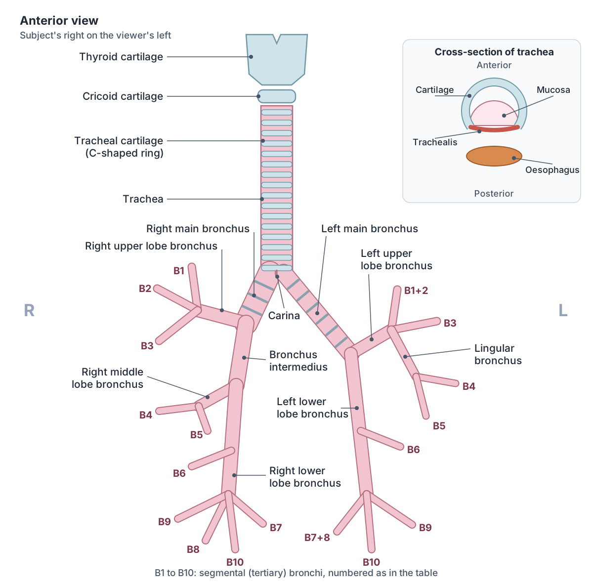
Segmental (tertiary) bronchi of each lung
| Lobe | Right lung (10 segments) | Left lung (usually 8 segments) |
|---|---|---|
| Upper lobe | Apical (B1), posterior (B2), anterior (B3) | Apicoposterior (B1+2), anterior (B3) |
| Middle lobe / lingula | Middle lobe: lateral (B4), medial (B5) | Lingula of upper lobe: superior lingular (B4), inferior lingular (B5) |
| Lower lobe | Superior (B6), medial basal (B7), anterior basal (B8), lateral basal (B9), posterior basal (B10) | Superior (B6), anteromedial basal (B7+8), lateral basal (B9), posterior basal (B10) |
Each segmental bronchus supplies one bronchopulmonary segment. On the left, B1 and B2, and B7 and B8, usually share a single segmental bronchus (Radiopaedia).
Trachea histology: the lining and the mucociliary escalator
The trachea is lined with pseudostratified ciliated columnar epithelium containing goblet cells. The goblet cells produce mucus that traps debris, and the cilia beat the mucus upwards towards the laryngopharynx, where it is swallowed down the oesophagus. (OpenStax Anatomy and Physiology 2e, 22.1: Organs and Structures of the Respiratory System.)
TeachMeAnatomy calls this combination of cilia and mucus the mucociliary escalator: it carries trapped particles and pathogens up out of the airways to be swallowed and destroyed. (TeachMeAnatomy: The Tracheobronchial Tree.)
Kenhub describes four layers in the tracheal wall: the mucosa (the epithelium on its lamina propria), the submucosa with mucous glands, vessels and nerves, the musculocartilaginous layer, and an outer fibroelastic adventitia. Radiopaedia groups them into three (mucous membrane, a fibromusculocartilaginous layer, and adventitia), which describes the same tissues counted differently. (Kenhub: Trachea.)
Going down the tree, cartilage, goblet cells and ciliated epithelium are present in the trachea and bronchi but absent in the bronchioles, where club cells are found instead. The alveoli at the end of the tree are lined by thin simple squamous epithelium. (TeachMeAnatomy: The Tracheobronchial Tree.)
Relations: what lies around the trachea
In the neck, the isthmus of the thyroid gland crosses the front of the trachea over rings 2 to 4, with the sternohyoid and sternothyroid muscles and the jugular venous arch in front. The oesophagus lies directly behind the trachea along its whole length. (Kenhub: Trachea.)
In the chest, the trachea has the vagus nerve, brachiocephalic vein, superior vena cava and azygos vein on its right, and the aortic arch, left common carotid artery, left subclavian artery and left recurrent laryngeal nerve on its left. (Kenhub: Trachea.)
Radiopaedia lists in front of the trachea the sternum, strap muscles, thyroid isthmus, ascending aorta, brachiocephalic artery, superior vena cava, inferior thyroid veins and left brachiocephalic vein. Beside it lie the lobes of the thyroid gland, the left common carotid artery, the arch of the azygos vein, and the right and left vagus and recurrent laryngeal nerves. (Radiopaedia: Trachea.)
Blood supply, lymph drainage and nerve supply
The upper half of the trachea is supplied by tracheo-oesophageal branches of the inferior thyroid artery. The lower half and the carina are supplied by the superior and middle bronchial arteries. Lymph drains to the pretracheal and paratracheal lymph nodes. (Radiopaedia: Trachea.)
Kenhub describes venous drainage through the inferior thyroid venous plexus to the brachiocephalic veins, and lymph passing from the pretracheal and paratracheal nodes on to the deep cervical nodes. (Kenhub: Trachea.)
TeachMeAnatomy gives tracheal venous drainage as the brachiocephalic, azygos and accessory hemiazygos veins. It describes sensory innervation of the trachea by the recurrent laryngeal nerve, a branch of the vagus, and supply of the bronchi by pulmonary branches of the vagus nerve (CN X). (TeachMeAnatomy: The Tracheobronchial Tree.)
Kenhub adds that the trachea receives parasympathetic fibres through the recurrent laryngeal nerves and sympathetic fibres from the sympathetic trunks. (Kenhub: Trachea.)
The main bronchi have their own bronchial vessels. The right bronchial vein drains to the azygos vein and the left bronchial vein to the accessory hemiazygos vein. The right main bronchus is usually supplied by one right bronchial artery and the left by two left bronchial arteries from the descending aorta. (Radiopaedia: Main bronchi.)
Development and variants
The trachea develops from the respiratory diverticulum, an outpouching of the foregut that appears in about the fourth week. A ridge called the tracheoesophageal septum separates it from the future oesophagus. If the septum forms abnormally, a tracheoesophageal fistula (an abnormal connection between trachea and oesophagus) results; affected babies may turn blue or bring milk back during feeds, and the fistula is repaired surgically. (TeachMeAnatomy: Development of the Respiratory System.)
The diverticulum then splits into two buds that become the main bronchi, and these keep dividing to form the secondary and tertiary bronchi. (TeachMeAnatomy: Development of the Respiratory System.)
About 2% of people have a tracheal bronchus ("pig bronchus"): an extra bronchus to the right upper lobe that arises from the trachea above the carina. It is usually harmless and found by chance, but it can complicate intubation and is linked with collapse and infection of the lobe it supplies. (Radiopaedia: Trachea.)
Clinical anatomy: intubation, tracheostomy and cricothyroidotomy
After intubation, the tip of an adult endotracheal tube should sit about 5 cm (plus or minus 2 cm) above the carina with the neck in a neutral position. Flexing the neck pushes the tip down towards the carina and extending it pulls the tip up. In children the target is about 1.5 cm above the carina because the trachea is shorter. (Radiopaedia: Evaluation of endotracheal tube position.)
A tube pushed in too far usually enters the right main bronchus, because it is the straighter path. The right lung is then overinflated and the left lung collapses. (Radiopaedia: Evaluation of endotracheal tube position.)
A tracheostomy is an opening made through the front of the trachea, usually for patients who need breathing support for a long time. In one clinical series of percutaneous tracheostomies, the trachea was opened at the level of the second or third tracheal ring rather than directly below the cricoid cartilage. (Thakur PK et al. Conventional Tracheostomy Versus Percutaneous Tracheostomy: A Retrospective Study (PMC).)
In an emergency, when the airway is blocked at or above the larynx and intubation has failed, a cricothyroidotomy is made instead: the thyroid cartilage is felt, the cricothyroid ligament just below it is found, and a small midline cut through this ligament allows a tube to be placed. It enters the airway above the trachea, between the thyroid and cricoid cartilages. (TeachMeAnatomy: Laryngeal Ligaments and Folds.)
Structures next to the trachea
The nearest named structures in the 3D model, measured from the centre of each part:
- Common carotid artery · cardiovascular
- Intervertebral disk of first thoracic vertebra · skeletal
- Inferior oblique part of longus colli · muscular
- Brachiocephalic artery · cardiovascular
- Intervertebral disk of second thoracic vertebra · skeletal
- Intervertebral disk of seventh cervical vertebra · skeletal
- Brachiocephalic vein · cardiovascular
- Sternothyroid · muscular
- Internal jugular vein · cardiovascular
- First rib · skeletal
Frequently asked questions
What is the trachea?
The trachea, or windpipe, is the airway tube that runs from the larynx to the lungs. It is about 10 to 13 cm long, held open by 16 to 20 C-shaped rings of hyaline cartilage, and lined by ciliated epithelium with mucus-secreting goblet cells. It begins at C6 below the cricoid cartilage and divides at the carina into the right and left main bronchi.
What is the carina of the trachea?
The carina is the ridge of cartilage at the bottom of the trachea where it splits into the right and left main bronchi. It lies at about the sternal angle and the T4 to T5 vertebral level. It is the most sensitive spot for the cough reflex and is the landmark used to check the position of a breathing tube on a chest X-ray.
Why are the tracheal rings C-shaped?
The cartilage keeps the trachea open, while the open back of each C is closed only by the trachealis muscle and elastic tissue. This soft back wall lets the oesophagus, which lies directly behind the trachea, bulge forwards when you swallow, and lets the fibroelastic membrane stretch slightly as you breathe.
Why do inhaled objects usually go into the right main bronchus?
The right main bronchus is wider, shorter and more vertical than the left: it runs at about 25 degrees to the midline, against about 40 degrees for the left. It is therefore the more direct continuation of the trachea, so inhaled objects, and breathing tubes pushed in too far, usually enter it.
What are tertiary bronchi?
Tertiary bronchi are the segmental bronchi. The main (primary) bronchi divide into lobar (secondary) bronchi, three on the right and two on the left, and each lobar bronchus divides into segmental (tertiary) bronchi. Each one supplies a bronchopulmonary segment: 10 on the right and usually 8 on the left.
Is the trachea in front of or behind the oesophagus?
The trachea is in front of the oesophagus. The oesophagus lies directly behind the trachea's soft back wall all the way from the neck to the carina. The recurrent laryngeal nerves run up in the groove between the two tubes.
Where is a tracheostomy made?
A tracheostomy is made through the front wall of the trachea in the lower neck, below the cricoid cartilage, typically at about the second or third tracheal ring. The isthmus of the thyroid gland lies over rings 2 to 4, so surgeons have to deal with it. An emergency cricothyroidotomy is different: it goes through the cricothyroid ligament, above the trachea.
References
- OpenStax. Anatomy and Physiology 2e, §22.1 Organs and Structures of the Respiratory System. Rice University, 2022.
- TeachMeAnatomy. The Tracheobronchial Tree.
- TeachMeAnatomy. Development of the Respiratory System.
- TeachMeAnatomy. Laryngeal Ligaments and Folds.
- Kenhub. Trachea.
- Radiopaedia. Trachea.
- Radiopaedia. Carina.
- Radiopaedia. Tracheal bifurcation angle.
- Radiopaedia. Main bronchi.
- Radiopaedia. Right main bronchus.
- Radiopaedia. Left main bronchus.
- Radiopaedia. Bronchopulmonary segmental anatomy.
- Radiopaedia. Evaluation of endotracheal tube position.
- Thakur PK, Jain AK, Khan TA, Jain S. Conventional Tracheostomy Versus Percutaneous Tracheostomy: A Retrospective Study. Indian J Otolaryngol Head Neck Surg. 2019;71(Suppl 1):459–464.
- Foundational Model of Anatomy: Trachea (FMA7394). EMBL-EBI Ontology Lookup Service.
- Mitsuhashi N. et al. (2009). BodyParts3D: 3D structure database for anatomical concepts. Nucleic Acids Research 37, D782–D785. Data © The Database Center for Life Science, CC BY 4.0.
Related anatomy
- LarynxLarynx anatomy with 3D model and diagrams: thyroid, cricoid, arytenoid and epiglottis cartilages, vocal folds…
- Esophagus (oesophagus)Esophagus (oesophagus) anatomy: its course and levels, four constrictions, upper and lower sphincters, muscle layers…
- Main bronchusMain bronchus: The left main (primary) bronchus, one of the two airways formed by the division of the trachea. See…
- Rib cageRib cage anatomy: 12 pairs of ribs, true, false and floating ribs, parts of a rib, sternum and jugular notch, costal…
- AortaAorta anatomy explained: ascending aorta, aortic arch and its three branches, thoracic aorta, and every abdominal…
Explore the whole body in the interactive 3D human anatomy model, or browse the A–Z list of human structures.