

What are the extraocular muscles?
The movements of the eyeball are controlled by the extrinsic eye muscles. They arise outside the eye, on the bones of the orbit, and insert on the outer surface of the eyeball, the white sclera. Because they lie inside the eye socket they cannot be seen on the surface of the eye. (OpenStax Anatomy and Physiology 2e, 11.3: Axial Muscles of the Head, Neck, and Back.)
Six skeletal muscles insert on the eye: four rectus muscles (superior, inferior, medial and lateral) and two obliques (superior and inferior). Levator palpebrae superioris is usually counted with them as a seventh extraocular muscle, although it moves the eyelid rather than the eye. A small muscle called orbitalis is sometimes listed as an eighth but is usually left out. (Radiopaedia: Extraocular muscles.)
The extraocular muscles are different from the intrinsic muscles of the eye, the ciliary muscle and the muscles of the iris, which sit inside the eyeball and are supplied by parasympathetic fibres travelling in the oculomotor nerve. (TeachMeAnatomy: The Oculomotor Nerve (CN III).)
The common tendinous ring (annulus of Zinn)
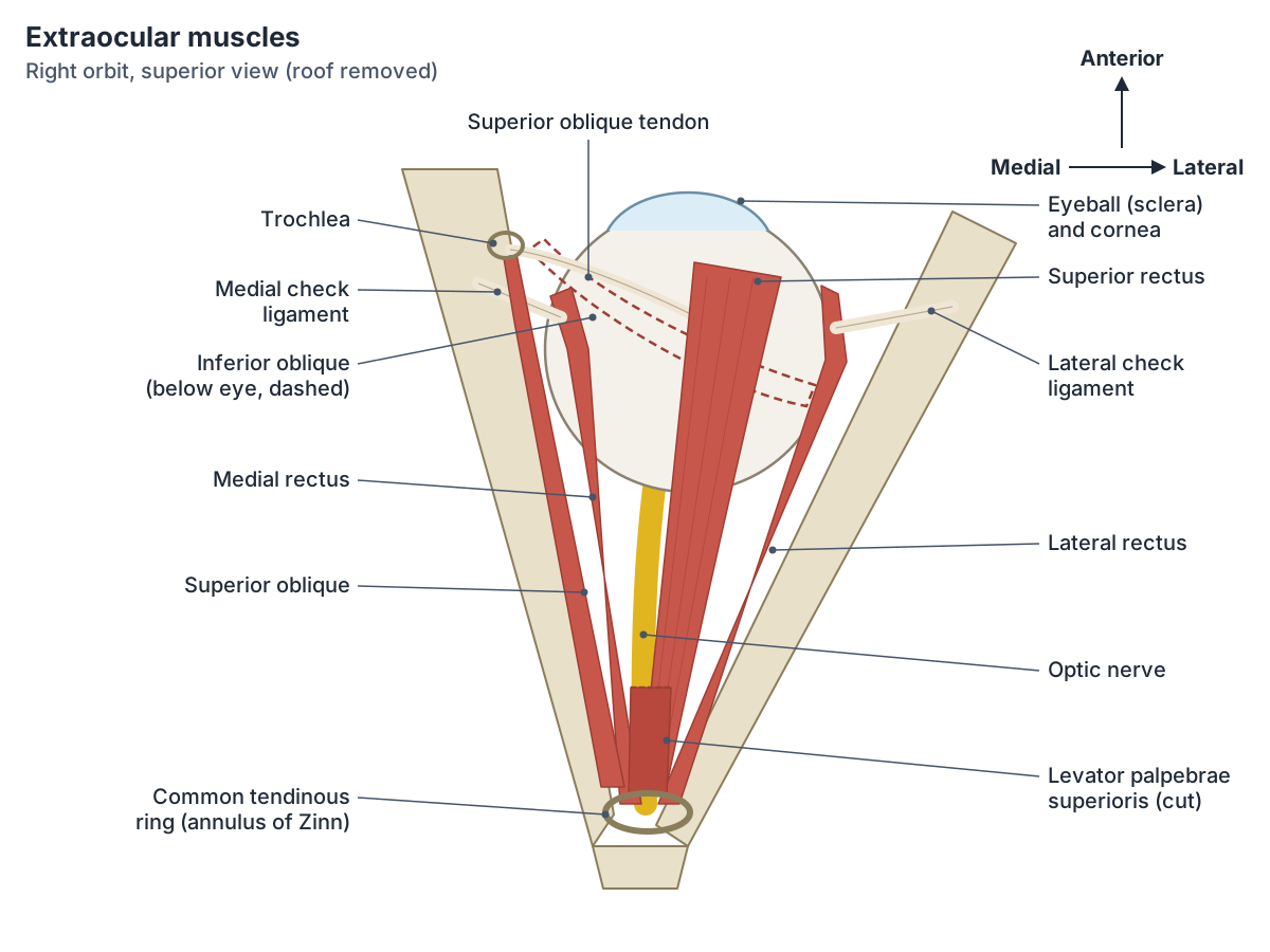
All four rectus muscles arise from the common tendinous ring, also called the annulus of Zinn: a ring of fibrous tissue that surrounds the optic canal at the back of the orbit. From this ring they spread forwards like the sides of a cone to the eyeball. (TeachMeAnatomy: The Extraocular Muscles.)
Superior rectus arises mainly from the upper part of the ring, with a small slip from the dura mater around the optic nerve, and inserts on the front half of the upper surface of the eyeball, just behind the corneoscleral junction. (Kenhub: Superior rectus muscle.)
The four rectus muscles: superior, inferior, medial and lateral rectus
Each rectus muscle takes a straight path forwards from the ring to the front half of the eyeball. The medial rectus inserts on the medial side of the sclera and adducts the eye (turns it towards the nose). The lateral rectus inserts on the lateral side and abducts it (turns it out towards the ear). These two act in the horizontal plane only. (TeachMeAnatomy: The Extraocular Muscles.)
The superior rectus inserts on the upper surface of the sclera and the inferior rectus on the lower surface. The superior rectus mainly elevates the eye and the inferior rectus mainly depresses it, but each also adducts the eye, and they rotate it in opposite directions: superior rectus rotates the top of the eye medially (intorsion) and inferior rectus rotates it laterally (extorsion). (TeachMeAnatomy: The Extraocular Muscles.)
These extra actions happen because the orbit points outwards while the eye looks straight ahead. Kenhub explains that the superior rectus runs at about 23 degrees to the median plane of the eye, so its pull lifts the eye and also turns it inwards and rotates it. When we look straight up, superior rectus normally contracts together with the inferior oblique, which cancels out the unwanted adduction and rotation. (Kenhub: Superior rectus muscle.)
Radiopaedia summarises the actions in neutral gaze: superior rectus elevates and adducts, inferior rectus depresses and adducts, medial rectus adducts and lateral rectus abducts. (Radiopaedia: Extraocular muscles.)
The superior and inferior oblique muscles
The superior oblique arises from the body of the sphenoid at the back of the orbit and runs forwards along the medial side of the orbit. Its tendon passes through a pulley called the trochlea, then turns backwards and laterally to insert on the sclera behind the superior rectus. It is supplied by the trochlear nerve (CN IV). (TeachMeAnatomy: The Extraocular Muscles.)
The inferior oblique (musculus obliquus inferior) differs from the other extraocular muscles, which arise at the back of the orbit: it starts on the anterior part of the orbital floor, runs backwards and laterally beneath the eyeball, and inserts on the sclera behind the lateral rectus. It is supplied by the oculomotor nerve (CN III). (TeachMeAnatomy: The Extraocular Muscles.)
Because both obliques approach the eye from the front and attach behind its equator, they act roughly opposite to the vertical recti. In TeachMeAnatomy's summary the superior oblique depresses, abducts and medially rotates the eyeball, and the inferior oblique elevates, abducts and laterally rotates it. (TeachMeAnatomy: The Extraocular Muscles.)
Radiopaedia gives their main actions more precisely: in neutral gaze the superior oblique mainly rotates the eye inwards (intorsion) and the inferior oblique outwards (extorsion); the superior oblique depresses the eye best when it is adducted, and the inferior oblique elevates it best when it is adducted. (Radiopaedia: Extraocular muscles.)
Kenhub offers a short rule: the obliques abduct and the recti adduct (except the lateral rectus); the superior muscles intort and the inferior muscles extort; and the recti act according to their names while the obliques act opposite to their names, so the superior oblique depresses and the inferior oblique elevates. (Kenhub: Extraocular muscles.)
Levator palpebrae superioris muscle
Levator palpebrae superioris arises from the underside of the lesser wing of the sphenoid, above and in front of the common tendinous ring, and lies immediately above the superior rectus. Its deep fibres attach to the front of the superior tarsal plate, the firm plate of connective tissue inside the upper eyelid; its superficial fibres reach the skin of the upper eyelid; and its lateral and medial fibres fan out to the orbital tubercle of the zygomatic bone and the medial palpebral ligament. (Kenhub: Levator palpebrae superioris muscle.)
It raises and retracts the upper eyelid and widens the gap between the lids. It is supplied by the superior division of the oculomotor nerve. It works with the superior rectus when we look up, and its steady tone balances orbicularis oculi, the facial muscle that closes the eye. (Kenhub: Levator palpebrae superioris muscle.)
The upper eyelid has five layers, from superficial to deep: skin, orbicularis oculi, the tarsal plate, the levator apparatus (levator palpebrae superioris and the superior tarsal muscle) and the conjunctiva. (TeachMeAnatomy: The Eyelids.)
Superior tarsal muscle (superior palpebral muscle, Müller muscle)
The superior tarsal muscle, also called Müller muscle and in Latin musculus tarsalis superior, is a thin slip that arises from the undersurface of levator palpebrae superioris and inserts on the upper margin of the superior tarsal plate. It helps to raise and retract the upper eyelid. (Radiopaedia: Superior tarsal muscle.)
Unlike the other eyelid muscles it is smooth muscle, so it is not under voluntary control. It is supplied by sympathetic fibres from the superior cervical ganglion, which travel along the internal carotid artery and then with the oculomotor nerve. Kenhub notes that it adds extra widening of the eyelids in fight-or-flight responses. (Kenhub: Levator palpebrae superioris muscle.)
Damage anywhere along the sympathetic pathway to the eye, as in Horner syndrome, weakens the superior tarsal muscle and causes a partial ptosis (a mildly drooping upper eyelid). Other signs of Horner syndrome usually help to tell this apart from other causes of ptosis. (Radiopaedia: Superior tarsal muscle.)
Nerve supply: the LR6 SO4 rule
Three cranial nerves supply the extraocular muscles. TeachMeAnatomy teaches the formula LR6 SO4 R3: the lateral rectus is supplied by the abducens nerve (CN VI), the superior oblique by the trochlear nerve (CN IV), and the rest by the oculomotor nerve (CN III). (TeachMeAnatomy: The Extraocular Muscles.)
The oculomotor nerve leaves the skull through the superior orbital fissure and splits into two branches. Its superior branch supplies the superior rectus and levator palpebrae superioris. Its inferior branch supplies the inferior rectus, the medial rectus and the inferior oblique, and carries parasympathetic fibres to the ciliary ganglion for the sphincter pupillae and ciliary muscle. (TeachMeAnatomy: The Oculomotor Nerve (CN III).)
The muscles receive blood from muscular branches of the ophthalmic artery. Their veins drain through the superior and inferior ophthalmic veins to the cavernous sinus and the pterygoid plexus. (Radiopaedia: Extraocular muscles.)
Extraocular muscles: origin, insertion, nerve and action
| Muscle | Origin | Insertion | Nerve | Actions (main action first) |
|---|---|---|---|---|
| Superior rectus | Upper part of the common tendinous ring | Superior sclera, just behind the corneoscleral junction | Oculomotor (CN III), superior division | Elevation; adduction; intorsion (medial rotation) |
| Inferior rectus | Lower part of the common tendinous ring | Inferior sclera, in front of the equator | Oculomotor (CN III), inferior division | Depression; adduction; extorsion (lateral rotation) |
| Medial rectus | Common tendinous ring | Medial sclera, behind the corneoscleral junction | Oculomotor (CN III), inferior division | Adduction |
| Lateral rectus | Common tendinous ring | Lateral sclera, in front of the equator | Abducens (CN VI) | Abduction |
| Superior oblique | Body of the sphenoid; tendon passes through the trochlea | Posterior superolateral sclera, behind the superior rectus | Trochlear (CN IV) | Intorsion; depression (mainly in adduction); abduction |
| Inferior oblique | Front of the orbital floor (maxilla) | Posterior inferolateral sclera, behind the lateral rectus | Oculomotor (CN III), inferior division | Extorsion; elevation (mainly in adduction); abduction |
| Levator palpebrae superioris | Lesser wing of the sphenoid, above and in front of the optic canal | Superior tarsal plate and skin of the upper eyelid | Oculomotor (CN III), superior division | Raises (elevates) the upper eyelid |
| Superior tarsal muscle (Müller muscle) | Underside of levator palpebrae superioris | Upper margin of the superior tarsal plate | Sympathetic fibres from the superior cervical ganglion | Helps raise and retract the upper eyelid |
Attachments from TeachMeAnatomy and Kenhub; actions from Radiopaedia and Kenhub. Sources describe the obliques' origins slightly differently.
Check ligaments and the suspensory ligament of the eye
The eyeball sits in a fascial sheath that runs from the corneoscleral junction to the optic nerve. The tendons of the recti and obliques pierce this sheath, and the sheath is reflected over them as a tube around each muscle. (Radiopaedia: Stability of the eye.)
The medial check ligament is the thickened sheath of the medial rectus, attached to the posterior lacrimal crest of the lacrimal bone. The lateral check ligament is the thickened sheath of the lateral rectus, attached to the marginal (Whitnall) tubercle on the orbital surface of the zygomatic bone. Together with the recti they help to prevent the eye being displaced backwards. (Radiopaedia: Stability of the eye.)
Between the two check ligaments a thickened part of the fascial sheath forms the suspensory ligament of Lockwood, a hammock that supports the eyeball above the orbital floor. Orbital fat around the muscle cone and the forward pull of the two obliques also keep the eye in place. (Radiopaedia: Stability of the eye.)
How the eye muscles are tested: the H test
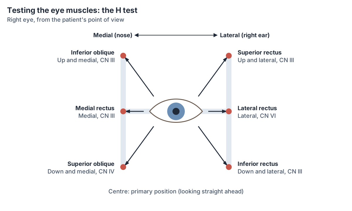
Clinicians test the extraocular muscles by asking the patient to follow a target, such as a finger, through the positions of gaze. Kenhub describes this as the H test, because the path traced looks like the letter H, and adds a convergence test in which the patient follows an object moved towards the middle of the face. (Kenhub: Extraocular muscles.)
EyeWiki explains that each of the six cardinal positions of gaze corresponds to the main field of action of one extraocular muscle, the muscle chiefly responsible for moving the eye into that position, so weakness of that muscle shows up most clearly there. (EyeWiki (American Academy of Ophthalmology): Sensory and Motor Testing.)
The cardinal positions are not the same as the muscles' actions from the straight-ahead position. EyeWiki notes that the obliques are tested in adduction and the vertical recti in abduction. In practice, for each eye, looking out tests the lateral rectus, looking in tests the medial rectus, up and out tests the superior rectus, down and out the inferior rectus, up and in the inferior oblique, and down and in the superior oblique. (EyeWiki (American Academy of Ophthalmology): Sensory and Motor Testing.)
The two eyes move together because yoke muscles, the pair that moves both eyes in the same direction (for example the right lateral rectus and the left medial rectus), receive equal nerve signals. This principle is called Hering's law. (EyeWiki (American Academy of Ophthalmology): Sensory and Motor Testing.)
Oculomotor, trochlear and abducens nerve palsies
In an oculomotor (CN III) palsy the upper eyelid droops (ptosis) because levator palpebrae superioris is paralysed. At rest the eye points down and out, pulled by the lateral rectus and superior oblique, which still work, and the patient cannot elevate, depress or adduct it. The pupil may be dilated because the parasympathetic fibres to the sphincter pupillae are lost. (TeachMeAnatomy: The Oculomotor Nerve (CN III).)
A trochlear (CN IV) palsy paralyses the superior oblique. The resting position of the eye may look almost normal, but the patient has double vision (diplopia) and may tilt the head away from the affected side. An abducens (CN VI) palsy paralyses the lateral rectus, so the affected eye is pulled inwards by the unopposed medial rectus. (TeachMeAnatomy: The Extraocular Muscles.)
The eye muscles in the 3D model
The BodyParts3D model includes all six extraocular muscles of each eye, levator palpebrae superioris and its tendon, the common tendinous ring, the trochlea of the superior oblique and the check ligaments of the medial and lateral rectus, so these can be rotated and viewed from any side. It does not include the superior tarsal muscle, which is too thin to model separately.
In the model, look at the superior oblique from above to see how its tendon bends sharply through the trochlea before running back to the eyeball, and look from below to find the inferior oblique, which starts at the front of the orbital floor and runs back beneath the eyeball. (TeachMeAnatomy: The Extraocular Muscles.)
Eye muscles in the 3D model
- Check ligament of lateral rectus
- A fibrous expansion of the sheath of the lateral rectus eye muscle.
- Check ligament of medial rectus
- A fibrous expansion of the sheath of the medial rectus eye muscle.
- Common tendinous ring
- A fibrous ring at the back of the orbit giving origin to the recti muscles of the eye.
- Inferior oblique
- An extraocular muscle that elevates and turns the eye outward (this name refers to the eye muscle, not the abdominal or suboccipital one).
- Inferior rectus
- An extraocular muscle on the floor of the orbit.
- Lateral rectus
- An extraocular muscle on the lateral wall of the orbit.
- Levator palpebrae superioris
- The muscle that lifts the upper eyelid.
- Medial rectus
- An extraocular muscle on the medial wall of the orbit.
- Superior oblique
- An extraocular muscle that passes through a pulley (trochlea) before reaching the eyeball.
- Superior rectus
- An extraocular muscle on the top of the eyeball.
- Tendon of levator palpebrae superioris
- The flattened tendon (aponeurosis) of the muscle that raises the upper eyelid.
- Trochlea of superior oblique
- A small fibrocartilaginous pulley in the orbit.
Frequently asked questions
How many muscles does the eye have?
Six muscles move each eyeball: the superior, inferior, medial and lateral rectus and the superior and inferior oblique. A seventh muscle in the orbit, levator palpebrae superioris, raises the upper eyelid. Inside the eyeball there are also intrinsic smooth muscles, the ciliary muscle and the iris muscles, which control focusing and pupil size.
What does LR6 SO4 R3 mean?
LR6 SO4 R3 is a memory aid for the nerves of the eye muscles. The lateral rectus (LR) is supplied by cranial nerve VI, the abducens nerve. The superior oblique (SO) is supplied by cranial nerve IV, the trochlear nerve. The remaining muscles, superior, inferior and medial rectus, inferior oblique and levator palpebrae superioris, are supplied by cranial nerve III, the oculomotor nerve.
What does the lateral rectus do?
The lateral rectus abducts the eye, turning it outwards towards the ear. It runs from the common tendinous ring at the back of the orbit to the lateral side of the sclera and is the only muscle supplied by the abducens nerve (CN VI). If that nerve is damaged, the eye is pulled inwards by the unopposed medial rectus and the patient sees double when looking to that side.
What does the inferior oblique muscle do?
The inferior oblique mainly rotates the eye outwards (extorsion) and elevates it, especially when the eye is turned in towards the nose; it also helps abduct the eye. It arises from the front of the orbital floor, runs back beneath the eyeball and inserts on the sclera behind the lateral rectus. It is supplied by the inferior division of the oculomotor nerve.
What is the difference between levator palpebrae superioris and the superior tarsal muscle?
Levator palpebrae superioris is a skeletal muscle under voluntary control, supplied by the oculomotor nerve, that does most of the work of lifting the upper eyelid. The superior tarsal (Müller) muscle is a thin smooth muscle that arises from its underside and attaches to the superior tarsal plate. It is supplied by sympathetic nerves and adds a little extra lift, which is lost in Horner syndrome.
What are the check ligaments of the eye?
The check ligaments are thickened parts of the sheaths of the medial and lateral rectus muscles. The medial one attaches to the lacrimal bone and the lateral one to the zygomatic bone at the marginal tubercle. With the suspensory ligament of Lockwood between them, they support the eyeball and help stop it being pushed backwards into the orbit.
References
- OpenStax. Anatomy and Physiology 2e, §11.3 Axial Muscles of the Head, Neck, and Back. Rice University, 2022.
- TeachMeAnatomy. The Extraocular Muscles.
- TeachMeAnatomy. The Oculomotor Nerve (CN III).
- TeachMeAnatomy. The Eyelids.
- Kenhub. Extraocular muscles (muscles of the orbit).
- Kenhub. Superior rectus muscle.
- Kenhub. Levator palpebrae superioris muscle.
- Radiopaedia. Extraocular muscles.
- Radiopaedia. Superior tarsal muscle.
- Radiopaedia. Stability of the eye.
- EyeWiki, American Academy of Ophthalmology. Sensory and Motor Testing.
- Mitsuhashi N. et al. (2009). BodyParts3D: 3D structure database for anatomical concepts. Nucleic Acids Research 37, D782–D785. Data © The Database Center for Life Science, CC BY 4.0.
Related anatomy
- Eye anatomyEyeball anatomy with a labeled 3D eye model: sclera, cornea, choroid, iris, lens, retina and chambers, the 3 layers…
- Cranial nervesThe 12 cranial nerves in order with a labelled diagram: type, skull foramen, functions and test for CN I to CN XII…
- Superior obliqueSuperior oblique: An extraocular muscle that passes through a pulley (trochlea) before reaching the eyeball. See where…
- Superior rectusSuperior rectus: An extraocular muscle on the top of the eyeball. See where it is in an interactive 3D human body…
- Facial musclesFace muscles labeled: labelled diagrams and a table of every muscle of facial expression, from procerus and…
- Skull bonesLearn the 22 bones of the skull, the 8 cranial bones of the neurocranium and the coronal, sagittal, lambdoid and…
Explore the whole body in the interactive 3D human anatomy model, or browse the A–Z list of human structures.