

What are the facial muscles?
The muscles of facial expression are unusual skeletal muscles: they attach to the skin rather than moving a joint. Their origins lie on the surface of the skull and stay fixed, while their insertions are in the skin, so contraction moves the skin of the face. (OpenStax Anatomy and Physiology 2e, 11.3: Axial Muscles of the Head, Neck, and Back.)
All of them develop from the second pharyngeal arch of the embryo. As the muscle cells migrate into the face they take their nerve with them, which is why every muscle of facial expression is supplied by branches of the facial nerve (cranial nerve VII). (TeachMeAnatomy: Muscles of Facial Expression.)
Besides producing expressions, these muscles act as sphincters and dilators of the openings of the face: they close and open the eyes, widen or narrow the nostrils and control the lips. Radiopaedia groups them by region into epicranial (scalp), circumorbital, nasal and buccolabial (mouth) muscles. (Radiopaedia: Muscles of facial expression.)
Two muscles near the face are often confused with this group. Levator palpebrae superioris, which lifts the upper eyelid, lies in the orbit and is supplied by the oculomotor nerve, and the chewing muscles such as masseter and temporalis are supplied by the trigeminal nerve. Neither is a muscle of facial expression in the strict sense. (TeachMeAnatomy: The Eyelids.)
Face muscles labelled: the main groups
TeachMeAnatomy divides the facial muscles into three main groups. The orbital group (orbicularis oculi and corrugator supercilii) works around the eye. The nasal group (nasalis, procerus and depressor septi nasi) acts on the nose. The oral group (orbicularis oris, buccinator and the small muscles that raise and lower the lips) acts on the mouth. (TeachMeAnatomy: Muscles of Facial Expression.)
The oral group is the largest. Its upper muscles are risorius, zygomaticus major, zygomaticus minor, levator labii superioris, levator labii superioris alaeque nasi and levator anguli oris. Its lower muscles are depressor anguli oris, depressor labii inferioris and mentalis. (TeachMeAnatomy: Muscles of Facial Expression.)
Kenhub adds the scalp muscle occipitofrontalis, the three auricular muscles around the ear and platysma in the neck, which are also supplied by the facial nerve. The anterior diagram shows the face in two layers so that the deeper muscles, buccinator, levator anguli oris and corrugator supercilii, are visible. (Kenhub: Facial muscles.)
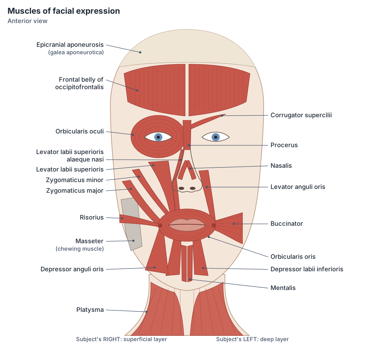
Epicranius and the epicranial aponeurosis (scalp muscles)
The epicranius is the muscle sheet of the scalp. Radiopaedia lists it as occipitofrontalis, made of the frontalis and occipitalis bellies, together with temporoparietalis, a thin muscle above the ear. (Radiopaedia: Muscles of facial expression.)
The frontal and occipital bellies of occipitofrontalis are joined by a thick, flat tendon called the epicranial aponeurosis or galea aponeurotica, which covers the top of the skull. The frontal belly has no bony attachment: it arises from the skin of the eyebrow and the muscles of the forehead. The occipital belly arises from the lateral two-thirds of the superior nuchal line of the occipital bone. Both bellies insert into the aponeurosis. (Kenhub: Occipitofrontalis muscle.)
The frontal belly raises the eyebrows and wrinkles the skin of the forehead, as in surprise; the occipital belly pulls the scalp backwards. The frontal belly is supplied by the temporal branches of the facial nerve and the occipital belly by the posterior auricular nerve, another branch of the facial nerve. (Kenhub: Occipitofrontalis muscle.)
The auricular muscles are small and largely rudimentary in humans. Auricularis anterior and auricularis superior arise from the epicranial aponeurosis and pull the ear forwards and upwards; auricularis posterior arises from the mastoid process and pulls the ear backwards. The anterior and superior muscles are supplied by temporal branches and the posterior one by the posterior auricular branch of the facial nerve. (Kenhub: Facial muscles.)
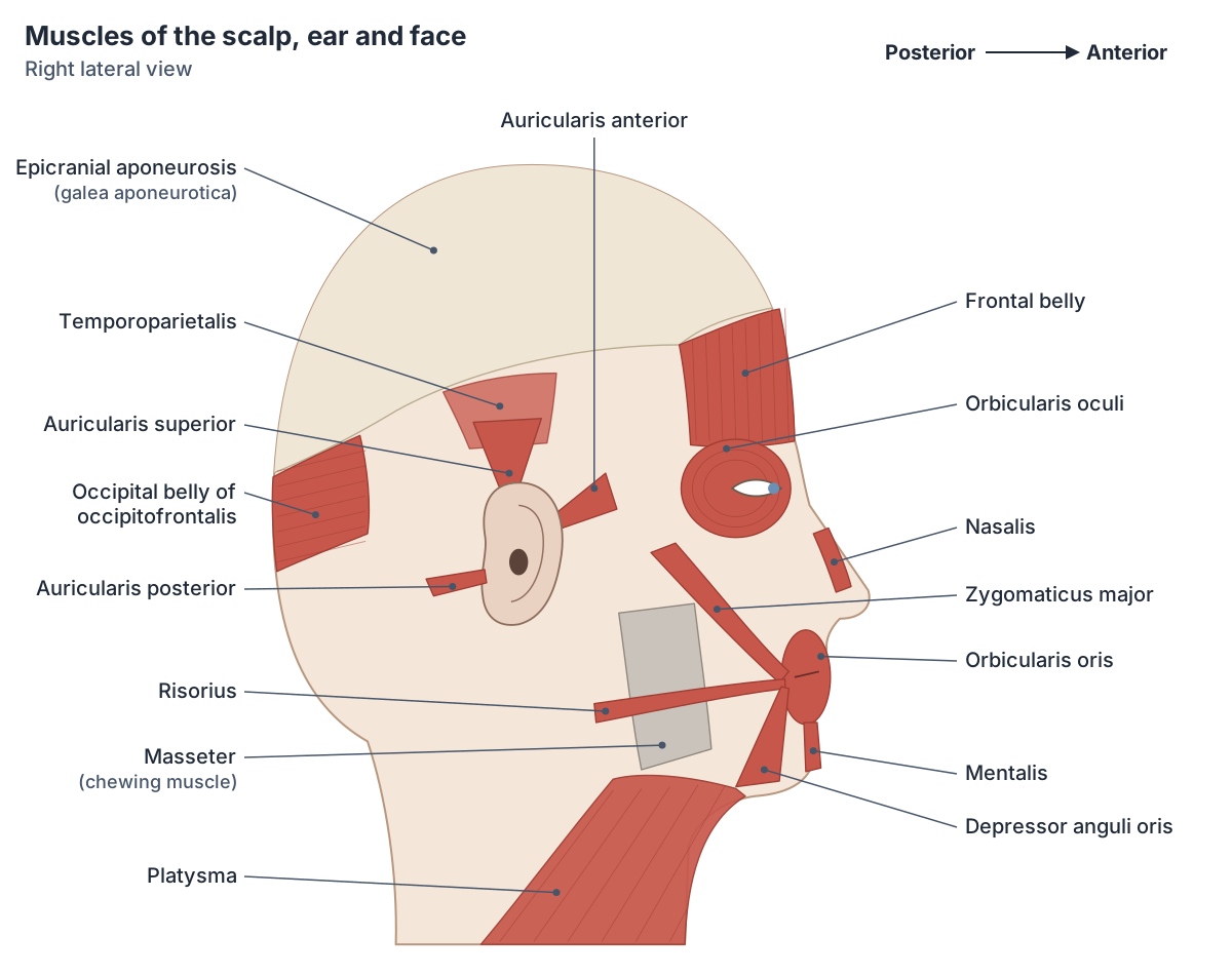
Muscles around the eye: orbicularis oculi, corrugator and procerus
Orbicularis oculi is a flat ring of muscle around the orbit and in the eyelids. Its palpebral part, inside the lids, closes the eyelids gently, as in blinking. Its orbital part, around the bony rim, closes them tightly. Its small lacrimal part helps to drain tears. It is supplied by the temporal and zygomatic branches of the facial nerve. (TeachMeAnatomy: Muscles of Facial Expression.)
The corrugator supercilii muscle sits at the inner end of the eyebrow. It arises from the medial end of the superciliary arch of the frontal bone and inserts into the skin of the eyebrow. It draws the eyebrows together and produces the vertical wrinkles on the bridge of the nose when we frown. It is supplied by the temporal branches of the facial nerve. (TeachMeAnatomy: Muscles of Facial Expression.)
Procerus is a small muscle over the top of the nose. It arises from the nasal bone and the upper part of the lateral nasal cartilage and inserts into the skin of the glabella, the area between the eyebrows. It pulls the inner ends of the eyebrows down and wrinkles the glabellar skin. Repeated frowning eventually leaves permanent glabellar lines, which is why procerus is a common target for cosmetic botulinum toxin injections. (Kenhub: Procerus muscle.)
Sources differ on the nerve to procerus: Kenhub lists the temporal, lower zygomatic or buccal branches of the facial nerve, while TeachMeAnatomy gives the buccal branches. (Kenhub: Procerus muscle.)
Nose muscles: nasalis, depressor septi nasi and the alar muscles
Nasalis has two parts. The transverse part arises from the maxilla just lateral to the nose and inserts into an aponeurosis across the bridge of the nose; it compresses the nasal opening. The alar part arises from the maxilla over the lateral incisor tooth and inserts into the alar cartilage, the cartilage of the wing (ala) of the nose; it widens the nostril. (TeachMeAnatomy: Muscles of Facial Expression.)
Depressor septi nasi arises from the maxilla above the central incisor and inserts into the nasal septum. It pulls the septum down and so widens the nostril. Nasalis, procerus and depressor septi nasi are supplied by the buccal branches of the facial nerve. (TeachMeAnatomy: Muscles of Facial Expression.)
The alar of the nose is therefore moved by three muscles: the alar part of nasalis and depressor septi nasi flare the nostril, and levator labii superioris alaeque nasi, described in the next section, lifts the ala as it raises the upper lip. (Kenhub: Facial muscles.)
Levator labii superioris alaeque nasi and the other upper lip muscles
Levator labii superioris alaeque nasi means the lifter of the upper lip and of the wing of the nose. It arises from the upper part of the frontal process of the maxilla, beside the nose, and runs down to insert into the skin and perichondrium over the major alar cartilage, with some fibres reaching the upper lip. It raises and everts the upper lip, flares the nostril and deepens the nasolabial furrow, as in a snarl. It is supplied by the zygomatic and buccal branches of the facial nerve. (Kenhub: Facial muscles.)
Levator labii superioris lies just lateral to it. It arises from the zygomatic process of the maxilla and the maxillary process of the zygomatic bone, just above the infraorbital foramen, and blends into the muscles of the upper lip. It raises the upper lip to show the upper teeth and deepens the nasolabial lines. Levator anguli oris lies lateral and deep to it, and the two muscles bound the canine space, a route by which dental infections can spread. (Kenhub: Levator labii superioris muscle.)
Zygomaticus major and zygomaticus minor both arise from the lateral surface of the zygomatic (cheek) bone. Zygomaticus minor inserts into the skin of the upper lip, medial to zygomaticus major, and raises the lip. Zygomaticus major runs further down to the angle of the mouth and pulls it upwards and outwards, the main movement of a smile. (Kenhub: Facial muscles.)
Levator anguli oris arises from the canine fossa of the maxilla and raises the corner of the mouth. Risorius has a variable origin from the fascia over the parotid gland, masseter or platysma and pulls the corner of the mouth sideways. Both insert into the modiolus at the angle of the mouth. (Kenhub: Facial muscles.)
Lower lip and chin: depressor labii inferioris, depressor anguli oris and mentalis
Depressor labii inferioris arises from the oblique line of the mandible, between the midline (symphysis) and the mental foramen, and inserts into the skin and submucosa of the lower lip. It pulls the lower lip down. Kenhub's article names the marginal mandibular branch of the facial nerve in its text but the cervical branch in its summary box, so its nerve is best remembered as the marginal mandibular branch, with cervical branch fibres in some people. (Kenhub: Depressor labii inferioris muscle.)
Depressor anguli oris arises from the oblique line and mental tubercle of the mandible and inserts into the modiolus. It pulls the corner of the mouth down, as in a frown or sad expression. Mentalis arises from the incisive fossa of the mandible and inserts into the skin of the chin; it wrinkles the chin and pushes the lower lip out. (Kenhub: Facial muscles.)
OpenStax describes the mentalis as the prime mover for pouting and the risorius for drawing the corners of the mouth sideways, while zygomaticus major is the main smiling muscle. (OpenStax Anatomy and Physiology 2e, 11.3: Axial Muscles of the Head, Neck, and Back.)
Orbicularis oris, buccinator and the modiolus
Orbicularis oris is the circular muscle of the lips. Its fibres come from the maxilla and from the neighbouring cheek muscles, and it inserts into the skin and mucous membrane of the lips. It closes the lips and narrows the mouth, and it shapes the lips during speech. TeachMeAnatomy gives its nerve supply as the buccal branches of the facial nerve; Kenhub adds the marginal mandibular branch. (TeachMeAnatomy: Muscles of Facial Expression.)
Buccinator forms most of the cheek. It arises from the alveolar processes of the maxilla and mandible opposite the molar teeth and, at the back, from the pterygomandibular raphe, a tendinous band that runs from the hamulus of the medial pterygoid plate to the mandible. The superior pharyngeal constrictor of the pharynx attaches to the back edge of the same raphe. (Radiopaedia: Pterygomandibular raphe.)
Buccinator's fibres converge on the angle of the mouth. It presses the cheek against the molar teeth, keeping food between the teeth during chewing, and helps in blowing, for example when playing a wind instrument. It is supplied by the buccal branches of the facial nerve. (Kenhub: Facial muscles.)
The modiolus is a knot of muscle fibres just lateral to each corner of the mouth. Fibres of many of the lip muscles pass into or through it. (Radiopaedia: Muscles of facial expression.)
Kenhub lists zygomaticus major, levator anguli oris, risorius, depressor anguli oris, buccinator, orbicularis oris and the lateral fibres of platysma as attaching to the modiolus, so the corner of the mouth can be pulled up, out, sideways or down depending on which muscles contract. (Kenhub: Facial muscles.)
Platysma: the facial muscle in the neck
Platysma is a broad, thin sheet in the subcutaneous tissue of the neck. It arises from the skin and fascia of the upper chest and shoulder and runs up the neck to insert into the lower border of the mandible, the skin of the lower lip and the modiolus. Its lateral fibres pull the corners of the mouth and the lower lip down, and its medial fibres help to depress the mandible. It is supplied by the cervical branch of the facial nerve. (Kenhub: Facial muscles.)
Platysma is the only muscle of facial expression present in the BodyParts3D model used on this site; it can be found on the automatic platysma page.
Facial nerve branches and which muscles they supply
After leaving the skull, the facial nerve gives the posterior auricular nerve, which supplies the auricular muscles and the occipital belly of occipitofrontalis. It then enters the parotid gland, which it passes through without supplying, and splits into five terminal branches. (TeachMeAnatomy: The Facial Nerve (CN VII).)
The five branches, from top to bottom, are temporal, zygomatic, buccal, marginal mandibular and cervical. In TeachMeAnatomy's summary the temporal branch supplies frontalis, orbicularis oculi and corrugator supercilii; the zygomatic branch supplies orbicularis oculi; the buccal branch supplies orbicularis oris, buccinator and the zygomaticus muscles; the marginal mandibular branch supplies depressor labii inferioris, depressor anguli oris and mentalis; and the cervical branch supplies platysma. (TeachMeAnatomy: The Facial Nerve (CN VII).)
The branches join each other in many places, and Kenhub lists several muscles as supplied by two branches, for example zygomaticus major by both the zygomatic and buccal branches. The table therefore gives the main branch or branches named in the sources rather than a single fixed answer. (Kenhub: Facial muscles.)
Muscles of facial expression: origin, insertion, action and nerve
| Muscle | Origin | Insertion | Main action | Facial nerve (CN VII) branch |
|---|---|---|---|---|
| Occipitofrontalis, frontal belly (frontalis) | Skin of the eyebrow and forehead muscles (no bony origin) | Epicranial aponeurosis | Raises the eyebrows, wrinkles the forehead | Temporal |
| Occipitofrontalis, occipital belly (occipitalis) | Lateral two-thirds of the superior nuchal line | Epicranial aponeurosis | Pulls the scalp backwards | Posterior auricular nerve |
| Auricularis anterior | Lateral border of the epicranial aponeurosis | Spine of the helix | Pulls the ear forwards (rudimentary) | Temporal |
| Auricularis superior | Epicranial aponeurosis | Upper part of the auricle | Pulls the ear upwards (rudimentary) | Temporal |
| Auricularis posterior | Mastoid process of the temporal bone | Back of the concha (eminentia conchae) | Pulls the ear backwards (rudimentary) | Posterior auricular nerve |
| Orbicularis oculi | Nasal part of the frontal bone, frontal process of the maxilla, medial palpebral ligament, lacrimal bone | Skin around the orbit, lateral palpebral ligament, tarsal plates | Closes the eyelids: gently (palpebral part) or tightly (orbital part); lacrimal part helps tear drainage | Temporal and zygomatic |
| Corrugator supercilii | Medial end of the superciliary arch of the frontal bone | Skin of the middle of the eyebrow | Draws the eyebrows together (vertical frown lines) | Temporal |
| Procerus | Nasal bone and upper lateral nasal cartilage | Skin over the glabella between the eyebrows | Pulls the inner ends of the eyebrows down (horizontal wrinkles over the nose) | Temporal, lower zygomatic or buccal (sources vary) |
| Nasalis, transverse part | Maxilla just lateral to the nose | Aponeurosis over the bridge (dorsum) of the nose | Compresses the nostril | Buccal |
| Nasalis, alar part | Maxilla over the lateral incisor | Alar cartilage of the nose | Widens (flares) the nostril | Buccal |
| Depressor septi nasi | Maxilla above the central incisor | Nasal septum | Pulls the septum down, widening the nostril | Buccal |
| Levator labii superioris alaeque nasi | Upper frontal process of the maxilla | Major alar cartilage and skin of the nose; upper lip | Raises the upper lip and flares the nostril; deepens the nasolabial furrow | Zygomatic and buccal |
| Levator labii superioris | Maxilla and zygomatic bone just above the infraorbital foramen | Muscles and skin of the upper lip | Raises and everts the upper lip, showing the upper teeth | Zygomatic and buccal |
| Zygomaticus minor | Lateral surface of the zygomatic bone | Skin of the upper lip, medial to zygomaticus major | Raises the upper lip | Zygomatic and buccal |
| Zygomaticus major | Lateral surface of the zygomatic bone | Angle of the mouth (modiolus) | Pulls the corner of the mouth up and out (smiling) | Zygomatic and buccal |
| Levator anguli oris | Canine fossa of the maxilla | Angle of the mouth (modiolus) | Raises the corner of the mouth | Zygomatic and buccal |
| Risorius | Variable: fascia over the parotid gland, masseter or platysma | Angle of the mouth (modiolus) | Pulls the corner of the mouth sideways | Buccal |
| Depressor anguli oris | Oblique line and mental tubercle of the mandible | Angle of the mouth (modiolus) | Pulls the corner of the mouth down (frowning) | Marginal mandibular and buccal |
| Depressor labii inferioris | Oblique line of the mandible, between the symphysis and the mental foramen | Skin and submucosa of the lower lip | Pulls the lower lip down | Marginal mandibular (some sources: cervical) |
| Mentalis | Incisive fossa of the mandible | Skin of the chin | Wrinkles the chin and pushes out the lower lip (pouting) | Marginal mandibular |
| Orbicularis oris | Modiolus and neighbouring muscles; maxilla | Skin and mucous membrane of the lips | Closes and purses the lips; shapes them in speech | Buccal and marginal mandibular |
| Buccinator | Alveolar processes of the maxilla and mandible opposite the molars; pterygomandibular raphe | Angle of the mouth (modiolus), blending with orbicularis oris | Presses the cheek against the teeth; blowing | Buccal |
| Platysma | Skin and fascia of the upper chest and shoulder | Lower border of the mandible, skin of the lower lip and modiolus | Pulls the corners of the mouth and lower lip down; tenses the skin of the neck | Cervical |
Attachments and actions follow Kenhub's facial muscles summary, with TeachMeAnatomy for nasalis and depressor septi nasi. Facial nerve branch assignments overlap and vary between people and between textbooks.
Facial palsy and Bell's palsy
Damage to the facial nerve after it leaves the skull is a lower motor neuron lesion. It causes paralysis or severe weakness of all the muscles of facial expression on that side. When no cause can be found the condition is called Bell's palsy. (TeachMeAnatomy: The Facial Nerve (CN VII).)
Bell palsy is the most common cause of facial palsy. In a lower motor neuron palsy the whole half of the face is weak, including the forehead. A lesion of the upper motor neurons in the brain, such as a stroke, typically spares the forehead on the affected side, so the patient can still wrinkle the forehead, raise the eyebrow and close the eye. The traditional explanation is that the upper face receives input from both sides of the cortex, although Radiopaedia notes that this explanation may not be fully supported by the literature. (Radiopaedia: Facial palsy.)
Orbicularis oculi, the muscle that closes the eyelids, is supplied by the temporal and zygomatic branches of the facial nerve, so eye closure is weak in a facial nerve palsy. Opening the eye is not affected, because levator palpebrae superioris is supplied by the oculomotor nerve. (TeachMeAnatomy: The Eyelids.)
Frequently asked questions
How many muscles are in the face?
There is no single agreed number because textbooks split and group the small muscles differently, but about twenty paired muscles of facial expression are usually described. They include occipitofrontalis, orbicularis oculi, corrugator supercilii, procerus, nasalis, the lip levators and depressors, zygomaticus major and minor, risorius, mentalis, orbicularis oris, buccinator and platysma. All are supplied by the facial nerve.
What does levator labii superioris alaeque nasi do?
Levator labii superioris alaeque nasi runs from the frontal process of the maxilla beside the nose down to the alar cartilage of the nose and the upper lip. It raises and everts the upper lip, flares the nostril and deepens the fold between the nose and the cheek, as in a snarl. It is supplied by the zygomatic and buccal branches of the facial nerve.
What is the difference between zygomaticus major and minor?
Both arise from the lateral surface of the zygomatic (cheek) bone. Zygomaticus major is longer and runs to the angle of the mouth (the modiolus); it pulls the corner of the mouth up and out and is the main smiling muscle. Zygomaticus minor lies medial to it and inserts into the skin of the upper lip, which it raises. Both are supplied by zygomatic and buccal branches of the facial nerve.
Which muscles do you use to smile and to frown?
Smiling mainly uses zygomaticus major, helped by levator anguli oris and risorius, which pull the corners of the mouth up and out. Frowning uses corrugator supercilii and procerus to pull the eyebrows together and down, and depressor anguli oris to turn the corners of the mouth down.
What is the epicranius muscle?
The epicranius is the muscle of the scalp. It consists of occipitofrontalis, whose frontal belly over the forehead and occipital belly over the back of the head are joined by the epicranial aponeurosis (galea aponeurotica), and a thin temporoparietalis above the ear. The frontal belly raises the eyebrows and wrinkles the forehead. Both bellies are supplied by branches of the facial nerve.
What nerve supplies the muscles of facial expression?
The facial nerve, cranial nerve VII, supplies all of them. Its posterior auricular branch supplies the ear muscles and the occipital belly of occipitofrontalis. Its five terminal branches in the face are temporal, zygomatic, buccal, marginal mandibular and cervical. The chewing muscles, such as masseter, are supplied by the trigeminal nerve instead.
Is buccinator a muscle of mastication?
No. Buccinator helps chewing by pressing the cheek against the teeth and keeping food between them, but it is a muscle of facial expression supplied by the buccal branches of the facial nerve. The four muscles of mastication, masseter, temporalis and the medial and lateral pterygoids, move the mandible and are supplied by the mandibular division of the trigeminal nerve.
References
- OpenStax. Anatomy and Physiology 2e, §11.3 Axial Muscles of the Head, Neck, and Back. Rice University, 2022.
- TeachMeAnatomy. The Muscles of Facial Expression.
- TeachMeAnatomy. The Facial Nerve (CN VII).
- TeachMeAnatomy. The Eyelids.
- Kenhub. Facial muscles.
- Kenhub. Occipitofrontalis muscle.
- Kenhub. Procerus muscle.
- Kenhub. Levator labii superioris muscle.
- Kenhub. Depressor labii inferioris muscle.
- Radiopaedia. Muscles of facial expression.
- Radiopaedia. Facial palsy.
- Radiopaedia. Pterygomandibular raphe.
Related anatomy
- Muscles of masticationThe muscles of mastication explained: masseter, temporalis, medial and lateral pterygoid, with labelled diagrams, the…
- Eye musclesEye muscles explained: the six extraocular muscles, levator palpebrae superioris and superior tarsal muscle, with the…
- PlatysmaPlatysma: A thin superficial sheet of muscle in the subcutaneous tissue of the neck. See where it is in an interactive…
- Cranial nervesThe 12 cranial nerves in order with a labelled diagram: type, skull foramen, functions and test for CN I to CN XII…
- Skull bonesLearn the 22 bones of the skull, the 8 cranial bones of the neurocranium and the coronal, sagittal, lambdoid and…
- Neck musclesNeck muscles explained: sternocleidomastoid, suprahyoid, infrahyoid, scalene, prevertebral and suboccipital groups…
Explore the whole body in the interactive 3D human anatomy model, or browse the A–Z list of human structures.