

Gallbladder at a glance
- What it is
- The gallbladder is a pear-shaped sac that stores and concentrates bile.
- Location
- It lies in the gallbladder fossa on the visceral surface of the liver in the right upper quadrant, between segments IVb and V, with its fundus often projecting at the tip of the 9th costal cartilage.
- Function
- It stores about 30 to 50 mL of bile, concentrates it by absorbing water, and releases it into the duodenum when stimulated by cholecystokinin.
- Body system
- Digestive · single, midline structure
- FMA ID
- FMA7202 (Foundational Model of Anatomy)
Key facts
- Parts are fundus, body, infundibulum (Hartmann pouch) and neck.
- Cystic artery, usually from the right hepatic artery, supplies it.
- Its wall has a mucosa with folds, a muscularis, and a serosa/adventitia; there is no submucosa.
Clinical relevance
Gallstones (cholelithiasis) and cholecystitis commonly cause right upper quadrant pain; Murphy sign is a classic finding.
Where is the gallbladder situated?
The gallbladder lies in the right hypochondrial region of the abdomen, the region under the right ribs, in the right upper quadrant. It sits against the inferior surface of the liver in the mid-clavicular line. (TeachMeAnatomy: The Gallbladder.)
More precisely, it lies in a shallow fossa on the undersurface of the liver, in line with the interlobar fissure of the liver. Its neck lies to the right of the porta hepatis (where the bile ducts and vessels enter the liver), and its fundus points towards the front edge of the right lobe. (Radiopaedia: Gallbladder.)
The usual clinical landmark for the fundus is the tip of the right 9th costal cartilage, where the lateral border of the right rectus abdominis meets the costal margin. (Kenhub: Gallbladder.)
The fundus of the gallbladder is also one of the structures traditionally placed on the transpyloric plane, at about the level of L1, along with the pylorus, the neck of the pancreas and the hila of the kidneys, although exact levels vary between people. (Radiopaedia: Transpyloric plane.)
Parts: fundus, body, neck and Hartmann pouch
The gallbladder is a pear-shaped muscular sac, 7 to 10 cm long and 3 to 4 cm across in adults. It is divided into the fundus, the body, the infundibulum (which contains the Hartmann pouch) and the neck, which joins the cystic duct. (Radiopaedia: Gallbladder.)
TeachMeAnatomy describes three parts: the fundus is the rounded far end, the body is the largest part, and the neck tapers into the cystic duct. A mucosal fold in the neck, Hartmann's pouch, is a common place for gallstones to lodge. (TeachMeAnatomy: The Gallbladder.)
A distended gallbladder holds about 30 to 50 mL of bile, but it can stretch to hold up to 300 mL when its outlet is blocked. Its wall has a single layer of mucosal cells, a lamina propria, a muscle layer and an outer serosa; unlike the rest of the gut, it has no submucosa or muscularis mucosae. (Radiopaedia: Gallbladder.)
Kenhub gives a smaller normal volume of 25 to 30 mL, rising to about 50 mL, so published capacities differ. Kenhub also lists the parts from lateral to medial as fundus, body and neck (infundibulum). (Kenhub: Gallbladder.)
Relations: what lies around the gallbladder
Above, the gallbladder touches the visceral surface of the liver and the front wall of the abdomen. Below lie the transverse colon and the second part of the duodenum (or the pylorus). Behind are the right kidney and duodenum. Medially lie the first part of the duodenum, the free edge of the lesser omentum and the epiploic foramen, and laterally the right lobe of the liver. (Radiopaedia: Gallbladder.)
The peritoneum covering the liver continues over and covers the free surface of the gallbladder. A folded fundus, called a Phrygian cap, is a harmless variant that can be confused with disease on scans. (Radiopaedia: Gallbladder.)
What does the gallbladder do?
The gallbladder is a muscular sac that stores bile made by the liver, concentrates it, and pushes it into the duodenum through the common bile duct when stimulated. Its mucosa absorbs water and ions from the bile, concentrating it by up to 10 times. (OpenStax Anatomy and Physiology 2e, 23.6: Accessory Organs in Digestion: The Liver, Pancreas, and Gallbladder.)
Biliary tree anatomy: from liver to duodenum
The biliary tree is the system of ducts that carries bile from the liver cells to the small intestine. Hepatocytes release bile into tiny bile canaliculi, which drain into bile ductules and then bile ducts. The ducts unite into the right and left hepatic ducts, which merge to leave the liver as the common hepatic duct. (OpenStax Anatomy and Physiology 2e, 23.6: Accessory Organs in Digestion: The Liver, Pancreas, and Gallbladder.)
The right and left hepatic ducts merge at the porta hepatis to form the common hepatic duct. The cystic duct from the gallbladder then joins the common hepatic duct to form the common bile duct. (Kenhub: Gallbladder.)
The common bile duct is about 8 cm long and usually less than 6 to 7 mm wide in adults. It starts in the free edge of the lesser omentum, with the hepatic artery on its left and the portal vein behind it. It passes behind the first part of the duodenum together with the gastroduodenal artery, then runs behind the pancreas, joins the pancreatic duct at the ampulla of Vater and opens into the second part of the duodenum through the major duodenal papilla. (Radiopaedia: Common bile duct.)
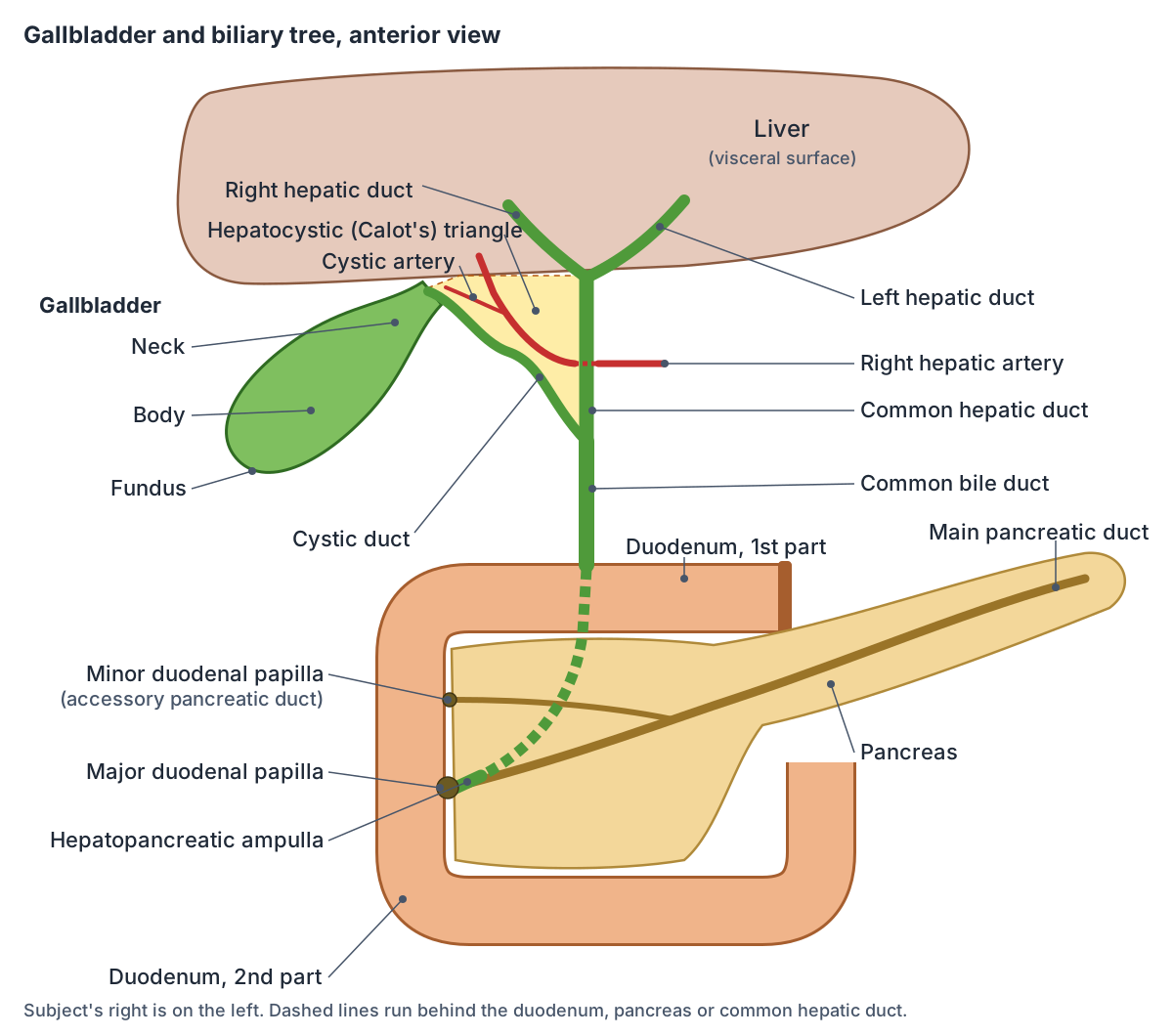
The biliary tree in order
| Duct | Formed by | Drains into |
|---|---|---|
| Bile canaliculi and ductules | Hepatocytes (liver cells) | Bile ducts |
| Right and left hepatic ducts | Bile ducts of each half of the liver | Common hepatic duct |
| Common hepatic duct | Right and left hepatic ducts | Joins the cystic duct |
| Cystic duct | Neck of the gallbladder | Joins the common hepatic duct (bile flows both ways) |
| Common bile duct | Common hepatic duct + cystic duct | Hepatopancreatic ampulla |
| Main pancreatic duct | Ducts of the pancreas | Hepatopancreatic ampulla |
| Hepatopancreatic ampulla | Common bile duct + main pancreatic duct | Second part of the duodenum at the major papilla |
| Accessory pancreatic duct | Ducts of the pancreatic head | Duodenum at the minor papilla |
Compiled from OpenStax, Kenhub and Radiopaedia (see references). Union patterns vary.
The cystic duct and the spiral folds of Heister
The cystic duct connects the neck of the gallbladder to the common hepatic duct. Radiopaedia gives its size as about 2 to 3 cm long and 2 to 3 mm wide (OpenStax gives a shorter 1 to 2 cm). Its lining forms multiple spiral mucosal folds, the folds (valves) of Heister. (Radiopaedia: Cystic duct.)
Kenhub notes that some anatomists think the spiral valves help keep the duct open. (Kenhub: Gallbladder.)
Bile flows both ways through the cystic duct: into the gallbladder for storage between meals and back out towards the common hepatic and common bile ducts when the gallbladder empties. (Radiopaedia: Gallbladder.)
Variants are common and matter in surgery. The cystic duct may join low down, into the lower third of the common hepatic duct (about 10%), join its left rather than right side (about 15%), or run parallel to it for at least 2 cm before joining (about 10%). (Radiopaedia: Cystic duct.)
Calot's triangle (hepatocystic triangle)
Calot's triangle is a small space near the porta hepatis with its apex pointing towards the liver. Its modern borders are the common hepatic duct medially, the cystic duct inferiorly and the inferior surface of the liver superiorly. (TeachMeAnatomy: Calot's Triangle.)
Calot's original 1891 description used the cystic artery as the upper border. The modern definition uses the liver instead because the course and origin of the cystic artery vary so much. Radiopaedia prefers the name hepatocystic triangle, also called the hepatobiliary or cystohepatic triangle. (Radiopaedia: Hepatocystic triangle.)
The triangle contains the right hepatic artery, the cystic artery (usually a branch of the right hepatic artery, crossing the triangle to the gallbladder), the cystic lymph node of Lund (the first lymph node of the gallbladder) and lymphatics. (TeachMeAnatomy: Calot's Triangle.)
During laparoscopic cholecystectomy (keyhole removal of the gallbladder) the surgeon dissects this triangle to identify the cystic duct and cystic artery before tying and cutting them, and to recognise the right hepatic artery first. If inflammation makes the triangle impossible to define, the surgeon may do a subtotal cholecystectomy or convert to open surgery. (TeachMeAnatomy: Calot's Triangle.)
Pancreatic duct and the ampulla of Vater
The main pancreatic duct (duct of Wirsung) runs through the centre of the pancreas from left to right, from the tail towards the duodenum. It normally measures up to 3 mm in the head, 2 mm in the body and 1 mm in the tail, and joins the common bile duct at about 60 degrees at the hepatopancreatic ampulla. (Radiopaedia: Main pancreatic duct.)
The hepatopancreatic ampulla (ampulla of Vater) is a cone-shaped structure where the common bile duct and pancreatic duct meet, opening on the major duodenal papilla on the medial wall of the descending (second part of the) duodenum. Smooth muscle of the sphincter of Oddi surrounds it. A shared channel is the standard pattern in about 60% of people; in about 38% the two ducts open side by side at the papilla, and in about 2% they open separately. (Radiopaedia: Ampulla of Vater.)
The sphincter of the hepatopancreatic ampulla controls the release of bile and pancreatic juice. A smaller accessory pancreatic duct (duct of Santorini) runs from the pancreas directly into the duodenum, about 1 inch (2.5 cm) above the ampulla. (OpenStax Anatomy and Physiology 2e, 23.6: Accessory Organs in Digestion: The Liver, Pancreas, and Gallbladder.)
Pancreas divisum is the commonest variant of the pancreatic ducts, present in about 5% of people (about 9% on MRCP). The dorsal and ventral pancreatic buds fail to fuse, so most of the gland drains through the accessory duct and minor papilla. Most people with it have no symptoms, although it is more common in people with idiopathic pancreatitis. (Radiopaedia: Pancreas divisum.)
The pancreas lies at the transpyloric plane (L1) and, except for its tail, is retroperitoneal. Gallstones are the first cause listed in the GET SMASHED mnemonic for the causes of pancreatitis. (TeachMeAnatomy: The Pancreas.)
Blood supply, lymph drainage and nerves
The gallbladder is supplied mainly by the cystic artery, a branch of the right hepatic artery. Cystic veins drain into the portal vein, usually its right branch, and other small veins pass directly into the liver tissue and then the hepatic veins. Sympathetic nerves reach it through the hepatic and coeliac plexuses, and vagal fibres through hepatic branches of the anterior vagal trunk. (Radiopaedia: Gallbladder.)
Lymph from the gallbladder drains first into the cystic lymph node and then into the hepatic and coeliac nodes. (TeachMeAnatomy: The Gallbladder.)
Gallstones, biliary colic and cholecystitis
Gallstones (cholelithiasis) occur in about 10% of people and are twice as common in women as in men, becoming more frequent with age. Risk factors for cholesterol stones include female sex, middle age, obesity, a family history and recent rapid weight loss. Only about 25% of gallstones cause symptoms. (Radiopaedia: Cholelithiasis.)
The commonest symptom is biliary colic: pain or discomfort in the right upper quadrant or epigastrium, especially after a fatty meal. Complications include cholecystitis, acute cholangitis, pancreatitis, Mirizzi syndrome and obstructive jaundice. (Radiopaedia: Cholelithiasis.)
Acute cholecystitis (inflammation of the gallbladder) is usually caused by a stone persistently blocking the cystic duct. Biliary colic comes first and can spread to the right shoulder or back; if the blockage lasts more than about six hours, the pain becomes constant and fever develops. Ultrasound is the first test, and a positive sonographic Murphy sign helps, although it has poor sensitivity (about 33%). (Radiopaedia: Acute cholecystitis.)
Cholangitis, inflammation of the bile ducts, is classically recognised by Charcot's triad of right upper quadrant pain, fever and jaundice. (TeachMeAnatomy: The Gallbladder.)
Structures next to the gallbladder
The nearest named structures in the 3D model, measured from the centre of each part:
- Hepatovenous segment V · digestive
- Musculophrenic artery · cardiovascular
- Cystic duct · digestive
- Stomach · digestive
- Common hepatic duct · digestive
- Gastro-epiploic artery · cardiovascular
- Gastroepiploic vein · cardiovascular
- Anterior superior pancreaticoduodenal artery · cardiovascular
- Hepatic artery proper · cardiovascular
- Trunk of gastroduodenal artery · cardiovascular
Frequently asked questions
Where is the gallbladder located in the body?
The gallbladder is in the right upper part of the abdomen, under the right ribs. It lies in a shallow hollow on the underside of the liver. Its rounded fundus usually reaches the edge of the liver near the tip of the right 9th costal cartilage, where the edge of the right rectus abdominis muscle meets the rib margin.
What is the biliary tree?
The biliary tree is the branching system of ducts that carries bile from the liver to the duodenum. Tiny canaliculi join into bile ducts, then the right and left hepatic ducts, which form the common hepatic duct. The cystic duct from the gallbladder joins it to make the common bile duct, which meets the pancreatic duct at the ampulla of Vater.
What are the borders of Calot's triangle?
In the modern definition, Calot's (hepatocystic) triangle is bounded by the common hepatic duct medially, the cystic duct inferiorly and the inferior surface of the liver superiorly. Calot's original 1891 version used the cystic artery as the upper border. The triangle contains the cystic artery, the right hepatic artery and the cystic lymph node of Lund.
What does the pancreatic duct do?
The main pancreatic duct collects pancreatic juice, which contains digestive enzymes, and carries it through the length of the pancreas to the duodenum. It usually joins the common bile duct at the hepatopancreatic ampulla, which opens at the major duodenal papilla. A smaller accessory duct opens at the minor papilla a little higher up.
Where does gallbladder pain radiate?
Biliary colic is felt in the right upper quadrant or the epigastrium, the upper middle part of the abdomen, especially after fatty meals. It can spread to the right shoulder or the back. If the pain becomes constant and is joined by fever, inflammation of the gallbladder (acute cholecystitis) is suspected.
How much bile does the gallbladder hold?
A distended gallbladder holds roughly 30 to 50 mL of bile, though Kenhub gives 25 to 30 mL under normal conditions. When its outlet is blocked it can stretch to hold up to about 300 mL. It concentrates the bile it stores by up to 10 times by absorbing water and ions through its lining.
References
- OpenStax. Anatomy and Physiology 2e, §23.6 Accessory Organs in Digestion: The Liver, Pancreas, and Gallbladder. Rice University, 2022.
- Jones O. The Gallbladder. TeachMeAnatomy.
- Jones O. Calot's Triangle. TeachMeAnatomy.
- Jones O. The Pancreas. TeachMeAnatomy.
- Gallbladder. Radiopaedia reference article.
- Hepatocystic triangle. Radiopaedia reference article.
- Common bile duct. Radiopaedia reference article.
- Cystic duct. Radiopaedia reference article.
- Ampulla of Vater. Radiopaedia reference article.
- Main pancreatic duct. Radiopaedia reference article.
- Pancreas divisum. Radiopaedia reference article.
- Cholelithiasis. Radiopaedia reference article.
- Acute cholecystitis. Radiopaedia reference article.
- Transpyloric plane. Radiopaedia reference article.
- Gallbladder. Kenhub anatomy library.
- Foundational Model of Anatomy: Gallbladder (FMA7202). EMBL-EBI Ontology Lookup Service.
- Mitsuhashi N. et al. (2009). BodyParts3D: 3D structure database for anatomical concepts. Nucleic Acids Research 37, D782–D785. Data © The Database Center for Life Science, CC BY 4.0.
Related anatomy
- Cystic ductCystic duct: The cystic duct connects the neck of the gallbladder to the common hepatic duct. See where it is in an…
- Common hepatic ductCommon hepatic duct: The common hepatic duct is formed by the union of the right and left hepatic ducts and carries…
- Pancreatic ductPancreatic duct: The main pancreatic duct (of Wirsung) collects pancreatic juice from the gland and carries it to the…
- DuodenumDuodenum: The duodenum is the first and shortest part of the small intestine, about 20 to 25 cm long and C-shaped. See…
- PancreasPancreas: The pancreas is a soft, elongated gland with both exocrine and endocrine functions. See where it is in an…
- Hepatic portal veinHepatic portal vein: The hepatic portal vein carries nutrient-rich blood from the gut and spleen to the liver. See…
Explore the whole body in the interactive 3D human anatomy model, or browse the A–Z list of human structures.