

What are the muscles of the hand?
The hand is moved by two sets of muscles. The extrinsic muscles start in the forearm and reach the hand by long tendons. The intrinsic muscles both originate and insert within the hand. OpenStax groups the intrinsic muscles into three sets: thenar muscles that act on the thumb, hypothenar muscles that act on the little finger, and intermediate muscles (the lumbricals and the palmar and dorsal interossei) that act on all the fingers. (OpenStax Anatomy and Physiology 2e, 11.5: Muscles of the Pectoral Girdle and Upper Limbs.)
Books differ on where adductor pollicis belongs. OpenStax counts it as a fourth thenar muscle, while Kenhub lists the thenar group as only abductor pollicis brevis, flexor pollicis brevis and opponens pollicis, and places adductor pollicis in a separate adductor compartment. Both agree on what it does. (Kenhub: Thenar muscles.)
TeachMeAnatomy adds palmaris brevis, a thin muscle in the skin over the hypothenar eminence, and treats it and adductor pollicis separately from the thenar and hypothenar groups. Most hand muscles are supplied by the ulnar nerve; the median nerve supplies the thenar muscles and the two lateral lumbricals. (TeachMeAnatomy: The Intrinsic Muscles of the Hand.)
The carpals, metacarpals and phalanges these muscles attach to. Hand bones.
Thenar eminence muscles
The thenar eminence (thenar prominence) is the fleshy mound at the base of the thumb on the palm. It is formed by three short muscles on the lateral (radial) side of the hand: abductor pollicis brevis, flexor pollicis brevis and opponens pollicis. All three arise from the flexor retinaculum and the tubercle of the trapezium; abductor pollicis brevis also arises from the tubercle of the scaphoid. (Kenhub: Thenar muscles.)
Abductor pollicis brevis inserts on the lateral side of the base of the proximal phalanx of the thumb, abducts the thumb and helps oppose it. It is supplied by the recurrent branch of the median nerve (C8 and T1). (University of Washington Musculoskeletal Atlas: Abductor pollicis brevis.)
Flexor pollicis brevis inserts on the base of the proximal phalanx of the thumb and flexes the thumb at its metacarpophalangeal joint. It has two heads. The superficial head is supplied by the recurrent branch of the median nerve; the deep head, which varies in size, is supplied by the deep branch of the ulnar nerve. (TeachMeAnatomy: The Intrinsic Muscles of the Hand.)
Opponens pollicis arises from the flexor retinaculum and the tubercle of the trapezium and inserts along the lateral side of the first metacarpal. It draws the first metacarpal across the palm and rotates it medially, so that the thumb pad can face the finger pads. This is opposition. It is supplied by the recurrent branch of the median nerve. (University of Washington Musculoskeletal Atlas: Opponens pollicis.)
Because the recurrent branch of the median nerve supplies the thenar muscles, compression of the median nerve in the carpal tunnel can, if untreated, cause weakness and wasting of the thenar eminence. (TeachMeAnatomy: The Carpal Tunnel.)
Hypothenar muscles
The hypothenar eminence is the smaller pad on the medial (ulnar) side of the palm. Its three muscles mirror the thenar group and act on the little finger: abductor digiti minimi, flexor digiti minimi brevis and opponens digiti minimi. All three are supplied by the deep branch of the ulnar nerve (C8 and T1) and receive blood from the ulnar artery. (Kenhub: Hypothenar muscles.)
Abductor digiti minimi arises from the pisiform and inserts on the medial side of the base of the proximal phalanx of the little finger, which it abducts (spreads away from the ring finger). (University of Washington Musculoskeletal Atlas: Abductor digiti minimi.)
Flexor digiti minimi brevis arises from the hook of the hamate and the flexor retinaculum and inserts on the medial side of the base of the proximal phalanx of the little finger. It flexes the little finger at the metacarpophalangeal joint. (University of Washington Musculoskeletal Atlas: Flexor digiti minimi brevis.)
Opponens digiti minimi, the deepest, also arises from the hook of the hamate and the flexor retinaculum but inserts along the medial border of the fifth metacarpal. It draws the fifth metacarpal forward and rotates it, bringing the little finger into opposition with the thumb. (University of Washington Musculoskeletal Atlas: Opponens digiti minimi.)
Palmaris brevis
Palmaris brevis is a small, thin muscle that lies in the superficial tissue over the hypothenar eminence. It arises from the palmar aponeurosis and the flexor retinaculum and inserts into the skin on the medial side of the palm. Kenhub places it in the hypothenar region but not in the hypothenar muscle group, because it does not move the little finger. (Kenhub: Hypothenar muscles.)
Palmaris brevis wrinkles the skin over the hypothenar eminence and deepens the hollow of the palm. It is the only hand muscle supplied by the superficial branch of the ulnar nerve. It is not modelled in BodyParts3D, so it does not appear in the 3D view. (TeachMeAnatomy: The Intrinsic Muscles of the Hand.)
Thenar and hypothenar muscles, adductor pollicis and palmaris brevis
| Muscle | Origin | Insertion | Innervation | Main action |
|---|---|---|---|---|
| Abductor pollicis brevis (thenar) | Flexor retinaculum; tubercles of scaphoid and trapezium | Lateral base of proximal phalanx of thumb | Recurrent branch of median nerve (C8–T1) | Abducts thumb; helps opposition |
| Flexor pollicis brevis (thenar) | Superficial head: flexor retinaculum, trapezium; deep head: trapezoid and capitate | Base of proximal phalanx of thumb | Superficial head: recurrent branch of median nerve; deep head: deep branch of ulnar nerve | Flexes thumb at MCP joint |
| Opponens pollicis (thenar) | Flexor retinaculum; tubercle of trapezium | Lateral side of 1st metacarpal | Recurrent branch of median nerve (C8–T1) | Opposes thumb |
| Adductor pollicis | Oblique head: capitate, bases of 2nd and 3rd metacarpals; transverse head: body of 3rd metacarpal | Medial base of proximal phalanx of thumb | Deep branch of ulnar nerve (C8–T1) | Adducts thumb |
| Abductor digiti minimi (hypothenar) | Pisiform | Medial base of proximal phalanx of little finger | Deep branch of ulnar nerve (C8–T1) | Abducts little finger |
| Flexor digiti minimi brevis (hypothenar) | Hook of hamate; flexor retinaculum | Medial base of proximal phalanx of little finger | Deep branch of ulnar nerve (C8–T1) | Flexes little finger at MCP joint |
| Opponens digiti minimi (hypothenar) | Hook of hamate; flexor retinaculum | Medial border of 5th metacarpal | Deep branch of ulnar nerve (C8–T1) | Opposes little finger |
| Palmaris brevis | Palmar aponeurosis; flexor retinaculum | Skin of medial palm | Superficial branch of ulnar nerve | Wrinkles hypothenar skin; deepens palm |
Sources: University of Washington Musculoskeletal Atlas; Kenhub; TeachMeAnatomy.
Adductor pollicis
Adductor pollicis lies deep in the palm and has two heads. The oblique head arises from the capitate and the bases of the second and third metacarpals; the transverse head arises from the front of the body of the third metacarpal. Both insert on the medial side of the base of the proximal phalanx of the thumb. (University of Washington Musculoskeletal Atlas: Adductor pollicis.)
Adductor pollicis pulls the thumb towards the index finger (adduction). It is supplied by the deep branch of the ulnar nerve, not the median nerve. The radial artery passes between its two heads to form the deep palmar arch. (TeachMeAnatomy: The Intrinsic Muscles of the Hand.)
Weakness of adductor pollicis is tested with Froment's sign. The patient holds a sheet of paper between the thumb and the side of the index finger. With an ulnar nerve lesion, the patient cannot adduct the thumb strongly and instead bends the thumb tip using flexor pollicis longus, which is supplied by the median nerve. (Kenhub: Hypothenar muscles.)
Lumbricals
The four lumbricals are small muscles in the palm, numbered 1 to 4 from the index finger side. Unusually, they arise from tendons rather than bone: from the tendons of flexor digitorum profundus. Lumbricals 1 and 2 are unipennate and arise from one tendon each; lumbricals 3 and 4 are bipennate and arise from two neighbouring tendons. (Kenhub: Lumbrical muscles of the hand.)
Each lumbrical inserts on the radial (lateral) side of the extensor expansion of a finger. Together they flex the metacarpophalangeal joints and extend the interphalangeal joints at the same time, the position used to hold a pen. Lumbricals 1 and 2 are supplied by the median nerve and 3 and 4 by the ulnar nerve; a short mnemonic is '1, 2 me; 3, 4 u'. (Kenhub: Lumbrical muscles of the hand.)
Palmar and dorsal interossei
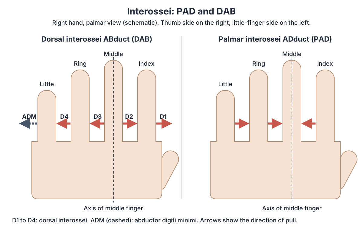
The interossei lie between the metacarpal bones. All of them are supplied by the deep branch of the ulnar nerve. Finger abduction and adduction are measured from the long axis of the middle finger: abduction spreads the fingers away from it and adduction brings them back. The rule to remember is PAD and DAB: Palmar interossei ADduct, Dorsal interossei ABduct. (Kenhub: Palmar interossei muscles.)
The University of Washington atlas describes three palmar interossei. They are unipennate and arise from the palmar surfaces of the second, fourth and fifth metacarpals, and insert into the extensor expansions and proximal phalanges of the index, ring and little fingers. They adduct these fingers towards the middle finger and help the lumbricals flex the metacarpophalangeal joints and extend the interphalangeal joints. (University of Washington Musculoskeletal Atlas: Palmar interossei.)
Some books count four palmar interossei by adding a small first palmar interosseous to the thumb, which arises from the base of the first metacarpal. Kenhub notes that this muscle is sometimes rudimentary or absent. The middle finger never has a palmar interosseous, because it is the axis that the other fingers move towards. (Kenhub: Palmar interossei muscles.)
The four dorsal interossei are bipennate: each arises from the facing sides of two neighbouring metacarpals. They insert into the extensor expansions and proximal phalanges of the index, middle and ring fingers and abduct them away from the axis of the middle finger. (University of Washington Musculoskeletal Atlas: Dorsal interossei.)
The first dorsal interosseous is the largest and strongest of the group and is sometimes called abductor indicis. It can be felt in the first web space, between the thumb and the index finger. The middle finger receives two dorsal interossei, one on each side, so it can be moved either way. The thumb and little finger have their own abductors (abductor pollicis brevis and longus, and abductor digiti minimi), so they have no dorsal interosseous. (Kenhub: Dorsal interossei muscles of the hand.)
Lumbricals and interossei
| Muscle | Origin | Insertion | Innervation | Main action |
|---|---|---|---|---|
| Lumbricals 1–2 (unipennate) | Radial side of FDP tendons to index and middle fingers | Radial side of extensor expansions of digits 2–3 | Median nerve | Flex MCP joints, extend IP joints |
| Lumbricals 3–4 (bipennate) | Adjacent sides of FDP tendons to middle, ring and little fingers | Radial side of extensor expansions of digits 4–5 | Deep branch of ulnar nerve | Flex MCP joints, extend IP joints |
| Palmar interossei (3; a 4th to the thumb in some texts) | Palmar surfaces of 2nd, 4th and 5th metacarpals | Extensor expansions and proximal phalanges of digits 2, 4 and 5 | Deep branch of ulnar nerve (C8–T1) | Adduct fingers (PAD); assist MCP flexion and IP extension |
| Dorsal interossei (4) | Adjacent sides of two metacarpals | Extensor expansions and proximal phalanges of digits 2–4 | Deep branch of ulnar nerve (C8–T1) | Abduct fingers (DAB); assist MCP flexion and IP extension |
FDP: flexor digitorum profundus. MCP: metacarpophalangeal. IP: interphalangeal. Sources: Kenhub; University of Washington Musculoskeletal Atlas.
Thumb abduction, adduction and opposition
The thumb's movements are named in its own planes. Abduction and adduction of the thumb happen in a plane perpendicular to the palm: abduction lifts the thumb forwards, away from the palm, and adduction brings it back. Flexion and extension happen in the plane of the palm. Opposition combines abduction and flexion with an automatic rotation of the thumb. (University of Oklahoma Health Sciences Center: Upper extremity anatomy and function (wrist and hand).)
Thumb abduction at the first carpometacarpal joint is produced mainly by abductor pollicis brevis with abductor pollicis longus from the forearm. Strong thumb adduction is produced by adductor pollicis. Opposition is produced by opponens pollicis, helped by the other thenar muscles and by adductor pollicis. (Kenhub: Thenar muscles.)
Which muscle makes a thumbs up?
A thumbs up is mainly thumb extension, done by forearm muscles rather than hand muscles. Extensor pollicis longus extends all the joints of the thumb, including the tip; its tendon winds around Lister's tubercle and forms the ulnar border of the anatomical snuffbox. Extensor pollicis brevis extends the thumb at its base and abductor pollicis longus abducts it. All three are supplied by the posterior interosseous branch of the radial nerve. (TeachMeAnatomy: Muscles in the Posterior Compartment of the Forearm.)
Extensor pollicis longus is the only thumb extensor that inserts on the base of the distal phalanx of the thumb, so it is the muscle that straightens the thumb tip into the upright 'thumbs up' curve. The curled fingers are held by the finger flexors. (University of Washington Musculoskeletal Atlas: Extensor pollicis longus.)
Nerve supply and nerve injuries
The ulnar nerve (C8 and T1) supplies most intrinsic hand muscles. Its deep branch supplies the hypothenar muscles, the medial two lumbricals, adductor pollicis and all the palmar and dorsal interossei; its superficial branch supplies palmaris brevis. An ulnar nerve injury at the wrist affects only these intrinsic hand muscles. (TeachMeAnatomy: The Ulnar Nerve.)
The median nerve supplies the thenar muscles through its recurrent branch and the lateral two lumbricals through its palmar digital branches. A median nerve injury at the wrist paralyses the thenar muscles, so thumb opposition is lost, and weakens the lateral two lumbricals. (TeachMeAnatomy: The Median Nerve.)
Long-standing deep ulnar nerve damage weakens finger abduction and wastes the interossei. It can produce an ulnar claw hand, with the metacarpophalangeal joints hyperextended and the interphalangeal joints flexed, mainly in the ring and little fingers, because their lumbricals (3 and 4) are ulnar-supplied while the lumbricals of the index and middle fingers are supplied by the median nerve. (Kenhub: Dorsal interossei muscles of the hand.)
The ulnar nerve can also be compressed in Guyon's canal at the wrist, for example by long pressure on bicycle handlebars. This causes pain and altered sensation on the little-finger side of the palm and, in advanced cases, weakness and wasting of the hypothenar and other ulnar-supplied hand muscles. (Kenhub: Hypothenar muscles.)
Hand muscles in the 3D model
- Abductor digiti minimi of hand
- The muscle forming the ulnar (little-finger) side of the hypothenar eminence.
- Abductor pollicis brevis
- A thenar muscle on the lateral side of the thenar eminence.
- Flexor digiti minimi brevis of hand
- A small hypothenar muscle.
- Flexor pollicis brevis
- A thenar muscle medial to abductor pollicis brevis.
- Oblique head of adductor pollicis
- The larger head of adductor pollicis in the palm.
- Opponens digiti minimi of hand
- A hypothenar muscle that deepens the hollow of the palm on the little finger side.
- Opponens pollicis
- A thenar muscle lying deep to abductor pollicis brevis that allows the thumb to oppose.
- Dorsal interossei of hand
- Four bipennate muscles between the metacarpals that spread the fingers.
- Lumbricals of hand
- Four small muscles of the palm that arise from the flexor digitorum profundus tendons.
- Palmar interossei of hand
- Small unipennate muscles in the palm that bring the fingers together.
- Superficial head of flexor pollicis brevis
- The larger, lateral head of flexor pollicis brevis in the thenar eminence.
- Transverse head of adductor pollicis
- The transverse head of adductor pollicis in the palm.
Frequently asked questions
What muscles make up the thenar eminence?
The thenar eminence is formed by three muscles: abductor pollicis brevis, flexor pollicis brevis and opponens pollicis. They arise from the flexor retinaculum and the scaphoid and trapezium and move the thumb. Some textbooks, including OpenStax, also count adductor pollicis as a thenar muscle. Most thenar muscles are supplied by the recurrent branch of the median nerve.
What are the hypothenar muscles?
The hypothenar muscles are abductor digiti minimi, flexor digiti minimi brevis and opponens digiti minimi. They form the pad on the little-finger side of the palm and abduct, flex and oppose the little finger. All three are supplied by the deep branch of the ulnar nerve. Palmaris brevis lies over them but is not counted in the group.
What is the difference between palmar and dorsal interossei?
Palmar interossei adduct the fingers towards the middle finger and dorsal interossei abduct them away from it (PAD and DAB). There are three palmar interossei (four if a small thumb slip is counted), each unipennate from one metacarpal, and four dorsal interossei, each bipennate from two metacarpals. All are supplied by the deep branch of the ulnar nerve.
Which muscle adducts the thumb?
Adductor pollicis. It has an oblique head from the capitate and the bases of the second and third metacarpals, and a transverse head from the third metacarpal, and both insert on the base of the proximal phalanx of the thumb. It is supplied by the deep branch of the ulnar nerve, and its weakness is tested with Froment's sign.
Which muscle makes a thumbs up?
Mainly extensor pollicis longus, a forearm muscle whose tendon reaches the base of the distal phalanx of the thumb and extends every thumb joint. Extensor pollicis brevis and abductor pollicis longus help at the base of the thumb. All three are supplied by the posterior interosseous branch of the radial nerve, while the fingers are curled by the long finger flexors.
What nerve supplies the muscles of the hand?
The ulnar nerve supplies most of them: the hypothenar muscles, adductor pollicis, all interossei, the medial two lumbricals, palmaris brevis and the deep head of flexor pollicis brevis. The median nerve supplies the thenar muscles (abductor pollicis brevis, opponens pollicis and the superficial head of flexor pollicis brevis) and the lateral two lumbricals.
What happens to the thenar eminence in carpal tunnel syndrome?
Carpal tunnel syndrome compresses the median nerve under the flexor retinaculum. Because the thenar muscles are supplied by the recurrent branch of the median nerve, untreated or severe cases can cause weakness and wasting of the thenar eminence, with difficulty abducting and opposing the thumb, as well as numbness, tingling and pain in the median nerve territory of the hand.
References
- OpenStax. Anatomy and Physiology 2e, §11.5 Muscles of the Pectoral Girdle and Upper Limbs. Rice University, 2022.
- TeachMeAnatomy. The Intrinsic Muscles of the Hand.
- TeachMeAnatomy. Muscles in the Posterior Compartment of the Forearm.
- TeachMeAnatomy. The Carpal Tunnel.
- TeachMeAnatomy. The Median Nerve.
- TeachMeAnatomy. The Ulnar Nerve.
- Kenhub. Thenar muscles.
- Kenhub. Hypothenar muscles.
- Kenhub. Lumbrical muscles of the hand.
- Kenhub. Palmar interossei muscles.
- Kenhub. Dorsal interossei muscles of the hand.
- University of Oklahoma Health Sciences Center. Upper extremity anatomy and function: wrist and hand (course notes, PHTH/OCTH 7143).
- University of Washington Department of Radiology. Musculoskeletal Atlas: Abductor pollicis brevis.
- University of Washington Department of Radiology. Musculoskeletal Atlas: Flexor pollicis brevis.
- University of Washington Department of Radiology. Musculoskeletal Atlas: Opponens pollicis.
- University of Washington Department of Radiology. Musculoskeletal Atlas: Adductor pollicis.
- University of Washington Department of Radiology. Musculoskeletal Atlas: Abductor digiti minimi.
- University of Washington Department of Radiology. Musculoskeletal Atlas: Flexor digiti minimi brevis.
- University of Washington Department of Radiology. Musculoskeletal Atlas: Opponens digiti minimi.
- University of Washington Department of Radiology. Musculoskeletal Atlas: Dorsal interossei.
- University of Washington Department of Radiology. Musculoskeletal Atlas: Palmar interossei.
- University of Washington Department of Radiology. Musculoskeletal Atlas: Extensor pollicis longus.
- Mitsuhashi N. et al. (2009). BodyParts3D: 3D structure database for anatomical concepts. Nucleic Acids Research 37, D782–D785. Data © The Database Center for Life Science, CC BY 4.0.
Related anatomy
- Hand bonesHow many bones are in the human hand? Learn the 8 carpal bones, 5 metacarpals and 14 phalanges by name, plus the thumb…
- Forearm musclesMuscles of the forearm explained: every anterior flexor and posterior extensor, with origin, insertion, nerve and…
- Upper limb nervesBrachial plexus diagram with roots, trunks, cords and branches; axillary, musculocutaneous, median, ulnar and radial…
- RadiusRadius labeled: head of radius, neck, radial tuberosity, shaft, Lister's tubercle, ulnar notch and styloid process…
- UlnaUlna labeled: olecranon, trochlear notch, coronoid process, radial notch, shaft, head and styloid process, how the…
Explore the whole body in the interactive 3D human anatomy model, or browse the A–Z list of human structures.