

Where do the nerves of the arm come from?
Spinal nerves leave the vertebral column at each level, but the nerves of the neck and limbs are reorganised in nerve plexuses first. Of the four plexuses, two are at the cervical level (the cervical and brachial plexuses), one at the lumbar level and one at the sacral level. (OpenStax Anatomy and Physiology 2e, 13.4: The Peripheral Nervous System.)
The cervical plexus is made of axons from spinal nerves C1 to C5 according to OpenStax, and its best-known branch is the phrenic nerve to the diaphragm. In the brachial plexus, spinal nerves C4 to T1 reorganise to give rise to the nerves of the arms. (OpenStax Anatomy and Physiology 2e, 13.4: The Peripheral Nervous System.)
TeachMeAnatomy, like most anatomy texts, uses narrower ranges: the cervical plexus is formed by the anterior rami of C1 to C4, although its phrenic nerve also receives fibres from C5, and the brachial plexus by the anterior rami of C5 to T1. These are the ranges used on the rest of this page. (TeachMeAnatomy: The Cervical Plexus.)
The thoracic spinal nerves T2 to T11 are not part of any plexus. Their ventral rami become the intercostal nerves, which run between the ribs. (OpenStax Anatomy and Physiology 2e, 13.4: The Peripheral Nervous System.)
Brachial plexus: roots, trunks, divisions and cords
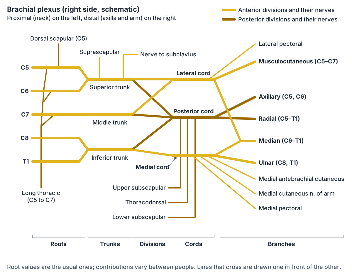
The brachial plexus is formed by the anterior rami of spinal nerves C5, C6, C7, C8 and T1. It has five parts, usually learned in order: roots, trunks, divisions, cords and branches. The roots pass between the anterior and middle scalene muscles to reach the base of the neck. (TeachMeAnatomy: The Brachial Plexus.)
At the base of the neck the roots join to form three trunks. C5 and C6 form the superior (upper) trunk, C7 continues alone as the middle trunk, and C8 and T1 form the inferior (lower) trunk. Kenhub describes the trunks crossing the base of the posterior triangle of the neck, between the scalene muscles and behind the subclavian artery, and passing over the apex of the lung and the first rib toward the clavicle. (Kenhub: Brachial plexus.)
Each trunk splits into an anterior and a posterior division near the middle third of the clavicle. The divisions pass down behind the clavicle into the axilla, where they form three cords around the second part of the axillary artery. (Kenhub: Brachial plexus.)
The cords are named by their position relative to the axillary artery. The lateral cord is formed by the anterior divisions of the superior and middle trunks. The posterior cord is formed by the posterior divisions of all three trunks. The medial cord is the continuation of the anterior division of the inferior trunk. (TeachMeAnatomy: The Brachial Plexus.)
The terminal branches come from the cords. The lateral cord continues as the musculocutaneous nerve. The posterior cord gives the axillary nerve and continues as the radial nerve. The medial cord continues as the ulnar nerve. The median nerve is formed by two roots, one from the lateral cord and one from the medial cord. (TeachMeAnatomy: The Brachial Plexus.)
Smaller branches of the brachial plexus
Several nerves leave the plexus before the terminal branches. From the roots come the dorsal scapular nerve (C5), which supplies levator scapulae and the rhomboids, and the long thoracic nerve (C5, C6, C7), which supplies serratus anterior. From the superior trunk come the suprascapular nerve (C5, C6) to supraspinatus and infraspinatus, and the nerve to subclavius. (Kenhub: Brachial plexus.)
The lateral cord gives the lateral pectoral nerve. The medial cord gives the medial pectoral nerve, the medial cutaneous nerve of the arm and the medial cutaneous nerve of the forearm. The posterior cord gives the upper subscapular, thoracodorsal and lower subscapular nerves. (TeachMeAnatomy: The Brachial Plexus.)
Kenhub gives the thoracodorsal nerve root values of C7 and C8 and states that it supplies latissimus dorsi. The upper (superior) subscapular nerve supplies the upper part of subscapularis, and the lower (inferior) subscapular nerve supplies the rest of subscapularis and teres major. The medial pectoral nerve supplies pectoralis minor and the lower sternocostal part of pectoralis major. Kenhub gives T1 alone as the root of the medial cutaneous nerve of the forearm, while Radiopaedia gives C8 and T1: small branches vary between people and sources. (Kenhub: Brachial plexus.)
Main nerves of the upper limb: root values, motor and sensory supply
| Nerve | Origin and roots | Main motor supply | Main skin supply |
|---|---|---|---|
| Musculocutaneous | Lateral cord; C5 to C7 | Coracobrachialis, biceps brachii, brachialis | Lateral forearm (lateral cutaneous nerve of forearm) |
| Axillary | Posterior cord; C5, C6 | Deltoid, teres minor | Skin over lower deltoid (regimental badge area) |
| Radial | Posterior cord; C5 to T1 | Triceps and posterior forearm extensors, brachioradialis | Posterior arm and forearm, lateral dorsum of hand |
| Median | Lateral and medial cords; C6 to T1 (sometimes C5) | Most forearm flexors, thenar muscles, lateral two lumbricals | Lateral palm and palmar lateral three and a half digits |
| Ulnar | Medial cord; C8, T1 | Flexor carpi ulnaris, medial half of FDP, most intrinsic hand muscles | Palmar medial one and a half fingers |
| Medial antebrachial cutaneous | Medial cord; C8, T1 (Kenhub: T1) | None (sensory) | Anterior and medial forearm to the wrist |
| Medial cutaneous nerve of arm | Medial cord; C8, T1 | None (sensory) | Lower medial arm |
| Dorsal scapular | C5 root | Levator scapulae, rhomboids | None |
| Long thoracic | C5 to C7 roots | Serratus anterior | None |
| Suprascapular | Superior trunk; C5, C6 | Supraspinatus, infraspinatus | None (sensory to shoulder and acromioclavicular joints) |
| Thoracodorsal | Posterior cord; C7, C8 (Kenhub) | Latissimus dorsi | None |
Root values from TeachMeAnatomy, Kenhub and Radiopaedia; they vary between people and between sources.
Musculocutaneous nerve
The musculocutaneous nerve (roots C5 to C7) is the terminal branch of the lateral cord. It pierces the coracobrachialis muscle and then runs between biceps brachii, above it, and brachialis, beneath it. (TeachMeAnatomy: The Musculocutaneous Nerve.)
It supplies the three muscles of the anterior compartment of the arm: coracobrachialis, biceps brachii and brachialis. It then continues as the lateral cutaneous nerve of the forearm, which supplies the skin on the lateral side of the forearm. Because it is well protected in the axilla, isolated injury is relatively uncommon. (TeachMeAnatomy: The Musculocutaneous Nerve.)
Axillary nerve
The axillary nerve (roots C5 and C6) arises from the posterior cord. In the axilla it lies behind the axillary artery and in front of subscapularis, and it leaves the axilla through the quadrangular space together with the posterior circumflex humeral artery and vein. The surgical neck of the humerus forms the lateral boundary of this space. (TeachMeAnatomy: The Axillary Nerve.)
The axillary nerve supplies the deltoid and teres minor muscles. It continues as the upper lateral cutaneous nerve of the arm, which supplies the skin over the lower part of the deltoid, the regimental badge area. Fracture of the surgical neck of the humerus and dislocation of the shoulder are common causes of injury, which weakens deltoid and teres minor and numbs the regimental badge area. (TeachMeAnatomy: The Axillary Nerve.)
Radial nerve
The radial nerve (roots C5 to T1) is the terminal continuation of the posterior cord. In the arm it runs in a shallow groove on the back of the humerus, the radial (spiral) groove, and then passes in front of the lateral epicondyle into the forearm, where it divides into a deep (motor) branch and a superficial (sensory) branch. After piercing supinator, the deep branch is called the posterior interosseous nerve. (TeachMeAnatomy: The Radial Nerve.)
The radial nerve supplies the muscles of the posterior arm, including the three heads of triceps brachii, and the muscles of the posterior forearm, including brachioradialis and extensor carpi radialis longus. Its skin territory includes the posterior arm (via the posterior cutaneous nerve of the arm), the posterior forearm and the lateral part of the dorsum of the hand. (TeachMeAnatomy: The Radial Nerve.)
Because the nerve lies in the spiral groove, it is vulnerable in fractures of the humeral shaft. Injury paralyses the wrist extensors, so unopposed flexion of the wrist produces wrist drop. (TeachMeAnatomy: The Radial Nerve.)
Median nerve
The median nerve is formed from the lateral and medial cords and carries fibres from C6 to T1, and in some people from C5. It runs down the arm lateral to the brachial artery, crosses it about halfway down to lie medially, and enters the forearm through the cubital fossa. It passes between the two heads of pronator teres and then runs between flexor digitorum superficialis and flexor digitorum profundus. (TeachMeAnatomy: The Median Nerve.)
In the forearm it supplies most of the anterior (flexor) muscles, except flexor carpi ulnaris and the medial half of flexor digitorum profundus, which belong to the ulnar nerve. It enters the hand through the carpal tunnel and supplies the thenar muscles and the lateral two lumbricals. Its skin territory is the lateral palm (through the palmar cutaneous branch, which does not pass through the carpal tunnel) and the palmar surface of the lateral three and a half digits. (TeachMeAnatomy: The Median Nerve.)
Compression of the median nerve in the carpal tunnel causes carpal tunnel syndrome: numbness, tingling and pain in the median nerve distribution of the fingers, often worse at night. The palm is usually spared because the palmar cutaneous branch does not pass through the tunnel. If untreated, the thenar muscles can weaken and waste. (TeachMeAnatomy: The Median Nerve.)
Ulnar nerve
The ulnar nerve (roots C8 and T1) is the continuation of the medial cord. It passes behind the elbow between the medial epicondyle and the olecranon, and it enters the hand through the ulnar canal (Guyon's canal). (TeachMeAnatomy: The Ulnar Nerve.)
In the forearm it supplies flexor carpi ulnaris and the medial half of flexor digitorum profundus, which flexes the ring and little fingers at the distal interphalangeal joints. In the hand its deep branch supplies most of the intrinsic hand muscles. Its skin territory is the palmar surface of the medial one and a half fingers. (TeachMeAnatomy: The Ulnar Nerve.)
Injuries at the medial epicondyle, such as fractures around the elbow, or cuts to the front of the wrist can damage the ulnar nerve. Loss of the interosseous muscles weakens abduction and adduction of the fingers. (TeachMeAnatomy: The Ulnar Nerve.)
Medial antebrachial cutaneous nerve and other skin nerves of the arm
The medial antebrachial cutaneous nerve, also called the medial cutaneous nerve of the forearm, arises from the medial cord of the brachial plexus with fibres from C8 and T1. It first runs alongside the ulnar nerve, then pierces the deep fascia at about mid-arm near the basilic vein and enters the subcutaneous tissue of the forearm. (Radiopaedia: Medial cutaneous nerve of the forearm.)
It divides into anterior (volar) and posterior (ulnar) branches. The anterior branch, usually the larger, passes in front of the basilic vein and runs down the anteromedial forearm; the posterior branch winds around the medial side of the basilic vein to the back of the forearm. Together they supply the skin of the anterior and medial forearm as far as the wrist. (Radiopaedia: Medial cutaneous nerve of the forearm.)
The upper arm has its own small cutaneous nerves. The medial cutaneous nerve of the arm (C8, T1) also comes from the medial cord and supplies the lower medial arm. (Kenhub: Brachial plexus.)
Brachial plexus injuries: Erb's palsy and Klumpke's palsy
Injury to the upper roots of the plexus, typically C5 and C6, is called Erb's palsy. It is often caused by stretching of the plexus, for example during a difficult delivery. The affected limb hangs limply, medially rotated by the unopposed pull of pectoralis major, in the waiter's tip position. (TeachMeAnatomy: The Brachial Plexus.)
Injury to the lower roots, typically C8 and T1, is called Klumpke's palsy. Because these roots supply many of the small muscles of the hand, the main feature is a clawed hand. (TeachMeAnatomy: The Brachial Plexus.)
Cervical plexus nerves (plexus cervicalis)
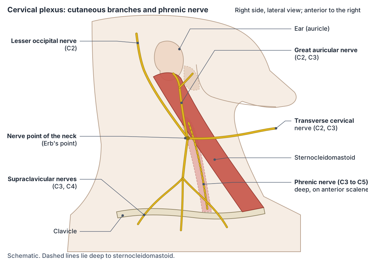
The cervical plexus (plexus cervicalis) is formed by the anterior rami of cervical spinal nerves C1 to C4. It lies in the posterior triangle of the neck, about halfway up sternocleidomastoid, within the prevertebral layer of fascia. (TeachMeAnatomy: The Cervical Plexus.)
Its four cutaneous (sensory) branches all emerge at the middle of the posterior border of sternocleidomastoid, an area called the nerve point of the neck or Erb's point. The lesser occipital nerve (C2, sometimes C3) supplies the skin of the posterosuperior scalp. The transverse cervical nerve (C2, C3) curves around sternocleidomastoid to supply the front of the neck. The supraclavicular nerves (C3, C4) supply the skin over the supraclavicular fossa and the clavicle region. (TeachMeAnatomy: The Cervical Plexus.)
The motor branches include the ansa cervicalis, a loop formed by C1 to C3 that supplies the infrahyoid (strap) muscles, and the phrenic nerve (C3 to C5) to the diaphragm. Fibres from C1 also travel with the hypoglossal nerve to reach geniohyoid and thyrohyoid. (TeachMeAnatomy: The Cervical Plexus.)
Great auricular nerve (greater auricular, nervus auricularis magnus)
The great auricular nerve, often searched as the greater auricular nerve and named nervus auricularis magnus in Latin, arises from the anterior rami of C2 and C3. It is the largest ascending branch of the cervical plexus. It curves around the posterior border of sternocleidomastoid, emerges onto the muscle's surface and runs upward beneath platysma toward the parotid gland. (Kenhub: Great auricular nerve.)
It ends in two branches. The anterior (facial) branch supplies the skin over the parotid gland and communicates with the facial nerve within the gland. The posterior (mastoid) branch supplies the skin over the mastoid process and the back of the auricle (outer ear). Kenhub links the facial nerve connection to Frey's syndrome, which can follow parotid surgery. (Kenhub: Great auricular nerve.)
TeachMeAnatomy summarises its territory as the external ear and the skin over the parotid gland. (TeachMeAnatomy: The Cervical Plexus.)
Phrenic nerve (nervio frénico)
The phrenic nerve (Spanish nervio frénico) arises from C3, C4 and C5, remembered as "C3, 4 and 5 keep the diaphragm alive". It begins at the lateral border of anterior scalene, descends over the front of the muscle deep to the prevertebral fascia, passes behind the subclavian vein and enters the thorax through the superior thoracic aperture. (TeachMeAnatomy: The Phrenic Nerve.)
In the thorax the right phrenic nerve runs along the right lung root and the pericardium over the right atrium; the left nerve crosses the aortic arch and descends in front of the left lung root along the pericardium over the left ventricle. Each nerve provides motor supply to its half of the diaphragm and sensory supply to the central diaphragm, the mediastinal pleura and the pericardium. (TeachMeAnatomy: The Phrenic Nerve.)
Damage, for example during surgery or from compression by a tumour, paralyses the diaphragm on that side. The paralysed half then moves paradoxically: upward during inspiration and downward during expiration. (TeachMeAnatomy: The Phrenic Nerve.)
Branches of the cervical plexus
| Branch | Roots | Type | Supply |
|---|---|---|---|
| Lesser occipital nerve | C2 (sometimes C3) | Sensory | Posterosuperior scalp |
| Great (greater) auricular nerve | C2, C3 | Sensory | External ear, skin over parotid gland and mastoid |
| Transverse cervical nerve | C2, C3 | Sensory | Skin of the front of the neck |
| Supraclavicular nerves | C3, C4 | Sensory | Skin over the supraclavicular fossa |
| Ansa cervicalis | C1 to C3 | Motor | Infrahyoid muscles |
| Phrenic nerve | C3 to C5 | Motor and sensory | Diaphragm; central diaphragm, mediastinal pleura and pericardium (sensory) |
Based on TeachMeAnatomy and Kenhub.
Intercostal nerves
The intercostal nerves are the anterior (ventral) rami of thoracic spinal nerves T1 to T11. The anterior ramus of T12 runs below the twelfth rib and is called the subcostal nerve. Most of the T1 ramus joins the brachial plexus, and only its smaller inferior branch becomes the first intercostal nerve. (Kenhub: Intercostal nerves.)
Each intercostal nerve runs between the internal intercostal and innermost intercostal muscles. Near the midaxillary line it gives off a lateral cutaneous branch, and it ends at the front of the chest or abdomen as an anterior cutaneous branch. The lower intercostal nerves continue into the abdominal wall, so they are sometimes called thoracoabdominal nerves. The lateral cutaneous branch of the second intercostal nerve is named the intercostobrachial nerve; it supplies the floor of the axilla and the upper posteromedial arm. (Kenhub: Intercostal nerves.)
Frequently asked questions
What are the five terminal branches of the brachial plexus?
The five terminal branches are the musculocutaneous nerve (lateral cord, C5 to C7), the axillary nerve (posterior cord, C5 and C6), the radial nerve (posterior cord, C5 to T1), the median nerve (lateral and medial cords, C6 to T1) and the ulnar nerve (medial cord, C8 and T1). Together they supply nearly all the muscles and skin of the upper limb.
How do you remember the brachial plexus?
Learn it in order: roots, trunks, divisions, cords, branches. Five roots (C5 to T1) form three trunks (superior C5 and C6, middle C7, inferior C8 and T1). Each trunk splits into anterior and posterior divisions. The three posterior divisions form the posterior cord, the upper two anterior divisions form the lateral cord, and the lower anterior division forms the medial cord.
What does the axillary nerve supply?
The axillary nerve (C5, C6) supplies two muscles, the deltoid and teres minor, and the skin over the lower deltoid, known as the regimental badge area, through the upper lateral cutaneous nerve of the arm. It winds around the surgical neck of the humerus, so it can be injured by fractures there or by shoulder dislocation.
What is the medial antebrachial cutaneous nerve?
The medial antebrachial cutaneous nerve, or medial cutaneous nerve of the forearm, is a purely sensory branch of the medial cord of the brachial plexus (C8, T1). It pierces the deep fascia near the basilic vein at about mid-arm and splits into anterior and posterior branches that supply the skin of the anterior and medial forearm down to the wrist.
What does the great auricular nerve supply?
The great auricular nerve (C2, C3), also called the greater auricular nerve, is a sensory branch of the cervical plexus. It curves around the posterior border of sternocleidomastoid and climbs toward the ear. Its anterior branch supplies skin over the parotid gland and its posterior branch supplies skin over the mastoid process and the back of the auricle.
Which nerve roots form the phrenic nerve?
The phrenic nerve comes from C3, C4 and C5, summed up as "C3, 4 and 5 keep the diaphragm alive". It runs down on the front of anterior scalene, enters the thorax behind the subclavian vein and passes in front of the lung root to the diaphragm. It provides the motor supply to its own half of the diaphragm.
Which nerve causes wrist drop?
Wrist drop is caused by injury to the radial nerve. The radial nerve supplies the extensor muscles of the posterior forearm, so when it is damaged, for example by a fracture of the humeral shaft where the nerve lies in the radial (spiral) groove, the wrist cannot be extended and falls into flexion.
References
- OpenStax. Anatomy and Physiology 2e, §13.4 The Peripheral Nervous System. Rice University, 2022.
- TeachMeAnatomy. The Brachial Plexus.
- TeachMeAnatomy. The Axillary Nerve.
- TeachMeAnatomy. The Musculocutaneous Nerve.
- TeachMeAnatomy. The Median Nerve.
- TeachMeAnatomy. The Ulnar Nerve.
- TeachMeAnatomy. The Radial Nerve.
- TeachMeAnatomy. The Cervical Plexus.
- TeachMeAnatomy. The Phrenic Nerve.
- Kenhub. Brachial plexus.
- Kenhub. Great auricular nerve.
- Kenhub. Intercostal nerves.
- Radiopaedia.org. Medial cutaneous nerve of the forearm.
Related anatomy
- Spinal cordSpinal cord anatomy with labelled diagrams: cross-section, grey matter horns, spinothalamic and dorsal column tracts…
- Lower limb nervesLower limb nerves explained: lumbar and sacral plexus diagram, femoral, saphenous, sciatic, tibial, common peroneal…
- Arm musclesArm muscles labeled in 3D: biceps brachii, brachialis, coracobrachialis, triceps brachii, deltoid and anconeus, with…
- Forearm musclesMuscles of the forearm explained: every anterior flexor and posterior extensor, with origin, insertion, nerve and…
- Hand musclesHand muscles anatomy: thenar eminence, hypothenar, adductor pollicis, lumbricals and palmar and dorsal interossei…
- Rotator cuffThe rotator cuff muscles explained: supraspinatus, infraspinatus, teres minor and subscapularis, with attachments…
Explore the whole body in the interactive 3D human anatomy model, or browse the A–Z list of human structures.