

Forearm anatomy: two compartments, five layers
The muscles of the forearm (antebrachium) form two groups. The anterior compartment holds the flexors and pronators: muscles that bend the wrist and fingers and turn the palm down. The posterior compartment holds the extensors: muscles that straighten the wrist and fingers, plus the supinator, which turns the palm up. (TeachMeAnatomy: Muscles in the Anterior Compartment of the Forearm.)
Anatomists split each compartment into layers. TeachMeAnatomy describes the anterior forearm in three layers, superficial, intermediate and deep, and the posterior forearm in two, superficial and deep, separated by a layer of fascia. Learning the muscles layer by layer is the easiest way to remember all of them. (TeachMeAnatomy: Muscles in the Posterior Compartment of the Forearm.)
Textbooks do not all count the same way. OpenStax lists flexor digitorum superficialis with the superficial flexors, and it lists brachioradialis and anconeus among the muscles that move the forearm at the elbow rather than with the wrist extensors. OpenStax also calls these long forearm muscles the extrinsic muscles of the hand, because they start in the forearm but act on the hand; the intrinsic muscles start and end within the hand. (OpenStax Anatomy and Physiology 2e, 11.5: Muscles of the Pectoral Girdle and Upper Limbs.)
At the front of the elbow the two groups frame the cubital fossa. Its lateral border is the medial edge of brachioradialis, its medial border is the lateral edge of pronator teres, and its floor is brachialis above and supinator below. The radial nerve, the biceps tendon, the brachial artery and the median nerve run through it, from lateral to medial. (TeachMeAnatomy: The Cubital Fossa.)
The lateral forearm bone: head, radial tuberosity, Lister's tubercle and styloid process. Radius.
Muscles of the anterior forearm (superficial layer)
The superficial anterior forearm muscles are pronator teres, flexor carpi radialis, palmaris longus and flexor carpi ulnaris, from lateral to medial. All four share a common flexor tendon that arises from the medial epicondyle of the humerus, the bony bump on the inner side of the elbow. (TeachMeAnatomy: Muscles in the Anterior Compartment of the Forearm.)
Pronator teres (musculus pronator teres) has two heads, one from the medial epicondyle and one from the coronoid process of the ulna. It inserts on the middle of the lateral surface of the radius, pronates the forearm and helps flex the elbow. It is supplied by the median nerve (C6 and C7). (University of Washington Musculoskeletal Atlas: Pronator teres.)
Flexor carpi radialis runs from the medial epicondyle to the base of the second metacarpal (TeachMeAnatomy adds the third). It flexes the wrist and, because it lies on the radial side, abducts it (bends it towards the thumb). It is supplied by the median nerve. (University of Washington Musculoskeletal Atlas: Flexor carpi radialis.)
Palmaris longus runs from the medial epicondyle to the flexor retinaculum and the palmar aponeurosis, the tough sheet of fascia in the palm. It flexes the wrist and tightens the palmar aponeurosis, and is supplied by the median nerve. (University of Washington Musculoskeletal Atlas: Palmaris longus.)
Palmaris longus is the forearm muscle most often missing. The figure usually quoted is about 15% of people, but a review of studies from many countries found absence rates from 1.5% to 63.9% depending on the population. To check for it (the Schaeffer test), touch the thumb to the little finger and flex the wrist: if the muscle is present, its tendon becomes visible and tense at the front of the wrist. Surgeons use the tendon as a graft in reconstructive hand and plastic surgery. (Dimitriou I et al. Palmaris Longus Muscle's Prevalence in Different Nations and Interesting Anatomical Variations. J Clin Med Res. 2015.)
Flexor carpi ulnaris is the most medial superficial flexor. Its humeral head arises from the medial epicondyle and its ulnar head from the olecranon and posterior border of the ulna. It inserts on the pisiform, the hook of the hamate and the fifth metacarpal, flexes and adducts the wrist (bends it towards the little finger), and is the only superficial flexor supplied by the ulnar nerve (C7, C8 and T1). (University of Washington Musculoskeletal Atlas: Flexor carpi ulnaris.)
Intermediate layer: flexor digitorum superficialis
Flexor digitorum superficialis is the only muscle of the intermediate layer. Its humero-ulnar head arises from the medial epicondyle, the ulnar collateral ligament and the coronoid process of the ulna; its radial head arises from the upper half of the anterior border of the radius. It ends in four tendons that insert on the middle phalanges of the index, middle, ring and little fingers. (University of Washington Musculoskeletal Atlas: Flexor digitorum superficialis.)
Flexor digitorum superficialis flexes the proximal interphalangeal (PIP) and metacarpophalangeal (MCP) joints of the four fingers and also flexes the wrist. It is supplied by the median nerve. The median nerve and ulnar artery pass between its two heads, and its four tendons pass through the carpal tunnel. (TeachMeAnatomy: Muscles in the Anterior Compartment of the Forearm.)
Deep anterior forearm muscles
The deep layer has three muscles: flexor digitorum profundus, flexor pollicis longus and pronator quadratus. Flexor digitorum profundus arises from the upper three-quarters of the anterior and medial surfaces of the ulna and the interosseous membrane. Its four tendons insert on the bases of the distal phalanges of the four fingers, so it is the only muscle that flexes the distal interphalangeal (DIP) joints, the joints nearest the fingertips. (University of Washington Musculoskeletal Atlas: Flexor digitorum profundus.)
Flexor digitorum profundus has a split nerve supply. The medial part, which moves the ring and little fingers, is supplied by the ulnar nerve; the lateral part, which moves the index and middle fingers, is supplied by the anterior interosseous branch of the median nerve. (TeachMeAnatomy: Muscles in the Anterior Compartment of the Forearm.)
Flexor pollicis longus lies lateral to flexor digitorum profundus. It arises from the anterior surface of the radius and the neighbouring interosseous membrane and inserts on the base of the distal phalanx of the thumb, so it bends the tip of the thumb. It is supplied by the anterior interosseous nerve (C8 and T1). (University of Washington Musculoskeletal Atlas: Flexor pollicis longus.)
Pronator quadratus is a flat, square muscle that runs across the lower quarter of the forearm, from the anterior surface of the ulna to the anterior surface of the radius, deep to the long flexor tendons. It pronates the forearm, and its deep fibres bind the radius and ulna together. It is also supplied by the anterior interosseous nerve. (University of Washington Musculoskeletal Atlas: Pronator quadratus.)
Anterior forearm muscles: origin, insertion, innervation and action
| Muscle (layer) | Origin | Insertion | Innervation | Main action |
|---|---|---|---|---|
| Pronator teres (superficial) | Medial epicondyle; coronoid process of ulna | Middle of lateral surface of radius | Median nerve (C6–C7) | Pronates forearm; helps flex elbow |
| Flexor carpi radialis (superficial) | Medial epicondyle | Base of 2nd metacarpal (and 3rd in some texts) | Median nerve (C6–C7) | Flexes and abducts wrist |
| Palmaris longus (superficial; often absent) | Medial epicondyle | Flexor retinaculum and palmar aponeurosis | Median nerve (C7–C8) | Flexes wrist; tenses palmar aponeurosis |
| Flexor carpi ulnaris (superficial) | Humeral head: medial epicondyle; ulnar head: olecranon and posterior border of ulna | Pisiform, hook of hamate, base of 5th metacarpal | Ulnar nerve (C7–T1) | Flexes and adducts wrist |
| Flexor digitorum superficialis (intermediate) | Medial epicondyle, ulnar collateral ligament, coronoid process; anterior border of radius | Middle phalanges of digits 2–5 | Median nerve (C7–T1) | Flexes PIP joints, then MCP joints and wrist |
| Flexor digitorum profundus (deep) | Anterior and medial ulna, interosseous membrane | Bases of distal phalanges of digits 2–5 | Lateral part: anterior interosseous nerve; medial part: ulnar nerve (C8–T1) | Flexes DIP joints (only muscle to do so); assists wrist flexion |
| Flexor pollicis longus (deep) | Anterior radius, interosseous membrane | Base of distal phalanx of thumb | Anterior interosseous nerve (C8–T1) | Flexes thumb, including its IP joint |
| Pronator quadratus (deep) | Distal quarter of anterior ulna | Distal quarter of anterior radius | Anterior interosseous nerve (C8–T1) | Pronates forearm; binds radius and ulna |
Sources: University of Washington Musculoskeletal Atlas (root values); TeachMeAnatomy. Root values vary slightly between textbooks.
Posterior forearm muscles (superficial layer)
TeachMeAnatomy lists seven superficial posterior forearm muscles: brachioradialis, extensor carpi radialis longus, extensor carpi radialis brevis, extensor digitorum, extensor digiti minimi, extensor carpi ulnaris and anconeus. Four of them (extensor carpi radialis brevis, extensor digitorum, extensor digiti minimi and extensor carpi ulnaris) share a common extensor tendon on the lateral epicondyle of the humerus. (TeachMeAnatomy: Muscles in the Posterior Compartment of the Forearm.)
Brachioradialis is the odd one out. It arises from the upper two-thirds of the lateral supracondylar ridge of the humerus, inserts on the lateral side of the distal radius and flexes the elbow, even though it sits in the posterior compartment and is supplied by the radial nerve. (University of Washington Musculoskeletal Atlas: Brachioradialis.)
Extensor carpi radialis longus (ECRL) arises from the lateral supracondylar ridge of the humerus and inserts on the base of the second metacarpal. It extends the wrist and abducts it towards the thumb side. The University of Washington atlas gives its nerve as the radial nerve (C6 and C7). (University of Washington Musculoskeletal Atlas: Extensor carpi radialis longus.)
Extensor carpi radialis brevis (ECRB) arises from the lateral epicondyle and inserts on the base of the third metacarpal. Its action is the same as the longus: extension and abduction of the wrist. It is supplied by the deep branch of the radial nerve (C7 and C8). (University of Washington Musculoskeletal Atlas: Extensor carpi radialis brevis.)
Extensor digitorum arises from the lateral epicondyle. In the distal forearm it splits into four tendons that insert into the extensor expansions (extensor hoods) of the index, middle, ring and little fingers, so it extends the fingers at the metacarpophalangeal and interphalangeal joints. Extensor digiti minimi, which lies medial to it and is sometimes fused with it, runs from the lateral epicondyle to the extensor hood of the little finger. (TeachMeAnatomy: Muscles in the Posterior Compartment of the Forearm.)
Extensor carpi ulnaris arises from the lateral epicondyle and the posterior border of the ulna and inserts on the base of the fifth metacarpal. It extends and adducts the wrist (towards the little finger). It is supplied by the posterior interosseous nerve (C7 and C8), the continuation of the deep branch of the radial nerve. (University of Washington Musculoskeletal Atlas: Extensor carpi ulnaris.)
Anconeus is a small muscle behind the elbow. It runs from the lateral epicondyle to the lateral surface of the olecranon and the upper posterior ulna, helps triceps extend the elbow, steadies the elbow joint and abducts the ulna during pronation. The atlas lists its nerve as the radial nerve (C7, C8 and T1). (University of Washington Musculoskeletal Atlas: Anconeus.)
Why an extensor-compartment muscle flexes the elbow, its relations and its reflex. Brachioradialis.
Deep posterior forearm muscles
The deep posterior layer has five muscles: supinator, abductor pollicis longus, extensor pollicis brevis, extensor pollicis longus and extensor indicis. Apart from supinator, all of them act on the thumb or the index finger. (TeachMeAnatomy: Muscles in the Posterior Compartment of the Forearm.)
Supinator wraps around the upper third of the radius. It arises from the lateral epicondyle, the radial collateral and annular ligaments and the supinator crest of the ulna, and it turns the palm up (supination). The atlas lists its nerve as the deep branch of the radial nerve (C5 and C6). (University of Washington Musculoskeletal Atlas: Supinator.)
Abductor pollicis longus arises from the posterior surfaces of the ulna, radius and interosseous membrane and inserts on the base of the first metacarpal. It abducts the thumb and extends it at the carpometacarpal joint. (University of Washington Musculoskeletal Atlas: Abductor pollicis longus.)
Extensor pollicis brevis arises from the posterior surfaces of the radius and interosseous membrane and inserts on the base of the proximal phalanx of the thumb, which it extends. (University of Washington Musculoskeletal Atlas: Extensor pollicis brevis.)
Extensor pollicis longus, which has a larger belly than extensor pollicis brevis, arises from the posterior ulna and the interosseous membrane and inserts on the distal phalanx of the thumb, so it extends every joint of the thumb. Its tendon winds around the dorsal tubercle of the radius (Lister's tubercle), which acts as a pulley. (TeachMeAnatomy: Muscles in the Posterior Compartment of the Forearm.)
The tendons of abductor pollicis longus and extensor pollicis brevis form the radial (outer) border of the anatomical snuffbox, the hollow on the back of the wrist at the base of the thumb, and the tendon of extensor pollicis longus forms its ulnar (inner) border. (TeachMeAnatomy: Muscles in the Posterior Compartment of the Forearm.)
Extensor indicis arises from the posterior ulna and interosseous membrane, below extensor pollicis longus, and inserts into the extensor hood of the index finger. It lets the index finger extend independently of the other fingers, as in pointing. Like the other deep extensors it is supplied by the posterior interosseous branch of the radial nerve. (TeachMeAnatomy: Muscles in the Posterior Compartment of the Forearm.)
Posterior forearm muscles: origin, insertion, innervation and action
| Muscle (layer) | Origin | Insertion | Innervation | Main action |
|---|---|---|---|---|
| Brachioradialis (superficial) | Upper two-thirds of lateral supracondylar ridge of humerus | Lateral distal radius, just above styloid process | Radial nerve (C5–C6; C7 in some texts) | Flexes elbow, strongest in mid-prone position |
| Extensor carpi radialis longus (superficial) | Lateral supracondylar ridge of humerus | Base of 2nd metacarpal | Radial nerve (C6–C7) | Extends and abducts wrist |
| Extensor carpi radialis brevis (superficial) | Lateral epicondyle | Base of 3rd metacarpal | Deep branch of radial nerve (C7–C8) | Extends and abducts wrist |
| Extensor digitorum (superficial) | Lateral epicondyle | Extensor expansions of digits 2–5 | Posterior interosseous nerve (C7–C8) | Extends fingers; helps extend wrist |
| Extensor digiti minimi (superficial) | Lateral epicondyle | Extensor expansion of little finger | Posterior interosseous nerve (C7–C8) | Extends little finger |
| Extensor carpi ulnaris (superficial) | Lateral epicondyle; posterior border of ulna | Base of 5th metacarpal | Posterior interosseous nerve (C7–C8) | Extends and adducts wrist |
| Anconeus (superficial) | Lateral epicondyle | Lateral olecranon and upper posterior ulna | Radial nerve (C7–T1) | Assists elbow extension; stabilises elbow |
| Supinator (deep) | Lateral epicondyle, radial collateral and annular ligaments, supinator crest of ulna | Proximal third of radius | Deep branch of radial nerve (C5–C6) | Supinates forearm |
| Abductor pollicis longus (deep) | Posterior ulna, radius and interosseous membrane | Base of 1st metacarpal | Posterior interosseous nerve (C7–C8) | Abducts thumb; extends it at CMC joint |
| Extensor pollicis brevis (deep) | Posterior radius and interosseous membrane | Base of proximal phalanx of thumb | Posterior interosseous nerve (C7–C8) | Extends thumb at MCP and CMC joints |
| Extensor pollicis longus (deep) | Middle third of posterior ulna, interosseous membrane | Base of distal phalanx of thumb | Posterior interosseous nerve (C7–C8) | Extends all thumb joints |
| Extensor indicis (deep) | Posterior ulna and interosseous membrane | Extensor expansion of index finger | Posterior interosseous nerve (C7–C8) | Extends index finger |
Sources: University of Washington Musculoskeletal Atlas (root values); TeachMeAnatomy. TeachMeAnatomy gives both radial wrist extensors an insertion on the bases of the 2nd and 3rd metacarpals.
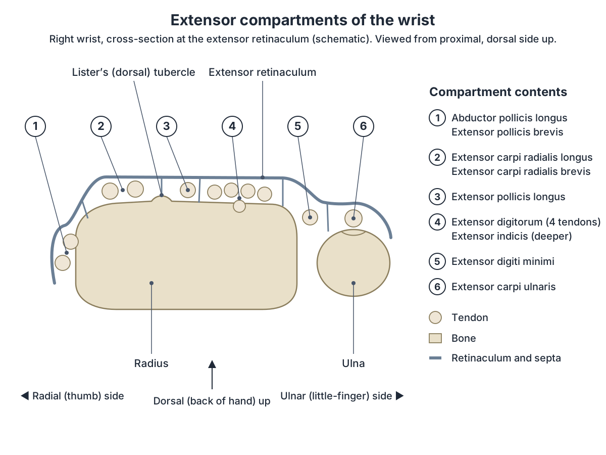
Extensor compartments of the wrist
At the wrist, the tendons are held down by bands of fascia called retinacula. The flexor retinaculum lies over the palmar side of the wrist and the extensor retinaculum over the back. (OpenStax Anatomy and Physiology 2e, 11.5: Muscles of the Pectoral Girdle and Upper Limbs.)
Under the extensor retinaculum the extensor tendons run in six separate tunnels, the dorsal extensor compartments, numbered from the radial to the ulnar side: (1) abductor pollicis longus and extensor pollicis brevis; (2) extensor carpi radialis longus and brevis; (3) extensor pollicis longus; (4) extensor digitorum and extensor indicis; (5) extensor digiti minimi; (6) extensor carpi ulnaris. Lister's tubercle on the radius separates compartments 2 and 3. (Radiopaedia: Extensor compartments of the wrist.)
On the front of the wrist, nine flexor tendons pass through the carpal tunnel under the flexor retinaculum: the single tendon of flexor pollicis longus and the four tendons each of flexor digitorum profundus and flexor digitorum superficialis. The median nerve passes through with them. (TeachMeAnatomy: The Carpal Tunnel.)
Which nerves supply the forearm muscles?
The anterior forearm muscles are supplied by the median nerve, with two exceptions: flexor carpi ulnaris and the medial half of flexor digitorum profundus, which are supplied by the ulnar nerve. All posterior forearm muscles are supplied by the radial nerve. The ulnar and radial arteries supply the anterior compartment. (TeachMeAnatomy: Muscles in the Anterior Compartment of the Forearm.)
The median nerve enters the forearm through the cubital fossa, passes between the two heads of pronator teres and then runs between flexor digitorum superficialis and flexor digitorum profundus. Its anterior interosseous branch supplies the three deep flexors: flexor pollicis longus, pronator quadratus and the lateral half of flexor digitorum profundus. (TeachMeAnatomy: The Median Nerve.)
The ulnar nerve (C8 and T1) supplies only one and a half forearm muscles, flexor carpi ulnaris and the medial half of flexor digitorum profundus, before continuing into the hand, where its deep branch supplies the hypothenar muscles, the interossei, the medial two lumbricals and adductor pollicis. (TeachMeAnatomy: The Ulnar Nerve.)
The radial nerve supplies brachioradialis and extensor carpi radialis longus before it divides near the elbow into a superficial branch (sensory, to the back of the hand) and a deep branch (motor). The deep branch supplies the remaining posterior forearm muscles and becomes the posterior interosseous nerve after it pierces supinator. (TeachMeAnatomy: The Radial Nerve.)
Which muscles pronate and supinate the forearm?
Pronation turns the palm down (or back) and supination turns it up (or forward). OpenStax names two pronators, pronator teres and pronator quadratus, and supinator as the muscle that turns the forearm so the palm faces forward. (OpenStax Anatomy and Physiology 2e, 11.5: Muscles of the Pectoral Girdle and Upper Limbs.)
Brachioradialis is an elbow flexor that also assists supination. TeachMeAnatomy notes that it is most visible when the forearm is half pronated (thumb up) and the elbow is flexed against resistance, as when lifting a heavy mug. (TeachMeAnatomy: Muscles in the Posterior Compartment of the Forearm.)
Tennis elbow, wrist drop and other forearm problems
Lateral epicondylitis (tennis elbow) is a painful overuse condition at the lateral epicondyle of the elbow. A 2022 review describes it as caused by repetitive microtrauma and overload of extensor carpi radialis brevis, states that it affects about 1 to 3% of the population, and notes that it usually affects adults aged 30 to 50 with no difference between men and women. (Karabinov V, Georgiev GP. Lateral epicondylitis: New trends and challenges in treatment. World J Orthop. 2022.)
Radial nerve injury above the elbow paralyses the extensors and causes wrist drop: the wrist hangs in flexion because the flexors, supplied by the median and ulnar nerves, are unopposed. The common sites are the axilla and the radial groove of the humerus, as in a fracture of the humeral shaft. (TeachMeAnatomy: Muscles in the Posterior Compartment of the Forearm.)
An injury to the deep branch or posterior interosseous nerve, for example with a radial head fracture, weakens most of the posterior forearm muscles but does not cause wrist drop, because extensor carpi radialis longus is supplied before the radial nerve divides. Sensation is preserved because the superficial branch is spared. (TeachMeAnatomy: The Radial Nerve.)
Median nerve injury at the elbow, for example in a supracondylar fracture, paralyses the forearm flexors and pronators, weakens wrist flexion and produces the 'hand of benediction' posture. (TeachMeAnatomy: The Median Nerve.)
Supracondylar fractures of the humerus, common in children after a fall on an outstretched hand, can also injure the anterior interosseous nerve. Its function is checked with the 'OK' sign, which tests flexor pollicis longus. (TeachMeAnatomy: The Cubital Fossa.)
Carpal tunnel syndrome is compression of the median nerve in the carpal tunnel. It causes numbness, tingling and pain in the median nerve territory of the hand, often worse at night, and if untreated can lead to weakness and wasting of the thenar muscles at the base of the thumb. (TeachMeAnatomy: The Carpal Tunnel.)
Forearm muscles in the 3D model
- Abductor pollicis longus
- A deep forearm extensor that forms the lateral border of the anatomical snuffbox.
- Anconeus
- A small triangular muscle on the posterior elbow.
- Brachioradialis
- An elbow flexor that forms the lateral border of the cubital fossa; unusually for a flexor, it is supplied by the radial nerve.
- Extensor carpi radialis brevis
- A wrist extensor in the posterior forearm.
- Extensor carpi radialis longus
- A wrist extensor lying just distal to brachioradialis.
- Extensor carpi ulnaris
- A wrist extensor on the ulnar side of the posterior forearm.
- Extensor digiti minimi
- An extensor of the little finger.
- Extensor digitorum
- The main extensor of the fingers.
- Extensor indicis
- A deep forearm muscle giving the index finger independent extension.
- Extensor pollicis brevis
- A deep thumb extensor forming the lateral border of the anatomical snuffbox.
- Extensor pollicis longus
- A thumb extensor whose tendon turns around the dorsal tubercle of the radius.
- Flexor carpi radialis
- A superficial anterior forearm flexor lateral to palmaris longus.
- Flexor digitorum profundus
- A deep forearm flexor that bends the distal phalanges.
- Flexor digitorum superficialis
- An intermediate-layer forearm flexor of the fingers.
- Flexor pollicis longus
- A deep forearm flexor of the thumb.
- Humeral head of flexor carpi ulnaris
- The humeral origin of flexor carpi ulnaris, the ulnar-side wrist flexor.
- Humeral head of pronator teres
- The larger, superficial head of pronator teres.
- Palmaris longus
- A slender forearm flexor with a short belly and a long tendon that fans into the palm.
- Pronator quadratus
- A square deep muscle on the front of the distal forearm.
- Supinator
- A deep forearm muscle wrapping around the upper radius.
- Ulnar head of flexor carpi ulnaris
- The ulnar origin of flexor carpi ulnaris.
- Ulnar head of pronator teres
- The smaller ulnar head of pronator teres.
Frequently asked questions
How many muscles are in the forearm?
Counting the scheme used on this page, there are 20 forearm muscles: 8 in the anterior compartment (4 superficial, 1 intermediate, 3 deep) and 12 in the posterior compartment (7 superficial, including brachioradialis and anconeus, and 5 deep). Some textbooks count brachioradialis and anconeus as elbow muscles instead, giving 18.
What are the anterior forearm muscles?
The anterior forearm muscles are pronator teres, flexor carpi radialis, palmaris longus and flexor carpi ulnaris (superficial layer), flexor digitorum superficialis (intermediate layer), and flexor digitorum profundus, flexor pollicis longus and pronator quadratus (deep layer). They flex the wrist and fingers and pronate the forearm. Most are supplied by the median nerve.
What are the posterior forearm muscles?
The superficial posterior forearm muscles are brachioradialis, extensor carpi radialis longus and brevis, extensor digitorum, extensor digiti minimi, extensor carpi ulnaris and anconeus. The deep layer is supinator, abductor pollicis longus, extensor pollicis brevis, extensor pollicis longus and extensor indicis. All are supplied by the radial nerve or its branches.
What are the forearm flexor muscles?
The wrist flexors are flexor carpi radialis, flexor carpi ulnaris and palmaris longus. The finger flexors are flexor digitorum superficialis, which bends the middle finger joints, and flexor digitorum profundus, which is the only muscle to bend the fingertip (DIP) joints. Flexor pollicis longus flexes the thumb. All start from the medial epicondyle or the front of the radius and ulna.
What muscle is missing in some people's forearm?
Palmaris longus. It is commonly said to be absent in about 15% of people, but rates in published studies range from 1.5% to 63.9% depending on the population. You can check by touching your thumb to your little finger and flexing the wrist; if present, a thin tendon stands out in the middle of the wrist.
What are the six extensor compartments of the wrist?
From the thumb side: 1, abductor pollicis longus and extensor pollicis brevis; 2, extensor carpi radialis longus and brevis; 3, extensor pollicis longus; 4, extensor digitorum and extensor indicis; 5, extensor digiti minimi; 6, extensor carpi ulnaris. They are tunnels under the extensor retinaculum, and Lister's tubercle separates compartments 2 and 3.
Which forearm muscle is affected in tennis elbow?
Tennis elbow (lateral epicondylitis) affects the common extensor origin on the lateral epicondyle, and extensor carpi radialis brevis is the muscle most often blamed. It results from repeated use of the wrist extensors, affects about 1 to 3% of people and is most common between 30 and 50 years of age.
References
- OpenStax. Anatomy and Physiology 2e, §11.5 Muscles of the Pectoral Girdle and Upper Limbs. Rice University, 2022.
- TeachMeAnatomy. Muscles in the Anterior Compartment of the Forearm.
- TeachMeAnatomy. Muscles in the Posterior Compartment of the Forearm.
- TeachMeAnatomy. The Cubital Fossa.
- TeachMeAnatomy. The Carpal Tunnel.
- TeachMeAnatomy. The Median Nerve.
- TeachMeAnatomy. The Ulnar Nerve.
- TeachMeAnatomy. The Radial Nerve.
- Radiopaedia. Extensor compartments of the wrist.
- University of Washington Department of Radiology. Musculoskeletal Atlas: Pronator teres.
- University of Washington Department of Radiology. Musculoskeletal Atlas: Flexor carpi radialis.
- University of Washington Department of Radiology. Musculoskeletal Atlas: Palmaris longus.
- University of Washington Department of Radiology. Musculoskeletal Atlas: Flexor carpi ulnaris.
- University of Washington Department of Radiology. Musculoskeletal Atlas: Flexor digitorum superficialis.
- University of Washington Department of Radiology. Musculoskeletal Atlas: Flexor digitorum profundus.
- University of Washington Department of Radiology. Musculoskeletal Atlas: Flexor pollicis longus.
- University of Washington Department of Radiology. Musculoskeletal Atlas: Pronator quadratus.
- University of Washington Department of Radiology. Musculoskeletal Atlas: Brachioradialis.
- University of Washington Department of Radiology. Musculoskeletal Atlas: Extensor carpi radialis longus.
- University of Washington Department of Radiology. Musculoskeletal Atlas: Extensor carpi radialis brevis.
- University of Washington Department of Radiology. Musculoskeletal Atlas: Extensor digitorum.
- University of Washington Department of Radiology. Musculoskeletal Atlas: Extensor digiti minimi.
- University of Washington Department of Radiology. Musculoskeletal Atlas: Extensor carpi ulnaris.
- University of Washington Department of Radiology. Musculoskeletal Atlas: Anconeus.
- University of Washington Department of Radiology. Musculoskeletal Atlas: Supinator.
- University of Washington Department of Radiology. Musculoskeletal Atlas: Abductor pollicis longus.
- University of Washington Department of Radiology. Musculoskeletal Atlas: Extensor pollicis brevis.
- University of Washington Department of Radiology. Musculoskeletal Atlas: Extensor pollicis longus.
- University of Washington Department of Radiology. Musculoskeletal Atlas: Extensor indicis.
- Dimitriou I, Katsourakis A, Natsis K, Kostretzis L, Noussios G. Palmaris Longus Muscle's Prevalence in Different Nations and Interesting Anatomical Variations: Review of the Literature. J Clin Med Res. 2015;7(11):825–830.
- Karabinov V, Georgiev GP. Lateral epicondylitis: New trends and challenges in treatment. World J Orthop. 2022;13(4):354–364.
- Mitsuhashi N. et al. (2009). BodyParts3D: 3D structure database for anatomical concepts. Nucleic Acids Research 37, D782–D785. Data © The Database Center for Life Science, CC BY 4.0.
Related anatomy
- BrachioradialisBrachioradialis anatomy: origin, insertion, radial nerve supply, why it flexes the elbow best with the thumb up, its…
- Hand musclesHand muscles anatomy: thenar eminence, hypothenar, adductor pollicis, lumbricals and palmar and dorsal interossei…
- Arm musclesArm muscles labeled in 3D: biceps brachii, brachialis, coracobrachialis, triceps brachii, deltoid and anconeus, with…
- RadiusRadius labeled: head of radius, neck, radial tuberosity, shaft, Lister's tubercle, ulnar notch and styloid process…
- UlnaUlna labeled: olecranon, trochlear notch, coronoid process, radial notch, shaft, head and styloid process, how the…
- Upper limb nervesBrachial plexus diagram with roots, trunks, cords and branches; axillary, musculocutaneous, median, ulnar and radial…
Explore the whole body in the interactive 3D human anatomy model, or browse the A–Z list of human structures.